Interviews are more than just a Q&A session—they’re a chance to prove your worth. This blog dives into essential Vascular Medicine interview questions and expert tips to help you align your answers with what hiring managers are looking for. Start preparing to shine!
Questions Asked in Vascular Medicine Interview
Q 1. Describe the pathophysiology of peripheral artery disease (PAD).
Peripheral artery disease (PAD) is a condition where atherosclerosis, the buildup of plaque in the arteries, narrows the arteries supplying blood to the limbs, most commonly the legs and feet. This reduced blood flow leads to symptoms like pain, numbness, and coldness in the affected areas, especially during exertion (claudication).
The pathophysiology involves several steps:
- Atherosclerosis: The initial process is the accumulation of cholesterol, fats, and other substances within the arterial wall, forming plaque. This plaque formation damages the endothelium (inner lining) of the artery, leading to inflammation.
- Inflammation: This inflammation attracts immune cells, further contributing to plaque buildup and thickening of the arterial wall.
- Narrowing of the artery (stenosis): As plaque accumulates, the artery lumen narrows, restricting blood flow.
- Ischemia: This reduced blood flow leads to ischemia (insufficient oxygen and nutrients) in the tissues downstream of the blockage.
- Tissue damage: If the ischemia is severe or prolonged, it can cause tissue damage, potentially leading to critical limb ischemia (CLI) requiring amputation.
Think of it like a clogged pipe – the plaque is the clog, reducing the flow of water (blood) through the pipe (artery).
Q 2. Explain the different types of aneurysms and their management.
Aneurysms are abnormal dilations or bulges in the wall of a blood vessel, typically an artery. Different types exist, primarily categorized by location and shape:
- Abdominal Aortic Aneurysm (AAA): A bulge in the aorta, the body’s largest artery, in the abdomen. This is the most common type and often asymptomatic until it ruptures, which is a life-threatening emergency.
- Thoracic Aortic Aneurysm (TAA): A bulge in the aorta located in the chest. TAAs can affect the ascending, arch, or descending aorta, each with different implications.
- Cerebral Aneurysm: A bulge in an artery in the brain. These aneurysms can rupture, causing a devastating subarachnoid hemorrhage (bleeding into the space surrounding the brain).
- Peripheral Aneurysm: Aneurysms in arteries of the limbs, typically the popliteal artery (behind the knee).
Management varies greatly depending on the type, size, and location of the aneurysm. Small, asymptomatic aneurysms may be monitored with regular imaging (ultrasound, CT scan). Larger aneurysms, particularly those at risk of rupture, may require surgical repair or endovascular intervention (stenting). For example, a large AAA is typically treated surgically to prevent rupture, while smaller AAAs might be monitored closely.
Q 3. What are the risk factors for deep vein thrombosis (DVT)?
Deep vein thrombosis (DVT) is the formation of a blood clot (thrombus) in a deep vein, usually in the leg. Several factors increase the risk:
- Virchow’s Triad: This classic framework summarizes the three primary contributors: stasis (slow blood flow), hypercoagulability (increased tendency for clotting), and endothelial injury (damage to the inner lining of the vein).
- Inherited clotting disorders: Genetic conditions that increase the risk of blood clot formation.
- Trauma or surgery: Injury or surgery can damage veins and promote clotting.
- Prolonged immobilization: Extended periods of immobility, such as long flights or bed rest, can slow blood flow and increase the risk of DVT.
- Cancer: Certain cancers and their treatments can increase clotting tendency.
- Pregnancy and postpartum period: Hormonal changes during pregnancy and the postpartum period increase the risk.
- Oral contraceptives: Some oral contraceptives can increase the risk of DVT.
- Obesity: Obesity is associated with increased risk of DVT.
- Age: The risk of DVT generally increases with age.
Imagine a slow-moving river (stasis); if the riverbed is damaged (endothelial injury) and the water is thick (hypercoagulability), a logjam (blood clot) is more likely to form.
Q 4. Discuss the diagnostic modalities used in evaluating vascular disease.
Evaluating vascular disease requires a multimodal approach, using various diagnostic tools:
- Doppler ultrasound: A non-invasive technique to assess blood flow in arteries and veins, identifying stenosis, aneurysms, and thrombi.
- Angiography: An invasive procedure involving injecting contrast dye into blood vessels to visualize them using X-rays. This provides detailed images of arteries and allows for intervention (angioplasty, stenting).
- Computed tomography angiography (CTA): A non-invasive imaging technique using CT scans and contrast dye to visualize blood vessels, offering a less invasive alternative to traditional angiography.
- Magnetic resonance angiography (MRA): A non-invasive imaging technique using MRI and contrast dye to visualize blood vessels, useful for certain applications where CTA is not suitable.
- Duplex ultrasound: Combines anatomical imaging with Doppler assessment of blood flow, providing comprehensive information about vessel structure and function.
- Ankle-brachial index (ABI): A non-invasive test comparing blood pressure in the ankle to blood pressure in the arm, useful in screening for PAD.
The choice of modality depends on the clinical question, patient factors, and availability of resources.
Q 5. Explain the principles of Doppler ultrasound in vascular assessment.
Doppler ultrasound uses sound waves to measure blood flow velocity within blood vessels. A handheld transducer emits ultrasound waves that reflect off moving red blood cells. The frequency shift of the reflected waves (Doppler effect) is proportional to the velocity of blood flow.
Principles:
- Doppler effect: The change in frequency of a wave due to the motion of the source or observer. Faster blood flow leads to a greater frequency shift.
- Spectral Doppler: Displays blood flow velocity as a waveform over time, providing information about flow direction, velocity, and presence of turbulence (indicating stenosis).
- Color Doppler: Displays blood flow direction and velocity as color-coded images on a grayscale ultrasound image, providing a visual representation of flow patterns.
For example, a narrowed artery will show a high-velocity jet of blood flow in the stenosis region. This is crucial for identifying arterial blockages, monitoring the effect of interventions, and assessing venous flow in DVT.
Q 6. What are the indications for angioplasty and stenting?
Angioplasty and stenting are endovascular procedures used to treat narrowed or blocked arteries. Angioplasty involves inflating a balloon catheter within the narrowed artery to widen the lumen. Stenting involves placing a small mesh tube (stent) within the artery to keep it open after angioplasty.
Indications for angioplasty and stenting include:
- Symptomatic PAD: Patients experiencing claudication or critical limb ischemia due to PAD.
- Coronary artery disease (CAD): Narrowing of the coronary arteries, leading to reduced blood flow to the heart.
- Carotid artery stenosis: Narrowing of the carotid arteries in the neck, increasing the risk of stroke.
- Renal artery stenosis: Narrowing of the renal arteries, affecting kidney function.
- Aneurysms: In some cases, endovascular stenting can be used to treat aneurysms.
The decision to proceed with angioplasty and stenting is based on several factors, including symptom severity, lesion location and characteristics, and patient risk profile.
Q 7. Describe the complications associated with endovascular procedures.
Endovascular procedures, while minimally invasive, carry potential complications:
- Bleeding or hematoma at the puncture site: The site where the catheter is inserted can bleed.
- Arterial dissection: A tear in the arterial wall can occur during catheterization or balloon inflation.
- Thrombosis (blood clot formation): A clot can form at the site of the stent, potentially blocking blood flow.
- Stent thrombosis: A clot forming within the stent itself.
- Embolization: Dislodged plaque or thrombus can travel downstream and block smaller arteries.
- Allergic reaction to contrast dye: Some patients may have allergic reactions to the contrast dye used in the procedure.
- Infection: Infection at the puncture site or elsewhere is a potential risk.
- Renal failure: Contrast dye can sometimes damage the kidneys, particularly in patients with pre-existing kidney disease.
These complications are relatively rare but need to be carefully considered when planning and performing endovascular procedures. Careful patient selection, meticulous technique, and close post-procedure monitoring can help minimize risks.
Q 8. How do you manage a patient with acute limb ischemia?
Acute limb ischemia (ALI) is a critical condition where blood flow to a limb is severely reduced, threatening tissue viability. Management hinges on rapid assessment and intervention. Think of it like a sudden blockage in a crucial water pipe – the downstream areas quickly run dry.
The first step is immediate assessment of the six Ps: Pain, Pallor, Pulselessness, Paresthesia (numbness or tingling), Paralysis, and Poikilothermia (coldness). We’ll also check for capillary refill time. Then, we need to determine the cause – is it an embolism (a clot traveling from elsewhere), thrombosis (a clot forming in the artery itself), or trauma?
- Revascularization is paramount. This could involve surgical procedures like embolectomy (removing the clot surgically) or angioplasty (using a balloon catheter to open the blocked artery), possibly followed by stent placement to keep the artery open. Thrombolysis (dissolving the clot with medication) is also an option, but carries significant risk.
- Anticoagulation is vital to prevent further clotting. Heparin, a fast-acting anticoagulant, is usually started immediately, followed by longer-term anticoagulation with warfarin or a direct oral anticoagulant (DOAC) after the acute phase.
- Pain management is crucial. Opioids may be necessary.
- Monitoring is continuous, including frequent assessments of the limb’s perfusion and overall patient status. We’ll closely monitor vital signs, looking for any signs of deterioration.
For instance, I recently managed a patient with ALI due to a cardiac embolism. After a rapid assessment confirming the six Ps, we performed an emergency embolectomy, followed by IV heparin, leading to a full recovery of limb function. Every minute counts in ALI, and swift action can be the difference between saving a limb and amputation.
Q 9. Explain the role of anticoagulation in venous thromboembolism.
Anticoagulation plays a critical role in venous thromboembolism (VTE), which encompasses deep vein thrombosis (DVT) and pulmonary embolism (PE). Think of it like preventing further damming up of a partially blocked vein. The goal is to inhibit the formation and extension of blood clots, thereby reducing the risk of complications.
Anticoagulation is usually achieved through the use of heparin (initially, for its rapid onset of action) followed by warfarin or a DOAC for long-term prophylaxis. The choice of anticoagulant depends on various factors, including patient-specific comorbidities, such as bleeding risk.
- Heparin: Rapid acting, frequently used initially to quickly reduce clotting risk. It’s usually given intravenously or subcutaneously.
- Warfarin: Acts by inhibiting vitamin K-dependent clotting factors. Requires regular monitoring of the international normalized ratio (INR) to ensure appropriate therapeutic range.
- Direct Oral Anticoagulants (DOACs): These are newer medications with a more predictable anticoagulant effect and less need for monitoring. Examples include apixaban, rivaroxaban, dabigatran, and edoxaban.
The duration of anticoagulation depends on the risk factors and the clinical presentation. For example, a patient with a proximal DVT will usually receive anticoagulation for at least three months, whereas those with recurrent VTE may require lifelong anticoagulation.
Failure to adequately anticoagulate can lead to potentially fatal complications, such as pulmonary embolism, which is why careful selection and monitoring of anticoagulants are crucial in VTE management.
Q 10. Discuss the various treatment options for varicose veins.
Treatment options for varicose veins vary depending on the severity of symptoms and the patient’s preferences. Think of varicose veins as overloaded, swollen veins that have lost their structural integrity. Treatments aim to improve venous flow and alleviate symptoms like pain, swelling, and cosmetic concerns.
- Lifestyle modifications: Weight management, regular exercise, and elevation of the legs can help improve symptoms.
- Compression stockings: These provide support and improve venous return.
- Sclerotherapy: Injection of a sclerosing agent into the vein to cause it to collapse and close.
- Endovenous ablation: Uses heat (radiofrequency or laser energy) to close the affected vein.
- Phlebectomy: A minimally invasive procedure to remove the varicose veins through small incisions.
- Surgical ligation and stripping: A more invasive procedure that involves tying off and removing the affected vein. Less commonly performed now due to minimally invasive alternatives.
The selection of the most appropriate treatment is based on a thorough assessment of the patient’s individual circumstances and the specific characteristics of their varicose veins. For example, a patient with small, superficial varicose veins might benefit from sclerotherapy, whereas a patient with large, symptomatic varicose veins might require endovenous ablation or even surgery.
Q 11. What are the contraindications for thrombolysis?
Thrombolysis, the process of dissolving blood clots using medications like tissue plasminogen activator (tPA), is a powerful technique, but it carries significant risks. It’s essential to carefully weigh the potential benefits against these risks before proceeding.
- Active bleeding: Thrombolysis should be avoided in patients with active bleeding or a high risk of bleeding. This is a major contraindication because it could lead to dangerous or life-threatening bleeding.
- Recent surgery or trauma: The increased risk of bleeding in the post-operative or post-trauma period makes thrombolysis risky.
- Uncontrolled hypertension: High blood pressure can increase the risk of bleeding complications.
- Recent stroke: Thrombolysis carries a significant risk of intracranial hemorrhage in patients who have recently experienced a stroke. This risk is exceptionally high within 3-4.5 hours of the onset of stroke symptoms.
- Aortic dissection or aneurysm: Thrombolysis in this situation can cause catastrophic outcomes due to the already compromised vessel integrity.
- Pregnancy: Increased risk of bleeding.
Prior to administering thrombolysis, a careful assessment of the patient’s medical history and current condition is critical to minimize the risks. A meticulous cost-benefit analysis needs to be performed, weighing the risk of bleeding against the potential benefits of restoring blood flow to the ischemic tissue. A detailed evaluation of possible alternative treatments should also be conducted.
Q 12. Describe the different types of vascular grafts.
Vascular grafts are artificial tubes used to replace or bypass diseased or damaged blood vessels. Think of them as plumbing replacements for the body’s circulatory system. They come in various types, each with its own advantages and disadvantages.
- Autologous vein grafts: These are harvested from the patient’s own veins (usually the saphenous vein). They are considered the gold standard because they have excellent long-term patency (they stay open).
- Synthetic grafts (polytetrafluoroethylene or PTFE, Dacron): These are artificial materials used to construct grafts. They’re often used when autologous veins are unavailable or unsuitable. However, they have a higher risk of thrombosis (clotting).
- Cryopreserved allografts: These are grafts harvested from human donors and preserved using cryopreservation techniques. They offer an alternative when autologous veins are unsuitable but may have higher rates of failure than autologous grafts.
The choice of graft material depends on various factors, including the location and diameter of the vessel to be replaced, patient’s overall health, and the availability of autologous vein. For instance, a small-diameter bypass might benefit from an autologous vein graft for optimal long-term results, while a large-diameter graft might utilize synthetic material due to the scarcity of appropriate autologous veins.
Q 13. Explain the principles of hemostasis during vascular surgery.
Hemostasis, the process of stopping bleeding, is fundamental to successful vascular surgery. Failure to achieve adequate hemostasis can lead to significant complications, including hemorrhage, hematoma formation, and even death. It’s essential to think of it like managing a critical plumbing leak.
Principles of hemostasis in vascular surgery involve a multi-pronged approach:
- Mechanical hemostasis: Direct pressure, surgical ligation (tying off vessels), and the use of vascular clips to close vessels.
- Pharmacological hemostasis: Use of topical hemostatic agents (e.g., thrombin, collagen sponges), and systemic administration of antifibrinolytic medications (e.g., tranexamic acid) to help prevent excessive bleeding.
- Electrocautery: Uses electrical energy to seal off small bleeding vessels.
- Vascular sealant: Special glues or sealants are used to close small vessels.
- Careful surgical technique: Minimizing tissue trauma during the procedure reduces bleeding.
The specific techniques used depend on the nature and extent of the bleeding, the location of the bleeding, and the surgical approach. For instance, direct pressure might suffice for minor bleeding while larger vessels require more robust techniques like ligation or the use of vascular clips. Meticulous attention to hemostasis is crucial for minimizing post-operative complications and ensuring patient safety.
Q 14. How do you interpret an arteriogram?
Interpreting an arteriogram, a type of angiogram focusing on the arteries, requires a systematic approach. It’s like reading a detailed map of the body’s arterial network. The goal is to identify areas of stenosis (narrowing), occlusion (blockage), aneurysm (bulging), or other abnormalities.
The interpretation involves analyzing several aspects:
- Vessel caliber: Assessment of the diameter of the vessels – is it normal, narrowed, or dilated?
- Vessel patency: Are there any blockages or areas of restricted flow?
- Collateral circulation: Are there alternative pathways for blood flow around a blocked area?
- Presence of aneurysms or dissections: Look for abnormal bulges or tears in the vessel wall.
- Vascular morphology: Assessment of the overall shape and structure of the arteries.
Advanced techniques such as digital subtraction angiography (DSA) allow for a clearer view of the vessels and enhance the diagnostic accuracy. For example, a significant stenosis in the left anterior descending coronary artery might be clearly visible on an arteriogram, which could lead to a timely intervention, such as angioplasty or stenting, to prevent a heart attack.
This requires extensive training and experience. Often a team effort, radiologists and vascular surgeons collaborate to accurately interpret the images and develop the best treatment plan.
Q 15. What are the signs and symptoms of venous insufficiency?
Venous insufficiency, essentially meaning the veins aren’t working efficiently, presents with a range of symptoms depending on the severity. Early stages might be subtle, with just aching, heaviness, or fatigue in the legs, particularly after prolonged standing or sitting. As the condition progresses, more noticeable signs appear.
- Leg Swelling (Edema): Fluid buildup in the legs and ankles is a common hallmark. Imagine a water balloon – the veins can’t efficiently return the blood to the heart, causing pooling.
- Varicose Veins: These are enlarged, twisted veins, often appearing blue or dark purple, usually visible just under the skin. They’re essentially the veins’ way of saying, ‘I’m overloaded!’
- Skin Changes: The skin on the legs can become dry, itchy, discolored (brownish or reddish), and thickened. In advanced cases, it might become fragile and prone to ulceration.
- Leg Pain: Pain can range from mild aching to severe, throbbing discomfort, often worse at the end of the day or after prolonged periods of standing. It can feel like a constant pressure.
- Leg Cramps: Night-time leg cramps are also a frequent complaint.
- Lipodermatosclerosis: This is a hardening and thickening of the skin due to chronic inflammation associated with venous insufficiency. It’s more severe and can significantly impact the patient’s quality of life.
It’s important to note that these symptoms can overlap with other conditions, so a proper diagnosis from a healthcare professional is crucial. Early intervention is key to preventing serious complications.
Career Expert Tips:
- Ace those interviews! Prepare effectively by reviewing the Top 50 Most Common Interview Questions on ResumeGemini.
- Navigate your job search with confidence! Explore a wide range of Career Tips on ResumeGemini. Learn about common challenges and recommendations to overcome them.
- Craft the perfect resume! Master the Art of Resume Writing with ResumeGemini’s guide. Showcase your unique qualifications and achievements effectively.
- Don’t miss out on holiday savings! Build your dream resume with ResumeGemini’s ATS optimized templates.
Q 16. Discuss the management of post-operative vascular complications.
Managing post-operative vascular complications requires a multi-faceted approach, focusing on early detection and prompt intervention. Complications can range from bleeding and hematoma formation at the surgical site to thrombosis (blood clot formation) in the veins or arteries. The specific management strategy will depend on the nature and severity of the complication.
- Hematoma Management: Small hematomas often resolve spontaneously. Larger ones might require aspiration (removal of the blood using a needle) or surgical evacuation to prevent pressure on surrounding structures and to reduce the risk of infection.
- Thrombosis Prevention and Treatment: Prophylactic measures like compression stockings, early ambulation (walking), and medications like anticoagulants (blood thinners) are crucial to prevent DVT (deep vein thrombosis). If a thrombus develops, treatment might involve anticoagulants, thrombolysis (dissolving the clot with medication), or even surgical intervention depending on the location and severity. We use imaging such as ultrasound to diagnose and monitor this effectively.
- Bleeding Control: Immediate pressure is the first step in managing post-operative bleeding. Surgical intervention may be necessary for uncontrolled bleeding. Often, we’ll use local compression dressings in conjunction with systemic measures.
- Infection Prevention and Management: Strict sterile technique during surgery, good wound care post-operatively and prompt treatment with antibiotics in the event of an infection is absolutely vital. We use cultures to guide antibiotic therapy and ensure the correct antibiotic is selected.
- Monitoring: Close monitoring of vital signs, including blood pressure and heart rate, and regular examination of the surgical site are essential to detect complications early. We commonly use Doppler ultrasound to evaluate blood flow post-operatively, especially in peripheral vascular procedures.
A multidisciplinary approach, involving surgeons, vascular specialists, and nurses, is critical for successful management of post-operative vascular complications. Early recognition, rapid assessment and prompt intervention are crucial to improve patient outcomes and reduce morbidity and mortality.
Q 17. Explain the role of vascular access in hemodialysis.
Vascular access is paramount in hemodialysis, providing the lifeline for removing waste products and excess fluid from the blood of patients with kidney failure. Efficient and reliable vascular access is crucial for ensuring adequate dialysis, maintaining patient health and preventing complications.
The access point needs to withstand repeated needle punctures over long periods without causing excessive damage or clotting. The aim is to create a robust and durable pathway for efficient blood flow into and out of the dialysis machine.
The location of the vascular access is strategically chosen to ensure good blood flow and to minimize potential complications. Careful consideration is given to the patient’s overall vascular health and the location that will ensure long term patency of the access.
Q 18. Describe the different types of vascular access devices.
Several types of vascular access devices are used in hemodialysis, each with its own advantages and disadvantages. The choice depends on factors like patient health, vein characteristics, and lifestyle.
- AV Fistula: This is considered the gold standard. It’s a surgically created connection between an artery and a vein, usually in the forearm or upper arm. The fistula matures over several weeks before it’s ready for use, allowing the vein to naturally strengthen and enlarge, making it ideal for repeated needle punctures.
- AV Graft: If a patient’s own veins aren’t suitable for fistula creation, a synthetic graft (a tube) is used to connect an artery and a vein. Graft patency tends to be shorter than fistulas, but it offers quicker access to dialysis than the native fistula.
- Central Venous Catheter (CVC): This is a temporary option, inserted into a large vein, typically in the neck, chest, or groin. It’s used as a short-term solution while awaiting fistula maturation or in cases of emergency dialysis needs. However, CVCs are associated with a higher risk of infection compared to fistulas and grafts.
Careful patient selection and consideration of individual factors are essential for choosing the optimal vascular access device to minimize complications and maximize dialysis effectiveness.
Q 19. What are the complications associated with vascular access devices?
Vascular access devices, while vital for hemodialysis, are prone to various complications that can compromise dialysis effectiveness and patient health. These can range from minor annoyances to life-threatening situations.
- Infection: Infection is a major concern, particularly with CVCs, potentially leading to bloodstream infections (bacteremia) and sepsis. It’s crucial for patients to maintain meticulous hygiene and to recognize signs of infection such as redness, swelling, tenderness, or purulent drainage.
- Thrombosis: Blood clots can form within the access, blocking blood flow. This can lead to loss of access function and necessitate intervention to restore patency (clearing the clot).
- Stenosis: Narrowing of the access due to scar tissue formation can impede blood flow. This can necessitate interventions such as angioplasty (ballooning) or stenting (placing a small mesh tube) to maintain adequate blood flow.
- Pseudoaneurysm: A false aneurysm (a bulging of the blood vessel wall) can form at the puncture site, often due to repeated needle punctures, potentially leading to rupture and hemorrhage.
- Bleeding: Bleeding at the puncture site can occur, ranging from minor oozing to more significant bleeds, especially in patients with clotting disorders. This necessitates prompt pressure to control the bleed.
- Aneurysm: A true aneurysm is a weakening of the blood vessel wall, which may occur at the site of the vascular access, which can rupture and lead to life threatening hemorrhage.
Regular monitoring of access patency, diligent hygiene practices, and prompt attention to any signs of complications are essential in preventing and managing these risks.
Q 20. How do you manage a patient with a bleeding vascular access site?
Managing a bleeding vascular access site requires prompt action to control the bleeding and prevent further complications. The initial steps depend on the severity of the bleed.
- Assess the Bleeding: Determine the rate and amount of bleeding. Minor oozing can often be managed with direct pressure. More significant bleeding requires immediate intervention.
- Direct Pressure: Apply firm, direct pressure to the bleeding site using a sterile gauze pad for at least 15-20 minutes. Do not interrupt pressure to check the bleeding site.
- Elevation: Elevating the affected limb can help reduce blood flow to the area and aid in clotting.
- Ice Pack: Applying an ice pack to the area can help constrict blood vessels and reduce swelling.
- Medical Attention: If the bleeding doesn’t stop with direct pressure or is significant (e.g., soaking through dressings, spurting blood), immediately seek medical attention. This might involve surgical repair, angiographic embolization (blocking the bleeding vessel), or other interventions depending on the cause and location of the bleed.
- Monitor for Signs of Shock: Watch for signs of hypovolemic shock (low blood volume) such as rapid heart rate, low blood pressure, and dizziness. This is especially crucial for extensive bleeds.
The specific management approach will be tailored to the individual patient, considering factors such as the type of vascular access, the underlying cause of the bleeding, and the patient’s overall health status.
Q 21. Discuss the use of compression therapy in venous disease.
Compression therapy plays a vital role in the management of venous disease, particularly chronic venous insufficiency (CVI). It works by improving venous return and reducing venous pressure in the legs.
Compression stockings or bandages apply external pressure to the legs, squeezing the veins and promoting blood flow back towards the heart. This helps prevent blood pooling in the lower extremities, reducing edema and alleviating symptoms such as leg pain, heaviness, and swelling. Imagine squeezing a toothpaste tube from the bottom – the pressure pushes the contents upwards.
The level of compression required varies depending on the severity of the venous disease. Different compression classes (e.g., class 1, class 2, class 3) are available, with higher classes providing stronger compression. A healthcare professional will assess the patient and prescribe the appropriate level of compression.
In addition to alleviating symptoms, compression therapy can also help prevent complications associated with CVI, such as skin changes, ulcers, and lipodermatosclerosis. It is crucial that the correct type and class of compression is applied correctly. Improper application can lead to complications such as compartment syndrome.
Regular assessment and monitoring of the patient’s skin are essential during compression therapy. Any signs of skin breakdown or excessive discomfort should be reported to the healthcare professional.
Q 22. Explain the role of lifestyle modifications in preventing vascular disease.
Lifestyle modifications are crucial in preventing vascular disease, primarily by mitigating risk factors like hypertension, dyslipidemia, and diabetes. Think of your arteries as roads; a healthy lifestyle keeps them clear and flowing smoothly, while unhealthy habits lead to blockages and damage.
Diet: A balanced diet low in saturated and trans fats, cholesterol, and sodium is paramount. Think of replacing red meat with lean protein sources like fish and poultry, and loading up on fruits, vegetables, and whole grains. This helps reduce LDL cholesterol (“bad” cholesterol) and blood pressure.
Exercise: Regular physical activity strengthens the heart, improves blood flow, and helps maintain a healthy weight. Aim for at least 150 minutes of moderate-intensity aerobic exercise per week. Imagine your heart as a muscle; regular exercise keeps it strong and efficient.
Smoking Cessation: Smoking is a major risk factor for vascular disease. Nicotine damages blood vessel linings, contributing to atherosclerosis (hardening of the arteries). Quitting smoking is one of the most impactful changes a person can make.
Weight Management: Maintaining a healthy weight reduces strain on the heart and blood vessels. Obesity is strongly linked to hypertension, dyslipidemia, and diabetes – all major contributors to vascular disease. Think of your weight as a stressor on your cardiovascular system; reducing weight alleviates this pressure.
Stress Management: Chronic stress can raise blood pressure and contribute to other risk factors. Techniques like yoga, meditation, or deep breathing can help manage stress levels. Finding healthy coping mechanisms is essential for overall cardiovascular health.
Q 23. What are the current guidelines for the management of abdominal aortic aneurysms?
The management of abdominal aortic aneurysms (AAAs) depends largely on the size of the aneurysm and the patient’s overall health. Guidelines generally recommend close monitoring for smaller aneurysms and surgical intervention for larger ones. Think of an AAA as a weak spot in a major artery; monitoring is like keeping an eye on the weak spot, while surgery is like repairing it.
Screening: Ultrasound screening is recommended for men aged 65-75 who have ever smoked. This helps detect aneurysms early.
Monitoring: Regular ultrasound surveillance is crucial to track the size of the aneurysm. If the aneurysm remains small and stable, monitoring may continue. The frequency of monitoring depends on the rate of growth.
Surgical Intervention: Surgical repair is generally recommended when the aneurysm reaches a certain size (usually 5.5 cm or larger, or smaller if rapidly expanding) or if symptoms develop. Two main approaches exist: Open surgical repair (OSR) and Endovascular aneurysm repair (EVAR).
Risk Factor Management: Managing risk factors like hypertension and smoking is crucial, even with closely monitored aneurysms. This aims to slow down the aneurysm’s growth.
Q 24. Discuss the role of imaging in the diagnosis and management of carotid artery disease.
Imaging plays a pivotal role in diagnosing and managing carotid artery disease (CAD). It helps visualize the degree of stenosis (narrowing) in the carotid arteries, which are major blood vessels supplying the brain. Imagine these arteries as pipes; imaging helps determine the extent of blockage in these pipes.
Carotid Ultrasonography: This is the most commonly used initial imaging modality. It’s non-invasive, relatively inexpensive, and provides real-time images of the carotid arteries, assessing the degree of stenosis and the presence of plaque.
Carotid Angiography: A more invasive procedure involving injecting contrast dye into the carotid arteries. It provides detailed images of the blood vessels, allowing precise assessment of stenosis and the planning of interventions like carotid endarterectomy or stenting.
Computed Tomography Angiography (CTA): A non-invasive technique using CT scans to visualize the carotid arteries. It’s faster than traditional angiography but involves radiation exposure.
Magnetic Resonance Angiography (MRA): A non-invasive technique using MRI to visualize the carotid arteries. It offers excellent image quality without radiation but may be more expensive and time-consuming.
The choice of imaging modality depends on factors such as the clinical presentation, accessibility, and availability of resources. The results guide treatment decisions, including medical management (e.g., antiplatelet therapy), carotid endarterectomy (surgical removal of plaque), or carotid artery stenting (placement of a stent to open the narrowed artery).
Q 25. Explain the different types of peripheral vascular bypass grafts.
Peripheral vascular bypass grafts are surgical procedures used to restore blood flow to the legs and feet in patients with peripheral artery disease (PAD). Imagine creating a detour around a blocked road to restore traffic flow. Different types of grafts utilize various materials and techniques.
Autogenous Vein Grafts: These are the gold standard, using the patient’s own veins (usually from the legs) to create the bypass. Because it’s the patient’s own tissue, the body is less likely to reject it.
Synthetic Grafts (e.g., PTFE, Dacron): These are artificial materials used to create the bypass. While convenient, they have a higher risk of clotting and infection compared to autogenous grafts.
In Situ Vein Grafts: In this technique, the vein is left in its original place, but its connections are surgically altered to create a bypass. It reduces the need for harvesting veins from another site.
Reversed Vein Grafts: The vein is harvested and reversed before being used as a graft, minimizing the risk of valve obstruction.
The choice of graft depends on factors such as the location and length of the blockage, the patient’s overall health, and the availability of suitable veins.
Q 26. Describe the techniques used in endovascular aneurysm repair (EVAR).
Endovascular aneurysm repair (EVAR) is a minimally invasive procedure for treating abdominal aortic aneurysms (AAAs). It involves placing a stent-graft inside the aneurysm to exclude it from the circulation. Think of it as placing a reinforced tube inside the weakened artery to prevent rupture.
Pre-operative Planning: This includes detailed imaging (CTA or MRA) to assess the aneurysm’s anatomy and suitability for EVAR. Careful selection of the appropriate stent-graft is crucial.
Femoral Artery Access: Small incisions are made in the groin to access the femoral arteries. Sheaths (tubes) are inserted to guide the stent-graft.
Stent-graft Deployment: The stent-graft is advanced through the sheath and deployed across the aneurysm neck, sealing it off from the blood flow. Fluoroscopy (real-time X-ray) guides the placement.
Post-operative Monitoring: Regular imaging studies are performed to ensure the stent-graft remains correctly positioned and the aneurysm is excluded from the circulation.
EVAR is less invasive than open surgical repair, resulting in reduced recovery time and lower risk of complications for suitable patients. However, it is not suitable for all patients, and careful patient selection is critical.
Q 27. What are the challenges in managing patients with complex vascular disease?
Managing patients with complex vascular disease presents unique challenges, often requiring a multidisciplinary approach. These patients often have multiple comorbidities and risk factors, making treatment more intricate. It’s like working on a complex machine with multiple interconnected parts.
Comorbidities: Patients frequently have diabetes, heart failure, chronic kidney disease, and other conditions that complicate vascular management. These conditions often impact treatment options and increase risks of complications.
Multiple Lesions: Patients may have blockages in multiple vessels (e.g., both carotid and leg arteries), requiring staged or simultaneous interventions. Determining the optimal sequence of procedures is critical.
Inadequate Run-off Vessels: In peripheral arterial disease, poor run-off vessels (small arteries distal to the blockage) can limit the success of bypass grafts. Alternative strategies may be needed, such as angioplasty and stenting.
High Surgical Risk: Patients with severe comorbidities may have a higher risk of surgical complications, requiring careful risk-benefit assessment of each procedure.
Patient Compliance: Adherence to medication regimens and lifestyle modifications is crucial for long-term success. Patient education and support are essential aspects of management.
Effective management requires collaboration between vascular surgeons, interventional radiologists, cardiologists, nephrologists, and other specialists, as well as close interaction with the patient and their support system.
Q 28. Discuss the future trends in vascular medicine.
Vascular medicine is rapidly evolving, with several promising trends on the horizon. These advancements aim to improve diagnosis, treatment, and patient outcomes.
Minimally Invasive Techniques: The focus will continue to shift towards less invasive procedures like EVAR and transcatheter therapies, leading to shorter recovery times and fewer complications.
Biomaterials and Tissue Engineering: Research into new biomaterials for grafts and stents aims to improve durability and reduce complications. Tissue engineering holds promise for creating living grafts.
Advanced Imaging: Improvements in imaging technologies like CTA, MRA, and molecular imaging will enhance diagnostic accuracy and treatment planning.
Personalized Medicine: Tailoring treatment strategies based on individual patient characteristics (e.g., genetics, comorbidities) will become increasingly important to optimize outcomes.
Artificial Intelligence (AI): AI has the potential to improve diagnostic accuracy, predict disease progression, and optimize treatment strategies.
These advancements promise to significantly improve the diagnosis and treatment of vascular diseases, leading to better patient outcomes and quality of life. The field is constantly innovating, pushing the boundaries of what’s possible.
Key Topics to Learn for Vascular Medicine Interview
- Peripheral Artery Disease (PAD): Understanding the pathophysiology, diagnostic approaches (ankle-brachial index, imaging modalities), and treatment strategies (lifestyle modifications, medical management, interventional procedures).
- Aortic Diseases: Knowledge of aortic aneurysms (abdominal and thoracic), dissections, and their management, including surgical and endovascular interventions. Practical application includes interpreting imaging studies and understanding risk stratification.
- Venous Disorders: Deep vein thrombosis (DVT), pulmonary embolism (PE), chronic venous insufficiency (CVI), and varicose veins. Focus on diagnostic workup (e.g., D-dimer, ultrasound), treatment algorithms (anticoagulation, compression therapy), and risk factor identification.
- Carotid Artery Disease: Understanding the significance of carotid stenosis, its impact on stroke risk, and the management options (medical therapy, carotid endarterectomy, angioplasty with stenting). Problem-solving skills are key here – interpreting ultrasound findings and determining optimal treatment strategies.
- Raynaud’s Phenomenon and other Vasospastic Disorders: Understanding the underlying mechanisms, clinical presentation, and management of these conditions.
- Vascular Access: Knowledge of various vascular access techniques (e.g., central venous catheters, arteriovenous fistulas) and their complications. This includes practical considerations for patient selection and procedural success.
- Non-invasive Vascular Diagnostics: Familiarity with various imaging techniques such as ultrasound, CT angiography, and MRI angiography and their applications in diagnosing vascular diseases.
- Medical Management of Vascular Disease: Thorough understanding of medications used in vascular disease, including antiplatelet agents, anticoagulants, and lipid-lowering drugs.
Next Steps
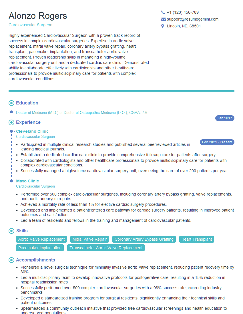
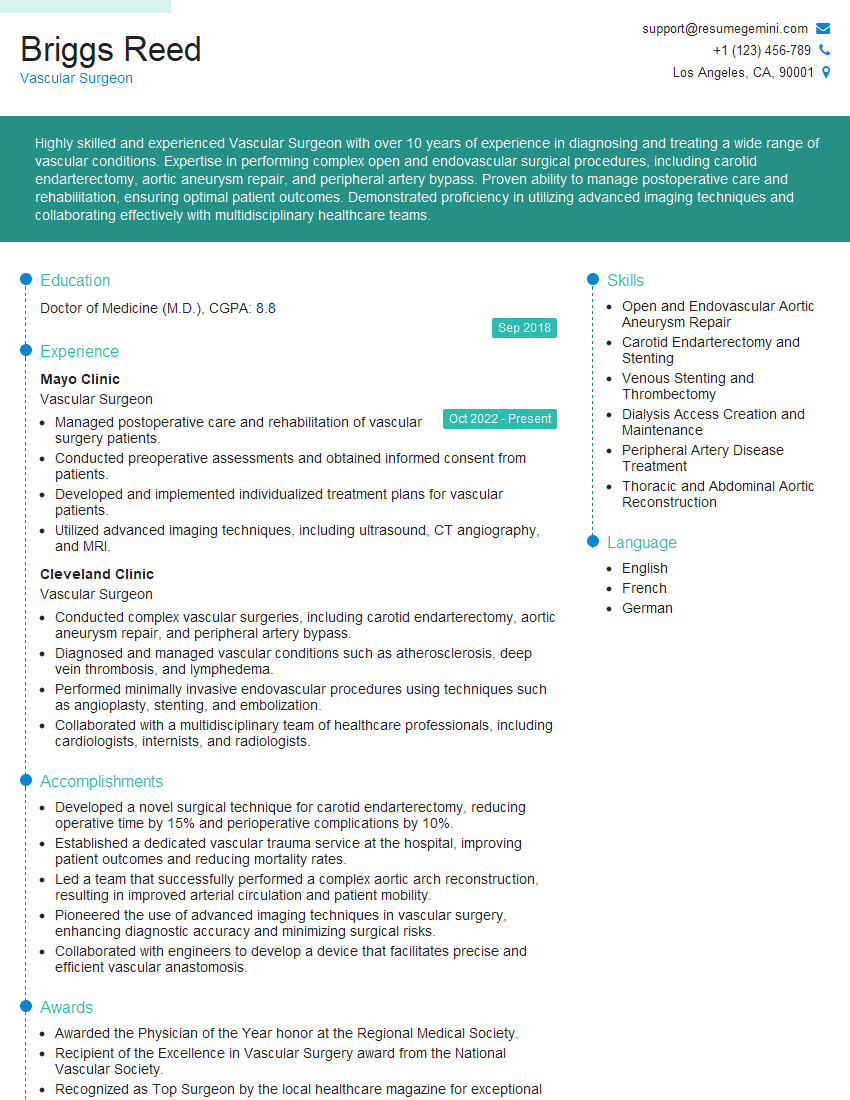
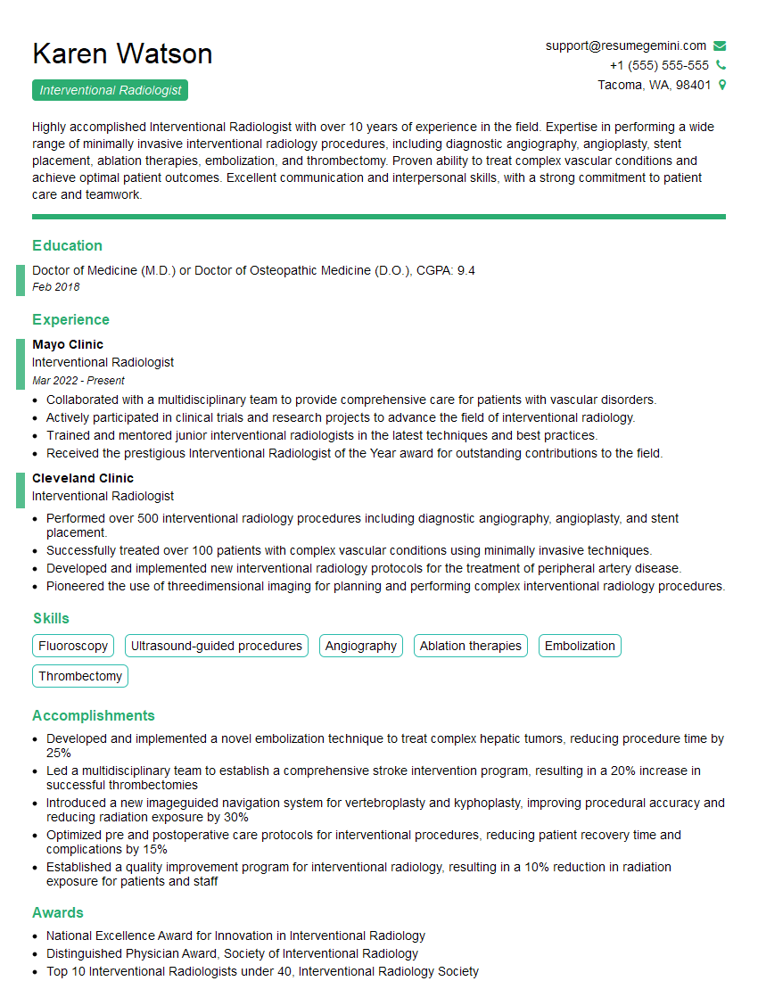
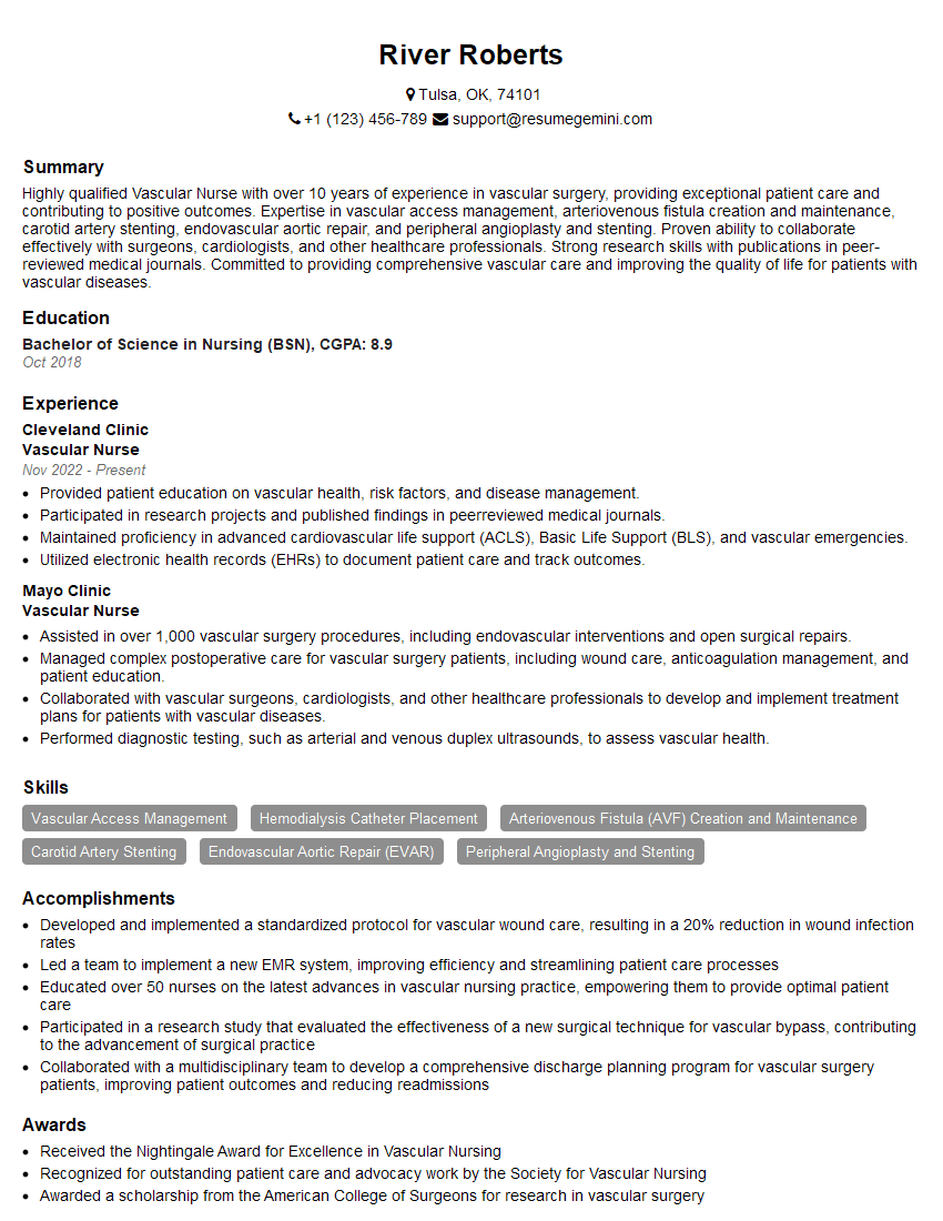
Mastering Vascular Medicine opens doors to a rewarding career with significant impact on patient lives. To enhance your job prospects, creating a strong, ATS-friendly resume is crucial. ResumeGemini is a trusted resource that can help you build a professional and impactful resume tailored to the specific requirements of Vascular Medicine positions. Examples of resumes tailored to Vascular Medicine are available to guide you through the process. Invest the time to craft a compelling resume – it’s your first impression with potential employers.
Explore more articles
Users Rating of Our Blogs
Share Your Experience
We value your feedback! Please rate our content and share your thoughts (optional).
What Readers Say About Our Blog
To the interviewgemini.com Webmaster.
Very helpful and content specific questions to help prepare me for my interview!
Thank you
To the interviewgemini.com Webmaster.
This was kind of a unique content I found around the specialized skills. Very helpful questions and good detailed answers.
Very Helpful blog, thank you Interviewgemini team.