Every successful interview starts with knowing what to expect. In this blog, we’ll take you through the top Biopsy interpretation and reporting interview questions, breaking them down with expert tips to help you deliver impactful answers. Step into your next interview fully prepared and ready to succeed.
Questions Asked in Biopsy interpretation and reporting Interview
Q 1. Describe the process of tissue fixation for optimal biopsy preservation.
Tissue fixation is the crucial first step in biopsy processing, aiming to preserve tissue architecture and cellular components as close as possible to their in-vivo state. This prevents autolysis (self-digestion) and putrefaction, ensuring accurate diagnostic interpretation.
The most common fixative is 10% neutral buffered formalin (NBF). The process involves submerging the tissue specimen in a sufficient volume of NBF (at least 10-20 times the tissue volume) to ensure complete penetration. Fixation time is generally 6-24 hours, depending on the tissue type and size; larger specimens require longer fixation times to ensure complete penetration. Inadequate fixation can lead to artifacts like tissue shrinkage, poor staining quality, and inaccurate diagnostic assessment. Conversely, over-fixation can also cause tissue hardening and loss of antigenicity, impacting immunohistochemical staining results. Proper handling and recording of fixation time is therefore critical for quality assurance in the pathology laboratory.
For example, a small skin biopsy might only need 6-8 hours of fixation, while a large lymph node might require a full 24 hours. Careful attention to the specimen’s size and the fixative’s volume and concentration guarantees optimum preservation.
Q 2. Explain the difference between H&E staining and special stains in biopsy analysis.
H&E staining (hematoxylin and eosin) is a routine staining technique used in virtually all biopsy analyses. Hematoxylin stains the cell nuclei blue/purple, highlighting their morphology and arrangement, while eosin stains the cytoplasm pink/red, providing contrast and allowing for evaluation of cellular differentiation. H&E staining is fundamental for assessing basic tissue architecture and identifying general cellular features.
Special stains, on the other hand, target specific cellular components or structures. They provide supplementary information that complements the H&E staining. Examples include:
- Periodic acid-Schiff (PAS): stains carbohydrates, useful for identifying fungi, glycogen, and mucin.
- Grocott-Gomori methenamine silver (GMS): highlights fungal elements, especially useful in infectious disease diagnostics.
- Immunohistochemistry (IHC): employs antibodies to detect specific proteins, crucial for identifying tumor markers and diagnosing certain cancers.
Imagine H&E as the basic blueprint of a house, showing the overall structure. Special stains are like specialized blueprints showing specific details, such as the plumbing or electrical systems. Both are needed for a complete understanding.
Q 3. What are the key microscopic features of dysplasia in a cervical biopsy?
Cervical dysplasia refers to abnormal cellular changes in the cervix, representing a precancerous condition. Microscopic features of dysplasia include:
- Nuclear atypia: enlarged, hyperchromatic (darkly stained) nuclei with irregular nuclear membranes and prominent nucleoli (structures within the nucleus).
- Increased nuclear-to-cytoplasmic ratio: the nucleus occupies a larger proportion of the cell compared to the cytoplasm.
- Architectural disorder: loss of normal cell arrangement and maturation, with cells showing haphazard organization.
- Mitotic figures: increased number of dividing cells, indicating rapid cellular proliferation.
The severity of dysplasia (mild, moderate, severe) is graded based on the extent of these abnormalities. Severe dysplasia is considered a high-grade lesion, with a significant risk of progressing to cervical carcinoma. The assessment of dysplasia relies on carefully evaluating these microscopic features within the context of the entire biopsy specimen.
Q 4. How would you differentiate between benign and malignant neoplasms in a breast biopsy?
Differentiating between benign and malignant neoplasms in a breast biopsy requires careful analysis of several architectural and cytological features. Benign lesions generally exhibit:
- Well-circumscribed borders: the lesion is clearly separated from the surrounding tissue.
- Uniform cellularity: cells are similar in size, shape, and staining characteristics.
- Low mitotic activity: few dividing cells are present.
- Absence of necrosis: no dead tissue is observed.
Malignant neoplasms, on the other hand, often demonstrate:
- Poorly defined borders: the lesion invades and infiltrates surrounding tissue.
- Pleomorphism: cells vary significantly in size, shape, and staining characteristics.
- High mitotic activity: numerous dividing cells are present.
- Presence of necrosis: areas of dead tissue are frequently observed.
- Nuclear atypia: similar to dysplasia description above but often more pronounced and pleomorphic.
It’s important to note that some lesions may present with overlapping features, requiring experienced pathologists to make the final diagnosis. Immunohistochemical stains can be essential for confirming the diagnosis in ambiguous cases.
Q 5. Interpret this image (showing a microscopic image – would need to be actually included in a real blog post): What is your diagnosis?
(This answer requires the microscopic image to be provided. A detailed interpretation can only be given upon visual examination of the image. The following is a placeholder for the actual diagnosis, which would be provided once the image is available.)
Placeholder Diagnosis: A detailed analysis of the provided microscopic image is needed to render a diagnosis. Features to be examined include cellular morphology, architectural pattern, presence of mitotic figures, inflammatory infiltrate, and any other relevant findings. Once the image is available, a precise diagnosis and justification can be provided.
Q 6. Describe the significance of inflammatory markers in biopsy interpretation.
Inflammatory markers in biopsy interpretation provide essential information about the underlying process driving the lesion. The type and intensity of inflammation can help in differentiating between various conditions, guiding diagnostic and therapeutic decisions.
For instance, a predominantly lymphocytic infiltrate (composed mainly of lymphocytes) might suggest a chronic inflammatory process, potentially associated with autoimmune disorders or certain infections. A neutrophilic infiltrate (rich in neutrophils) might indicate an acute inflammatory reaction, often seen in bacterial infections or acute tissue injury. Eosinophils are associated with allergic reactions or parasitic infections.
The presence and type of inflammatory cells provide valuable context to the overall histological picture. For example, the presence of significant inflammation around a tumor might indicate a host response to the malignancy. Conversely, the absence of a significant inflammatory reaction in a suspicious lesion may raise concerns about the nature of the lesion.
Q 7. How do you approach the interpretation of a biopsy with areas of necrosis?
Necrosis, or the death of tissue, is a significant finding in biopsy interpretation. Its presence warrants careful evaluation and correlation with the other microscopic features to determine the underlying cause and its significance.
The approach to interpreting a biopsy with areas of necrosis includes:
- Assessing the pattern of necrosis: Is it coagulative (cell outlines preserved), liquefactive (cell outlines lost), caseous (cheese-like), or another type? The pattern of necrosis can provide clues about the underlying cause (e.g., ischemia, infection, malignancy).
- Evaluating the surrounding tissue: Examining the tissue immediately adjacent to the necrotic areas is critical. Is there an inflammatory reaction? Are there atypical cells? This helps determine whether the necrosis is secondary to another process, such as inflammation or tumor invasion.
- Considering the clinical context: The patient’s clinical history, imaging findings, and other laboratory results should be considered in conjunction with the histopathological features.
For example, necrosis in a breast biopsy could signify tumor necrosis in a malignant process, ischemic damage due to lack of blood supply, or an infectious process. Careful analysis and integration of all available information are crucial for accurate interpretation.
Q 8. What are the common pitfalls in biopsy interpretation, and how can they be avoided?
Biopsy interpretation, while a crucial diagnostic tool, is prone to several pitfalls. These can range from sampling errors to subtle microscopic features that require extensive experience to correctly identify.
- Sampling error: This is perhaps the most significant pitfall, where the biopsy sample doesn’t accurately represent the entire lesion. For instance, a small biopsy from a large, heterogeneous tumor might miss areas of aggressive growth or important diagnostic features. Avoiding this requires careful selection of the biopsy site, guided by imaging (e.g., ultrasound, CT, MRI) and clinical examination, to obtain a representative sample. Sometimes multiple biopsies are necessary.
- Interpretation bias: Our pre-existing knowledge or expectations can unconsciously influence our interpretation. A clinician strongly suspecting malignancy, for example, might interpret ambiguous findings more aggressively than necessary. Systematic review of the entire slide, careful comparison to control tissues and using standardized reporting templates can mitigate this bias.
- Technical artifacts: Processing artifacts during tissue preparation (e.g., crush artifact, sectioning issues) can mimic pathological findings. Careful examination of the slide and considering the processing history are crucial to avoid misinterpretation.
- Inter-observer variability: Different pathologists might have slightly different interpretations of subtle microscopic features. Standardization of reporting, peer review, and participation in quality assurance programs help to minimize this variability.
By employing rigorous protocols, continuous learning and incorporating advanced techniques such as immunohistochemistry and molecular diagnostics, we can significantly reduce these pitfalls and increase the accuracy of biopsy interpretation.
Q 9. Explain the importance of proper sampling techniques in biopsy procedures.
Proper sampling techniques are paramount in biopsy procedures as they directly impact the accuracy and reliability of the subsequent diagnosis. A poorly obtained sample can lead to an inconclusive or even erroneous result, delaying treatment or causing unnecessary anxiety.
For instance, a needle biopsy targeting a deep-seated lesion requires precise needle placement, guided by imaging, to obtain a representative tissue sample from the target area. Incisional biopsies, which involve removing a small portion of the lesion, require careful selection of the site to capture the most characteristic histological features. Excisional biopsies, aiming to remove the entire lesion, necessitate precise surgical excision to ensure complete removal of the suspected pathology.
Key considerations include:
- Lesion size and location: Choosing the appropriate biopsy type (needle, incisional, excisional) based on the size, location, and accessibility of the lesion.
- Imaging guidance: Using ultrasound, CT, or MRI to guide needle biopsies for precise targeting, especially for deep-seated lesions.
- Adequate tissue sampling: Obtaining a sufficient amount of tissue to allow for thorough microscopic evaluation and ancillary testing (e.g., IHC, molecular studies).
- Proper tissue handling: Using appropriate fixation techniques to preserve the tissue architecture and prevent degradation, ensuring optimal sample quality for assessment.
Ultimately, meticulous attention to detail at each stage of the sampling process is crucial for generating reliable diagnostic information.
Q 10. Describe your experience with different types of biopsies (e.g., needle, incisional, excisional).
Throughout my career, I’ve had extensive experience with various biopsy types. Each has unique challenges and advantages.
- Needle biopsies: These are minimally invasive, ideal for deep-seated lesions. I’ve performed and interpreted numerous fine-needle aspiration (FNA) biopsies for thyroid nodules, lymph node evaluation and masses in organs such as the liver or kidney. The key here is interpreting the cellular material which can be less informative than tissue cores. Core needle biopsies, providing tissue fragments, are often preferred for diagnostic accuracy.
- Incisional biopsies: These are useful when complete excision isn’t immediately feasible or desirable, or when a large lesion needs to be sampled representatively. My experience includes interpreting numerous incisional biopsies from skin lesions, breast masses and GI tract lesions. They offer a larger amount of tissue for assessment but don’t provide complete information about the lesion’s margins.
- Excisional biopsies: These involve the complete removal of the lesion. This offers the most definitive diagnosis as the entire lesion is available for analysis. I regularly interpret excisional biopsies for skin lesions, breast biopsies, and biopsies from superficial organs. The evaluation of margins – the presence or absence of tumor cells at the edges of the resected tissue – is critical in these cases.
My expertise extends to interpreting the results from each type, understanding their limitations, and correlating them with clinical information to provide the most accurate diagnosis.
Q 11. How do you ensure accuracy and consistency in your biopsy reporting?
Maintaining accuracy and consistency in biopsy reporting is crucial for optimal patient care. I achieve this through a multi-faceted approach:
- Standardized reporting templates: I utilize standardized reporting templates that ensure all essential elements are included consistently across all reports. This includes detailed descriptions of the microscopic findings, measurements of lesion size, assessment of margins (if applicable), and a concise, clear diagnosis.
- Quality assurance: I actively participate in internal and external quality assurance programs that help to ensure consistency and identify areas for improvement in interpretation. Regular review of cases with colleagues, especially in complex or unusual cases, helps refine my interpretations.
- Continuing education: Pathology is a field with constant advancements. I actively participate in continuous learning opportunities – attending conferences, workshops, and reviewing recent literature – to maintain my knowledge of the latest diagnostic techniques and guidelines.
- Use of digital pathology: Using digital pathology systems enhances the accuracy and efficiency of my work. They allow for easier image analysis, quantification of features, consultation with specialists, and standardization of measurements.
By adhering to these practices, I strive to produce high-quality, reliable reports that are consistent and informative for clinicians.
Q 12. How would you manage a discrepancy between the clinical findings and your biopsy interpretation?
Discrepancies between clinical findings and biopsy interpretation are not uncommon and necessitate a careful and systematic approach. They often prompt further investigation rather than an immediate dismissal of either finding.
My approach includes:
- Reviewing the clinical history and imaging data: A thorough reassessment of the patient’s clinical history and radiological findings, if available, is the first step. This includes re-examining the imaging to correlate the biopsy site with the lesion.
- Rechecking the biopsy sample and processing: I would scrutinize the biopsy block and slides for adequacy and any potential processing artifacts. If necessary, I might request additional sections or special stains.
- Consultations: Consulting with other pathologists and clinicians is important to gain different perspectives and consider alternative interpretations. This might involve a multidisciplinary meeting involving surgeons, oncologists, or radiologists.
- Further Investigations: In some cases, additional investigations, such as repeat biopsies, different biopsy techniques, or molecular tests, might be necessary to clarify the diagnosis. For example, a negative biopsy in the presence of strong clinical suspicion might warrant a repeat biopsy targeting a different area or using a different technique.
The goal is to arrive at a consensus diagnosis that is consistent with all available information. Open communication with the referring clinician is essential throughout this process, to ensure the patient receives the most appropriate care.
Q 13. Explain your understanding of IHC (immunohistochemistry) and its role in biopsy analysis.
Immunohistochemistry (IHC) is a crucial technique used in biopsy analysis. It involves using antibodies that bind to specific proteins in tissue sections, allowing us to identify and localize these proteins within cells and tissues. This provides valuable diagnostic information that isn’t always evident with standard histological staining.
For example, in diagnosing cancer, IHC helps to determine the type of tumor (e.g., differentiating between different types of lymphoma), assess the aggressiveness of the tumor, and predict its response to specific therapies. In breast cancer, IHC for ER, PR, and HER2 receptors guides treatment decisions. In melanoma diagnosis, S100 and HMB45 are commonly used IHC markers to confirm the diagnosis.
IHC is particularly useful in:
- Tumor classification: Distinguishing between different types of tumors with similar histological features.
- Grading of tumors: Assessing the aggressiveness of tumors based on the expression of specific proteins.
- Predicting response to therapy: Identifying biomarkers that predict the likely response of a tumor to specific treatments.
- Improving diagnostic accuracy: Confirming a diagnosis when histological features are inconclusive.
By incorporating IHC results into the interpretation, I can enhance the diagnostic accuracy and provide clinicians with more comprehensive information for effective patient management.
Q 14. What are the ethical considerations related to biopsy interpretation and reporting?
Ethical considerations in biopsy interpretation and reporting are paramount. Our interpretations directly influence patient care, and it’s crucial to act with integrity, responsibility, and a patient-centered approach.
- Accuracy and objectivity: We must strive for accuracy and objectivity in our interpretations, avoiding bias and ensuring that our reports are based solely on the evidence available.
- Confidentiality: Maintaining patient confidentiality is essential. All information regarding a patient’s biopsy results must be handled according to strict confidentiality guidelines and regulations.
- Transparency and communication: Clear and timely communication with the referring clinician is essential, providing them with the information they need to make informed decisions regarding patient care. I believe in providing not just a diagnosis, but also explaining my reasoning, the limitations of the findings, and any uncertainties.
- Avoiding conflicts of interest: It is vital to avoid any conflicts of interest that might compromise our objectivity. This might involve disclosing any potential financial or personal relationships that could influence our interpretation.
- Continuing professional development: Maintaining competency through ongoing education and professional development is an ethical responsibility to provide the best possible care.
Ultimately, our ethical responsibility is to ensure that our work serves the best interests of the patient, promoting their well-being and providing them with the highest quality of care.
Q 15. How familiar are you with different types of microscopes used in pathology?
My expertise in biopsy interpretation is deeply rooted in a thorough understanding of microscopy. I’m proficient with both light microscopy, the cornerstone of pathology, and advanced techniques like fluorescence microscopy and confocal microscopy. Light microscopy, using brightfield, phase-contrast, and polarized light techniques, allows for the detailed examination of tissue architecture, cellular morphology, and staining patterns. For instance, using Hematoxylin and Eosin (H&E) staining, a standard procedure, I can assess the cellular nuclei and cytoplasm for features indicative of disease. Fluorescence microscopy, on the other hand, utilizes fluorescent dyes or antibodies to visualize specific cellular components or biomarkers, providing invaluable insights into disease processes. Confocal microscopy allows for the creation of high-resolution 3D images of tissues, particularly useful when dealing with complex structures.
- Brightfield Microscopy: Standard technique for examining stained tissue sections.
- Fluorescence Microscopy: Used for visualizing specific molecules or structures using fluorescent probes.
- Confocal Microscopy: Provides high-resolution 3D images by eliminating out-of-focus light.
Career Expert Tips:
- Ace those interviews! Prepare effectively by reviewing the Top 50 Most Common Interview Questions on ResumeGemini.
- Navigate your job search with confidence! Explore a wide range of Career Tips on ResumeGemini. Learn about common challenges and recommendations to overcome them.
- Craft the perfect resume! Master the Art of Resume Writing with ResumeGemini’s guide. Showcase your unique qualifications and achievements effectively.
- Don’t miss out on holiday savings! Build your dream resume with ResumeGemini’s ATS optimized templates.
Q 16. How do you handle situations where the biopsy is fragmented or inadequate?
Receiving a fragmented or inadequate biopsy is a common challenge in pathology. My approach involves a systematic strategy. First, I meticulously assess the available tissue fragments, noting their size, quality, and the representation of different tissue components. If sufficient representative tissue is present, I proceed with the analysis, carefully documenting the limitations in the report. If the sample is severely inadequate and does not allow for a definitive diagnosis, I clearly state this in the report, emphasizing the need for a repeat biopsy with improved tissue sampling. This includes clearly explaining the limitations of interpretation caused by the inadequate sample to the clinician and suggesting possible options for improving the biopsy collection technique for a repeat attempt.
For example, in a suspected case of prostate cancer, a fragmented needle biopsy might only show a few atypical glands. This insufficient sampling may hinder the accurate determination of Gleason grade and stage, necessitating a repeat biopsy to obtain a more representative sample for proper diagnosis and treatment planning.
Q 17. Describe your proficiency in using digital pathology software.
I am highly proficient in using various digital pathology software platforms. My skills encompass whole slide imaging (WSI) review, image analysis tools, and telepathology capabilities. I routinely use these tools for quantitative analysis, such as measuring tumor area or counting mitotic figures, which can significantly enhance diagnostic accuracy and reproducibility. For instance, using image analysis tools, I can objectively measure the nuclear-to-cytoplasmic ratio in a tissue sample, a critical feature in differentiating benign and malignant cells. Furthermore, my expertise extends to using digital platforms for remote consultation and case review, enabling collaborations with colleagues and experts across different geographical locations.
I’m familiar with platforms such as Aperio, 3DHISTECH, and others, and I regularly utilize their features for efficient case management, image annotation, and report generation.
Q 18. How do you stay updated on the latest advancements in biopsy techniques and interpretation?
Staying current in this rapidly evolving field is crucial. I actively engage in continuing medical education (CME) activities, attending conferences like the United States and Canadian Academy of Pathology (USCAP) meetings, and participating in online courses and webinars. I also regularly review peer-reviewed journals such as the American Journal of Clinical Pathology and Modern Pathology to stay abreast of the latest research findings on biopsy techniques and interpretation. Participation in professional organizations, such as the College of American Pathologists (CAP), provides access to valuable resources, guidelines, and networking opportunities to further enhance my knowledge.
Moreover, I actively seek out and participate in case conferences and multidisciplinary tumor boards, where I discuss challenging cases with other specialists, learning from diverse perspectives and gaining valuable insights.
Q 19. Explain your experience with different types of biopsies (e.g., fine needle aspiration, core needle biopsy).
My experience encompasses a wide range of biopsy techniques. I am adept at interpreting results from fine-needle aspiration (FNA) biopsies, core needle biopsies, incisional biopsies, and excisional biopsies. FNA biopsies are particularly useful for obtaining samples from lesions that are difficult to access surgically, and I am skilled in recognizing the cytological features of various malignancies and benign conditions in FNA specimens. Core needle biopsies, which yield larger tissue samples than FNA, provide better tissue architecture assessment crucial for accurate grading and staging, especially in cancers like breast and prostate. Incisional biopsies, involving the removal of a portion of a suspicious lesion, provide more tissue for evaluation than FNA or core needle biopsies. Excisional biopsies involve the complete removal of a lesion, often providing the most comprehensive tissue assessment possible.
The interpretation of each biopsy type requires a distinct approach, considering factors such as the cellularity, tissue architecture, and the presence of specific diagnostic features, such as mitotic figures or nuclear atypia. Understanding these nuances is crucial for accurate interpretation.
Q 20. Discuss your understanding of the different grading systems used for various types of cancer.
My understanding of cancer grading systems is comprehensive and encompasses various tumor types. For instance, in breast cancer, the Nottingham grading system is commonly used, evaluating features like nuclear grade, tubule formation, and mitotic rate to assign a grade from 1 to 3. Similarly, prostate cancer is graded using the Gleason grading system, which categorizes tumor patterns based on their architectural complexity. For colorectal cancers, the Dukes staging system is often used to assess tumor staging, classifying cancers based on the extent of invasion and the presence of regional lymph node or distant metastases. Accurate grading and staging are essential for treatment planning and prognostication, significantly impacting patient outcomes. These grading systems, however, are only one element of a complete assessment, often requiring the integration of molecular findings as well.
It’s crucial to remember that the interpretation of these systems needs to be done within the context of the patient’s clinical presentation and additional testing such as immunohistochemistry and molecular studies.
Q 21. How do you incorporate clinical information into your biopsy interpretation?
Clinical information is absolutely integral to accurate biopsy interpretation. The pathologist’s report is not just a description of the tissue, it’s a synthesis of the tissue findings and the clinical context. For example, a patient’s age, gender, medical history, symptoms, and imaging studies provide critical context for interpreting biopsy findings. A nodule on a mammogram that appears suspicious on imaging might require a lower threshold for diagnosis of malignancy versus a similarly appearing nodule found incidentally during a surgical procedure in a young asymptomatic female. Integrating clinical data allows us to refine our differential diagnosis, assess the likelihood of specific conditions, and make more informed and accurate diagnoses, guiding appropriate management strategies.
Failure to consider clinical information can lead to misinterpretations and potentially inappropriate patient care. The collaborative communication between the clinician and pathologist is paramount in achieving optimal patient outcomes.
Q 22. Describe your experience in interpreting biopsies from different organ systems.
My experience in biopsy interpretation spans a wide range of organ systems, including but not limited to gastrointestinal, hepatobiliary, pulmonary, renal, and gynecological systems. I’ve had extensive training and practical experience in analyzing biopsies from each of these areas, focusing on microscopic examination to identify cellular architecture, inflammatory patterns, and any evidence of neoplasia or other pathology. For instance, in gastrointestinal biopsies, I’m adept at differentiating inflammatory bowel disease from infectious colitis, or identifying early signs of dysplasia in colorectal polyps. In pulmonary biopsies, my expertise lies in distinguishing between various types of pneumonia, interstitial lung diseases, and lung cancers. My experience extends to interpreting biopsies from a variety of specimens, including core needle biopsies, endoscopic biopsies, and surgical resection specimens, each requiring a slightly different approach to interpretation.
This broad experience allows me to correlate microscopic findings with the clinical presentation of the patient, providing a more comprehensive and accurate diagnosis. I’m constantly updating my knowledge through professional development activities, including participation in continuing medical education courses and review of relevant literature.
Q 23. What is your approach to reporting complex or unusual biopsy findings?
Reporting complex or unusual biopsy findings requires a structured and meticulous approach. My strategy begins with a thorough review of the entire microscopic slide, noting any atypical or unexpected findings. I then systematically correlate these findings with the clinical history provided by the referring physician. This clinical context is crucial in understanding the significance of the findings. Immunohistochemistry (IHC) and other ancillary studies, such as molecular testing, are often essential for clarifying ambiguous results. For example, a biopsy showing atypical cells may require IHC to determine the origin and subtype of the cells.
My reports are written in clear and concise language, avoiding jargon whenever possible. I use standardized terminology, in compliance with recognized guidelines, to avoid ambiguity. The report outlines all significant findings, including both positive and negative results. Crucially, I highlight any limitations of the biopsy, such as small sample size or tissue artifact, and discuss the implications of the findings, explaining potential diagnoses and their clinical relevance. The report always includes recommendations for further investigation or management, if appropriate. In cases of highly unusual or inconclusive findings, I consult with colleagues or specialists to ensure accuracy and arrive at a consensus interpretation. This collaborative approach minimizes errors and strengthens the reporting process.
Q 24. How do you prioritize your workload effectively when dealing with multiple biopsies?
Prioritizing my workload effectively is critical in providing timely and accurate reports. I utilize a combination of strategies, including a prioritization system that prioritizes urgent cases (e.g., biopsies suspected of malignancy) over routine cases. I use a digital pathology system that allows for efficient case management and tracking. This system allows me to efficiently manage multiple biopsies, utilizing tools like automated case reminders and batch processing. I also practice time management techniques such as time blocking and creating daily to-do lists. This organized approach ensures that I am focusing on the most urgent cases first, while still meeting the deadlines for all biopsies. This combined approach allows me to handle a high volume of biopsies while maintaining high quality and accuracy in reporting.
Q 25. What quality control measures do you implement in your daily practice?
Quality control is paramount in my practice. I implement several measures to ensure the accuracy and reliability of my biopsy interpretations. These include regular internal quality control checks by reviewing my own reports and comparing my findings with those of other experienced pathologists, particularly in ambiguous cases. Participation in proficiency testing programs and external quality assurance programs provides external validation of my interpretive skills. This allows for objective assessment of performance and identification of areas needing improvement. In addition, I meticulously document my methodology, including all staining techniques and interpretations, for complete transparency and traceability. Furthermore, I maintain updated knowledge of current diagnostic criteria and best practices through continuous medical education and review of scientific literature. These measures help to ensure consistency and accuracy in my diagnostic process.
Q 26. Describe a challenging case you encountered in biopsy interpretation, and how you resolved it.
One particularly challenging case involved a renal biopsy from a patient presenting with rapidly progressive glomerulonephritis. The initial microscopic findings were ambiguous, showing features suggestive of both membranoproliferative glomerulonephritis and anti-glomerular basement membrane (anti-GBM) disease. These two conditions have very different prognoses and treatment approaches. To resolve this ambiguity, I ordered additional immunofluorescence staining to detect immunoglobulin and complement deposits. The immunofluorescence findings were crucial; it indicated strong linear staining for IgG along the glomerular basement membrane, confirming the diagnosis of anti-GBM disease. This precise diagnosis was crucial because anti-GBM disease requires immediate and specific treatment with plasmapheresis and immunosuppressive therapy. This case highlighted the importance of utilizing ancillary testing to reach definitive diagnoses, particularly in complex cases with overlapping features.
Q 27. What are your strengths and weaknesses in biopsy interpretation and reporting?
My strengths lie in my broad experience across multiple organ systems, my meticulous attention to detail, my ability to utilize ancillary studies effectively, and my clear communication skills in reporting findings. I’m adept at integrating clinical information into my interpretation and am comfortable handling challenging and ambiguous cases. I also strive to maintain a high degree of professional collaboration and continuously update my knowledge.
A potential area for development could be further specialization in a specific area, such as molecular pathology or digital pathology. While my current knowledge is broad, focusing on a specific area could further enhance my expertise and efficiency. However, I am proactively working on this through participation in relevant workshops and continuing education programs.
Key Topics to Learn for Biopsy Interpretation and Reporting Interview
- Microscopic Anatomy: Understanding normal tissue architecture and cellular morphology across various organ systems. Practical application: Differentiating benign from malignant changes based on cellular features and tissue organization.
- Immunohistochemistry (IHC) & Special Stains: Interpreting IHC results to identify specific markers and their significance in diagnosis and prognosis. Practical application: Using IHC results to subtype cancers and guide treatment decisions.
- Molecular Pathology Techniques: Familiarity with techniques like FISH and PCR and their application in biopsy interpretation. Practical application: Understanding the implications of molecular findings in the context of the histopathological findings.
- Diagnostic Terminology and Reporting: Accurate and concise communication of findings using standardized terminology. Practical application: Writing clear and comprehensive biopsy reports that effectively communicate diagnostic information to clinicians.
- Quality Assurance and Quality Control in Pathology: Understanding the processes and procedures involved in maintaining quality in a pathology lab. Practical application: Recognizing and addressing potential sources of error in biopsy interpretation and reporting.
- Case Correlation & Clinical Context: Integrating clinical information (patient history, imaging) with microscopic findings. Practical application: Understanding how clinical information influences biopsy interpretation and report generation.
- Tumor Grading and Staging: Applying established systems (e.g., TNM staging) to characterize tumor behavior and prognosis. Practical application: Accurately assessing the aggressiveness and extent of disease based on histopathological features.
Next Steps
Mastering biopsy interpretation and reporting is crucial for career advancement in pathology and related fields. A strong understanding of these concepts opens doors to specialized roles, increased responsibility, and higher earning potential. To maximize your job prospects, it’s essential to present your skills and experience effectively. Creating an ATS-friendly resume is key to getting your application noticed by recruiters. ResumeGemini is a trusted resource to help you build a professional, impactful resume that showcases your expertise. Examples of resumes tailored to biopsy interpretation and reporting are available to guide you in this process.
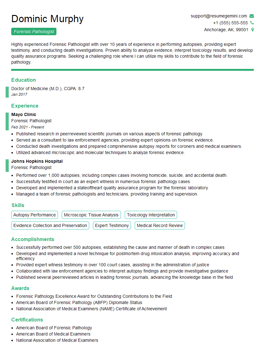
Forensic Pathologist Resume Sample
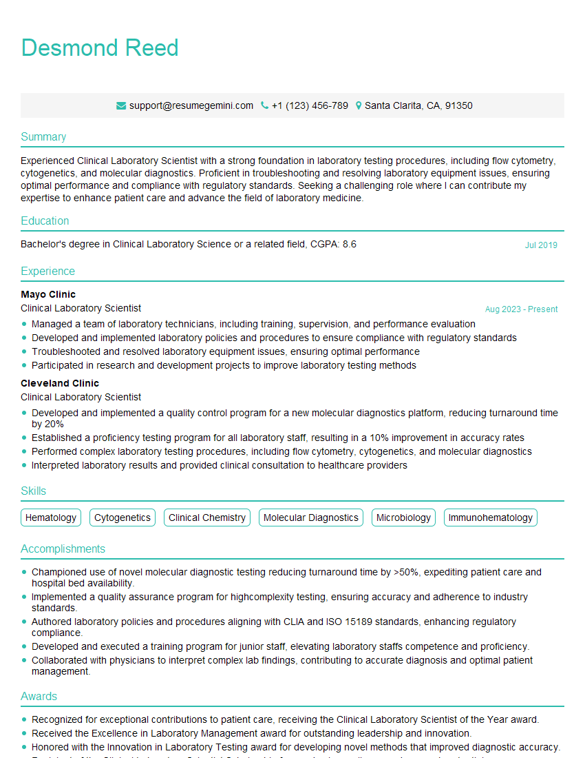
Clinical Laboratory Scientist Resume Sample
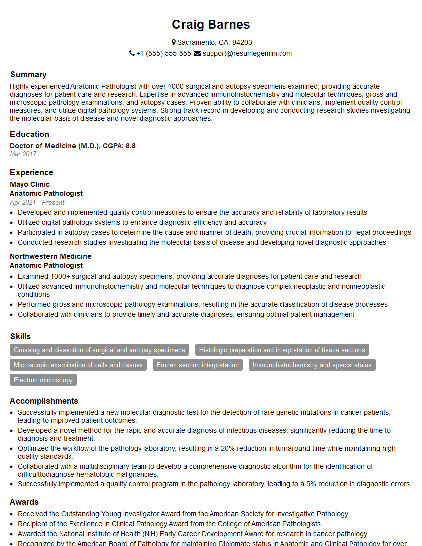
Anatomic Pathologist Resume Sample
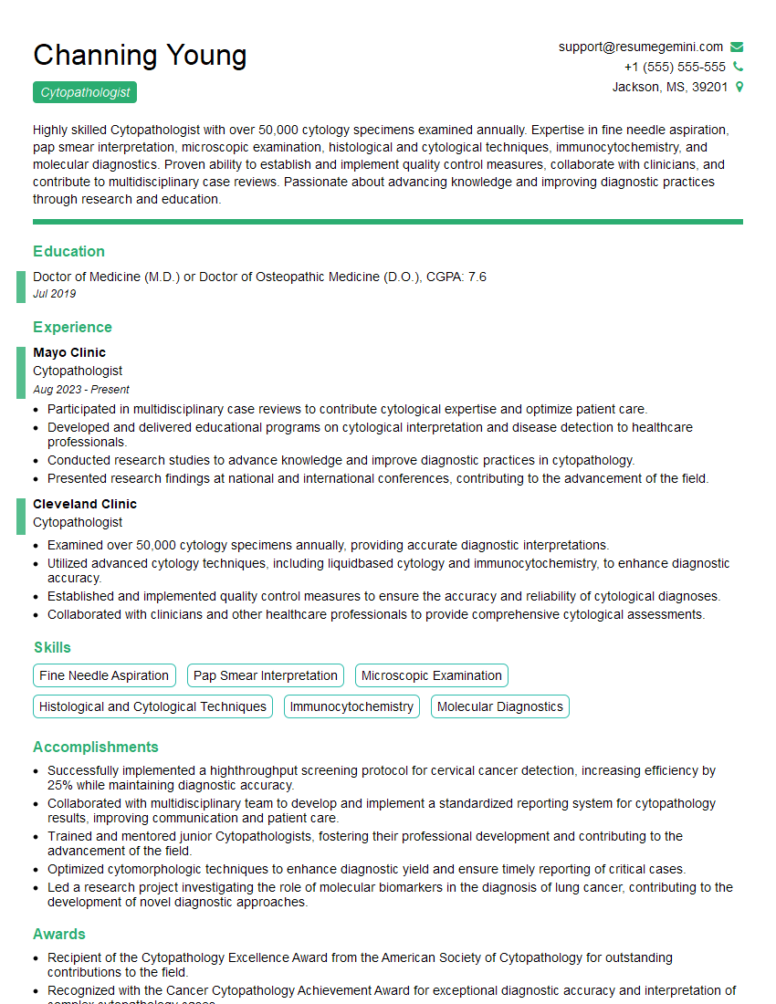
Cytopathologist Resume Sample
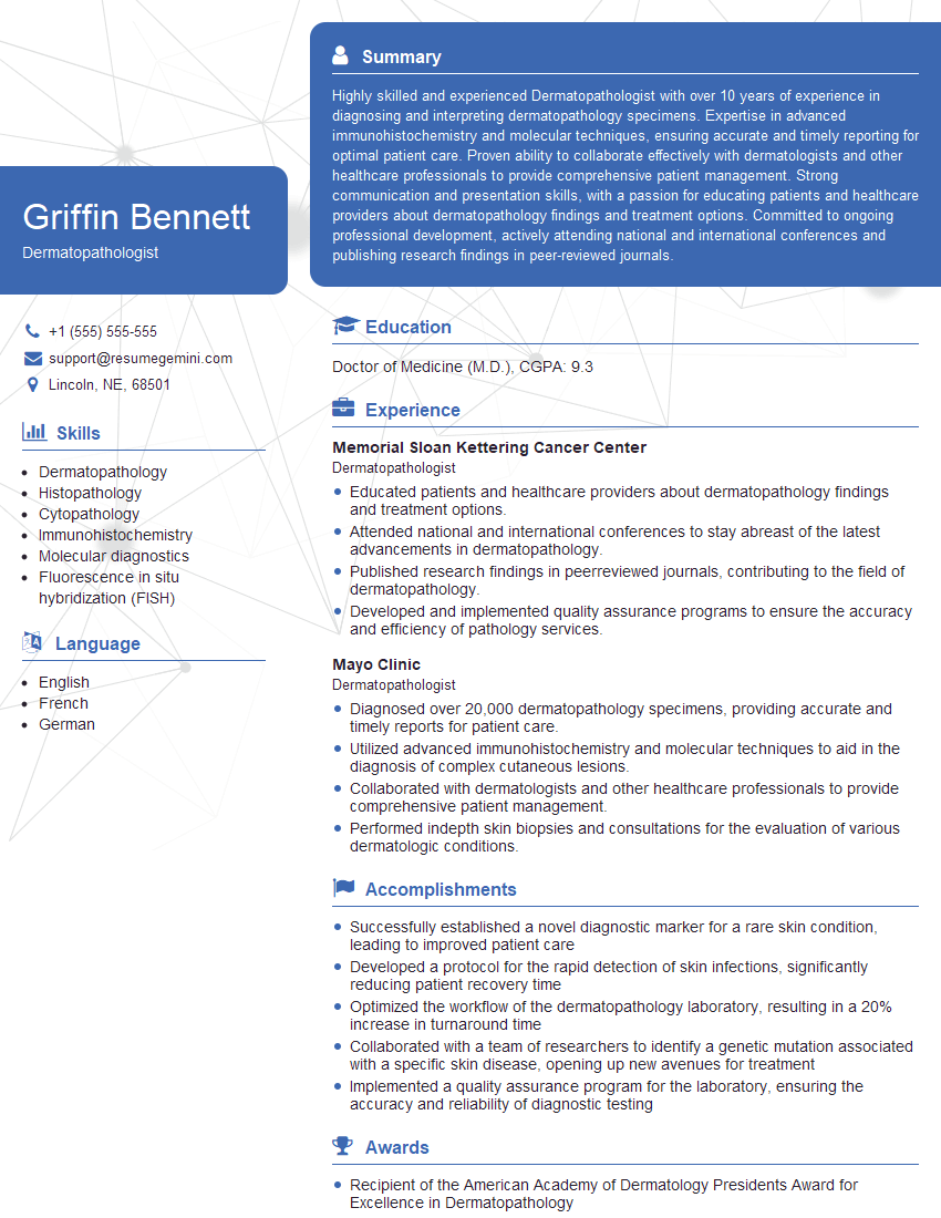
Dermatopathologist Resume Sample
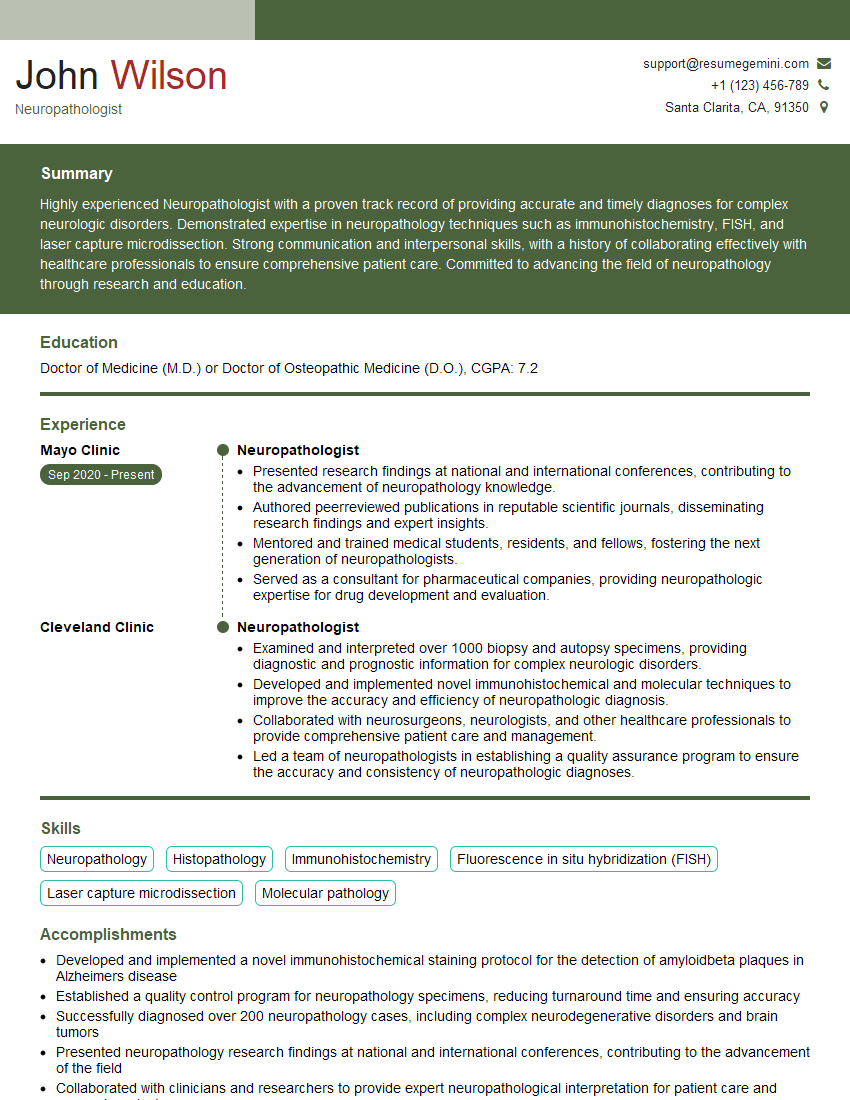
Neuropathologist Resume Sample
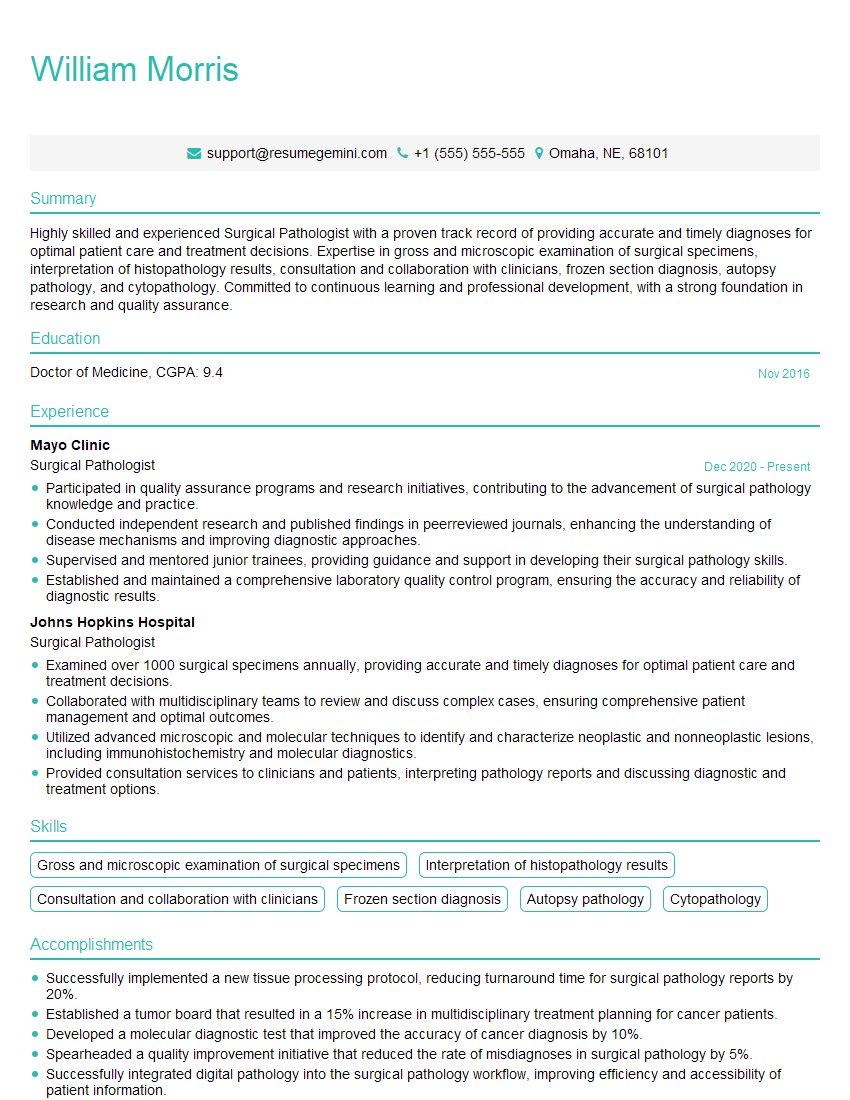
Surgical Pathologist Resume Sample
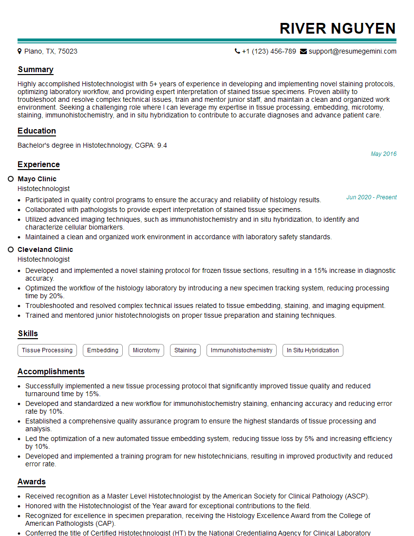
Histotechnologist Resume Sample
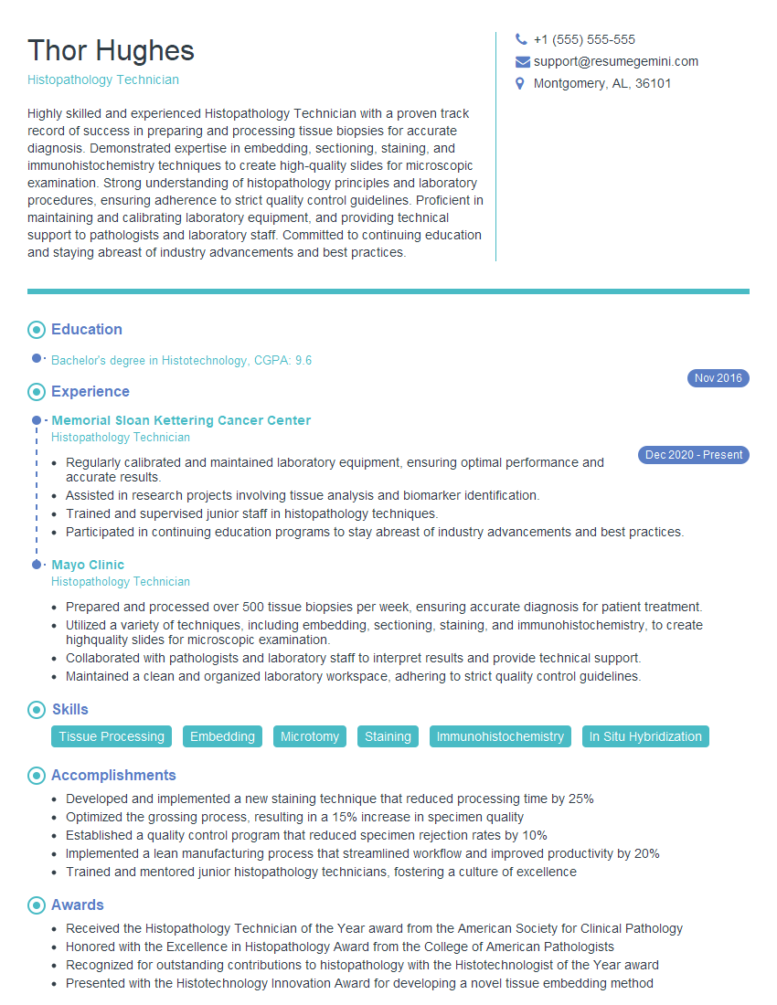
Histopathology Technician Resume Sample
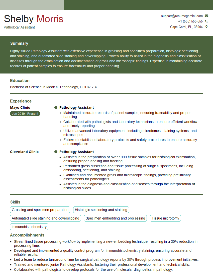
Pathology Assistant Resume Sample
Explore more articles
Users Rating of Our Blogs
Share Your Experience
We value your feedback! Please rate our content and share your thoughts (optional).
What Readers Say About Our Blog
To the interviewgemini.com Webmaster.
Very helpful and content specific questions to help prepare me for my interview!
Thank you
To the interviewgemini.com Webmaster.
This was kind of a unique content I found around the specialized skills. Very helpful questions and good detailed answers.
Very Helpful blog, thank you Interviewgemini team.