Unlock your full potential by mastering the most common CT-Guided Injections interview questions. This blog offers a deep dive into the critical topics, ensuring you’re not only prepared to answer but to excel. With these insights, you’ll approach your interview with clarity and confidence.
Questions Asked in CT-Guided Injections Interview
Q 1. Describe the process of performing a CT-guided biopsy.
A CT-guided biopsy involves using a computed tomography (CT) scanner to precisely guide a needle to a suspicious area within the body to obtain a tissue sample. The process is minimally invasive and allows for targeted sampling, maximizing the chances of obtaining a representative tissue sample.
Step-by-step process:
- Patient Preparation: The patient is positioned on the CT table, and the area of interest is identified. Sometimes, a local anesthetic is administered to numb the area.
- Imaging: A CT scan is performed to pinpoint the target location. This scan provides detailed cross-sectional images of the body.
- Needle Insertion: Using the CT images as a guide, a radiologist or interventionalist inserts a biopsy needle into the target area. Real-time CT imaging allows for precise needle placement and monitoring during the procedure.
- Tissue Collection: The needle is used to collect a tissue sample (either a core needle biopsy or a fine-needle aspiration biopsy). The type of needle depends on the size and location of the target lesion.
- Needle Removal and Imaging: Once the sample is obtained, the needle is removed, and a final CT scan is often performed to assess for any bleeding or complications.
- Sample Processing: The collected tissue sample is then sent to a pathology lab for analysis.
Example: I recently performed a CT-guided biopsy on a patient with a suspicious lung nodule. The CT scan showed the nodule’s exact location, enabling accurate needle placement and successful retrieval of tissue for diagnosis.
Q 2. Explain the different types of needles used in CT-guided injections.
The type of needle used in CT-guided injections depends on the specific procedure and target. Several needle types exist, each designed for particular purposes:
- Fine-needle aspiration (FNA) needles: These are small-gauge needles used to aspirate fluid or cells from a lesion. They are ideal for collecting cytology specimens, minimizing tissue trauma.
- Core biopsy needles: These are larger-gauge needles that extract a cylindrical tissue core. They are preferred when a larger tissue sample is needed for histological examination.
- Tru-cut needles: A type of core biopsy needle known for its ability to obtain larger tissue samples with minimal trauma. They are particularly useful for larger lesions.
- Vertebroplasty/Kyphoplasty needles: Specialized needles used to deliver bone cement into fractured vertebrae, aiding in the stabilization of compression fractures.
- Specialized injection needles: Various other needles are designed to deliver specific medications or contrast agents into different anatomical areas. For example, longer needles might be used for deep-seated lesions.
Example: For a liver lesion suspected of being cancerous, a core biopsy needle, such as a Tru-cut needle, would likely be used to obtain a substantial sample for histopathological analysis. In contrast, if fluid aspiration was the goal, an FNA needle would be the appropriate choice.
Q 3. What are the safety precautions taken during a CT-guided injection procedure?
Safety is paramount in CT-guided injections. We adhere to strict protocols to minimize risks:
- Sterile Technique: Strict adherence to aseptic techniques is crucial to prevent infection. This includes the use of sterile gloves, gowns, drapes, and equipment.
- Image Guidance: Real-time CT imaging ensures accurate needle placement, reducing the risk of damage to surrounding tissues and organs.
- Radiation Safety: Radiation exposure is minimized by using appropriate shielding and adhering to ALARA principles (As Low As Reasonably Achievable). Patient dose is carefully monitored.
- Contrast Agent Selection: The choice of contrast agent depends on the patient’s medical history and allergies. Appropriate pre-injection testing and medication are used to prevent or manage allergic reactions.
- Monitoring Vital Signs: Vital signs are carefully monitored throughout the procedure to detect any adverse reactions promptly.
- Emergency Preparedness: We ensure that emergency medications and equipment are readily available to manage any complications that might arise.
Example: Before any contrast agent is administered, we always assess the patient’s history for allergies. If the patient has a known contrast allergy, alternative procedures or medications are considered. If there’s no alternative, we might use pre-medication to decrease the risk of an allergic reaction.
Q 4. How do you determine the appropriate injection site for a CT-guided injection?
Determining the appropriate injection site is critical for the success and safety of a CT-guided injection. The site is selected based on several factors:
- Imaging Findings: CT scans provide crucial information about the lesion’s location, size, and relationship to surrounding structures. The injection site should be chosen to allow optimal access to the target while minimizing the risk of injury to adjacent organs or vessels.
- Patient Anatomy: Patient-specific anatomical variations influence the choice of approach.
- Lesion Characteristics: The nature of the lesion (e.g., size, density, vascularity) dictates the appropriate needle approach and type.
- Procedural Goals: The goal of the injection (e.g., biopsy, therapeutic injection) influences the choice of injection site.
Example: When performing a CT-guided injection into a spinal lesion, the approach must be carefully planned to avoid damage to the spinal cord or nerve roots. The precise location of the lesion, and its proximity to these sensitive structures, dictates the optimal injection site and approach angle.
Q 5. What are the potential complications of a CT-guided injection?
While CT-guided injections are generally safe, potential complications exist:
- Bleeding (Hemorrhage): Needle puncture of a blood vessel can cause bleeding, ranging from minor to life-threatening.
- Infection: Infection at the injection site is possible, though uncommon due to strict sterile techniques.
- Nerve Damage: Accidental needle puncture of a nerve can cause pain, numbness, or weakness.
- Pneumothorax (Collapsed Lung): This can occur during procedures near the lungs.
- Allergic Reactions to Contrast Agents: Reactions can range from mild to severe, potentially including anaphylaxis.
- Pain at the injection site: This is usually mild and self-limiting but can be managed with analgesics.
- Organ Damage: Accidental damage to other organs (e.g., kidney, liver, spleen) can occur.
Example: In a liver biopsy, the proximity of major blood vessels increases the risk of bleeding. Careful technique and meticulous attention to detail are essential to minimize this complication.
Q 6. How do you manage a patient who experiences an adverse reaction during a CT-guided injection?
Managing an adverse reaction during a CT-guided injection requires a prompt and decisive response. The approach depends on the nature and severity of the reaction:
- Mild Reactions: These may include mild nausea, itching, or a slight drop in blood pressure. Treatment might involve observation, supportive care, and possibly antihistamines.
- Severe Reactions: Severe reactions, such as anaphylaxis (life-threatening allergic reaction), require immediate attention. This includes stopping the procedure, administering oxygen, providing IV fluids, and potentially epinephrine, followed by transferring the patient to intensive care. We are always prepared for managing these types of emergencies.
Example: If a patient experiences significant hypotension (low blood pressure) during a procedure, we immediately stop the injection, administer intravenous fluids, and monitor the patient’s vital signs closely. If the hypotension persists despite these measures, we may administer medication to increase the blood pressure.
Q 7. Describe your experience with different contrast agents used in CT-guided procedures.
I have extensive experience with various contrast agents used in CT-guided procedures. The selection of a contrast agent is carefully considered based on the patient’s clinical condition, renal function, and the specific imaging needs.
- Iodinated Contrast Agents: These are commonly used in CT to enhance the visualization of tissues and organs. I have experience with different formulations, including ionic and non-ionic contrast media. Non-ionic agents are generally preferred because they are associated with a lower risk of allergic reactions.
- Gadolinium-based contrast agents: While less commonly used in CT compared to MRI, they can be useful in specific situations. I am aware of the potential risks associated with these agents, including nephrogenic systemic fibrosis (NSF), particularly in patients with impaired renal function. Appropriate precautions are always taken.
Example: For a patient with a history of severe allergic reactions to iodinated contrast, we carefully consider alternative imaging strategies or pre-medicate them to minimize the risk. We always ensure that the patient’s renal function is assessed before administering any contrast agent, especially gadolinium-based agents, to avoid potential nephrogenic systemic fibrosis. The selection of the agent is always a careful balance between image quality and safety considerations.
Q 8. Explain the role of image guidance in minimizing complications during CT-guided injections.
Image guidance in CT-guided injections is paramount for minimizing complications. Think of it like using a GPS for a precise destination – instead of blindly navigating, we get real-time visualization of the needle’s path, ensuring it reaches the exact target. This precision drastically reduces the risk of damaging surrounding tissues, nerves, or blood vessels.
CT scans provide cross-sectional images, allowing us to see the target area (e.g., a spinal disc, joint, or tumor) in three dimensions. This detailed visualization helps us plan the optimal needle trajectory and monitor its advancement in real-time, making adjustments as needed. For example, if we’re injecting medication near a major blood vessel, we can precisely steer clear of it, avoiding potential hemorrhages. Without image guidance, such accuracy would be impossible, leading to increased risks of complications.
Q 9. How do you interpret CT images to guide needle placement during injections?
Interpreting CT images for needle placement involves a systematic approach. First, we identify the target anatomy, using the images to precisely locate its position and size. We then assess surrounding structures, including critical organs, nerves, and vessels. This helps determine a safe injection path, avoiding potential hazards. We plan the trajectory by mentally visualizing a line from the entry point to the target on the images. During the injection, we continually monitor the needle’s position on the CT images, comparing its actual path to the planned trajectory. We use this to make adjustments in real-time, ensuring the needle remains in the desired location. Think of it like following a map – we don’t just look at the map once, we constantly check our progress to make sure we’re on the right path.
We use various imaging techniques such as bone window settings to clearly visualize bony landmarks and soft tissue windows for better visualization of soft tissue structures like muscles and nerves. Contrast agents are often used to better highlight the target structure and enhance visualization. For example, if we’re injecting into a specific facet joint in the spine, we would use both bone and soft tissue windows to optimize the visualization of both the bone and the soft tissue surrounding the joint. We might also use contrast media to better delineate the joint cavity.
Q 10. What are the contraindications for CT-guided injections?
Contraindications for CT-guided injections are situations where the procedure’s risks outweigh its benefits. These can include:
- Severe allergies to contrast media: Some patients are allergic to iodine-based contrast agents, which are often used in CT scans to enhance visualization. We carefully assess for allergy history and may use alternative non-iodinated agents or modify the approach if needed.
- Severe renal insufficiency: Contrast agents can be harmful to the kidneys, especially in patients with pre-existing kidney disease. We need to carefully weigh the benefits against the risks, often obtaining renal function tests beforehand.
- Pregnancy: Radiation exposure during CT scans should be avoided during pregnancy if possible, due to potential risks to the fetus. Alternative techniques might be considered.
- Uncooperative patients: The procedure requires the patient to remain still during the injection, so an uncooperative patient may not be a suitable candidate.
- Severe bleeding disorders: This increases the risk of bleeding complications at the injection site.
Each case is assessed individually to determine if the benefits of the procedure outweigh the potential risks. Alternatives might be explored, such as ultrasound-guided injections, if CT guidance is contraindicated.
Q 11. Describe your experience with different types of CT scanners used for guided injections.
My experience encompasses a range of CT scanners, from older generation multi-slice scanners to the latest multi-detector CT systems with advanced image reconstruction capabilities. The newer scanners offer improved image resolution, faster scan times, and reduced radiation doses. This translates to more precise needle placement and greater patient comfort. For instance, the faster scan times are particularly beneficial for procedures requiring real-time guidance and multiple needle adjustments, reducing the total radiation exposure time.
The difference in image quality is also significant. Higher-resolution images provided by modern scanners enable us to visualize even subtle anatomical details, enhancing accuracy. Advanced reconstruction algorithms allow for the creation of 3D images, further improving our visualization and planning capabilities. For example, in spine interventions, the ability to create 3D reconstructions of the vertebrae allows us to plan trajectories that avoid important neural structures with greater accuracy.
Q 12. How do you ensure patient comfort and safety during a prolonged CT-guided injection procedure?
Ensuring patient comfort and safety during prolonged procedures is a top priority. We start by providing detailed explanations of the procedure, addressing any concerns and anxieties. We use appropriate positioning techniques to maximize comfort and minimize stress. For extended procedures, we offer breaks to adjust position and address any discomfort. We monitor vital signs throughout the procedure to ensure the patient’s well-being. For example, we monitor heart rate, blood pressure, and oxygen saturation. Analgesia or sedation may be used to further enhance comfort, but these are carefully considered and adjusted depending on individual needs.
Effective communication is key. We maintain regular communication with the patient throughout the procedure to gauge comfort levels and address any issues. Pain management strategies are implemented proactively, with the patient’s feedback constantly considered and adjusted. Proper aseptic technique is used to maintain sterility and prevent infection. Every effort is made to streamline the procedure, keeping the duration as short as possible without compromising accuracy.
Q 13. What is your approach to managing pain in patients undergoing CT-guided injections?
Pain management is an integral part of CT-guided injections. Our approach is multimodal and tailored to each patient’s needs and pain tolerance. This may include local anesthetic infiltration at the injection site to numb the area before needle insertion. We might also administer systemic analgesics (pain relievers) intravenously or orally before or after the procedure, depending on the patient’s pain level and the anticipated discomfort. Sedation can be used, particularly for anxious patients or procedures anticipated to be lengthy or uncomfortable.
We assess the patient’s pain level before, during and after the procedure using a standardized pain scale. This allows us to monitor the effectiveness of our pain management strategy and make necessary adjustments. Post-procedural pain management is also important and includes providing the patient with adequate analgesia for home use, along with clear instructions on how to manage any residual pain or discomfort.
Q 14. Explain your understanding of radiation safety protocols and ALARA principles in CT procedures.
Radiation safety is paramount in CT procedures, guided by the ALARA principle – As Low As Reasonably Achievable. This means we strive to minimize radiation exposure to both patients and staff while maintaining diagnostic image quality. We use several strategies to achieve this, including:
- Collimation: This precisely limits the X-ray beam to the area of interest, reducing unnecessary radiation exposure.
- Optimized scan parameters: We select appropriate scan parameters (kVp, mAs) to obtain optimal images with the lowest possible radiation dose. This might involve utilizing iterative reconstruction techniques which reduce noise at a given dose, enabling us to lower the mAs without compromising image quality.
- Radiation dose monitoring: We constantly monitor radiation dose using built-in dose monitoring systems on CT scanners and utilize dose-tracking software to manage radiation exposure.
- Protective shielding: We use lead aprons and shields to protect patients and staff from scattered radiation.
- Image post-processing: Techniques like iterative reconstruction algorithms can reduce noise and artifacts, potentially allowing for a reduction in the radiation dose needed to achieve comparable image quality.
Moreover, we adhere to all institutional radiation safety protocols and ensure that all staff involved in the procedure are properly trained and understand the risks associated with ionizing radiation. We believe that a combination of robust techniques and a commitment to ALARA is essential for ensuring patient and staff safety.
Q 15. What is your experience with post-procedural care and patient follow-up after CT-guided injections?
Post-procedural care and follow-up after CT-guided injections are crucial for patient safety and optimal outcomes. My approach begins with a thorough assessment of the patient’s vital signs and injection site immediately after the procedure. This includes checking for bleeding, swelling, hematoma formation, or any signs of infection. I provide detailed instructions regarding activity restrictions, pain management (including prescribed analgesics and ice application), and signs and symptoms to watch for, such as increased pain, fever, or excessive swelling.
Follow-up typically involves scheduling a phone call within 24-48 hours to address any immediate concerns and then a scheduled appointment for a physical examination and assessment of the injection site. Imaging follow-up (e.g., an X-ray or repeat CT scan) might be necessary depending on the procedure and patient response. Detailed documentation is maintained throughout the process, including any adverse events, patient responses to treatment, and the effectiveness of the injection. I strive to provide personalized care, addressing each patient’s unique needs and concerns. For example, patients with mobility limitations might require home-healthcare visits, while others might only need phone consultations. A clear communication plan is established, ensuring the patient understands the follow-up schedule and knows how to contact me if needed.
Career Expert Tips:
- Ace those interviews! Prepare effectively by reviewing the Top 50 Most Common Interview Questions on ResumeGemini.
- Navigate your job search with confidence! Explore a wide range of Career Tips on ResumeGemini. Learn about common challenges and recommendations to overcome them.
- Craft the perfect resume! Master the Art of Resume Writing with ResumeGemini’s guide. Showcase your unique qualifications and achievements effectively.
- Don’t miss out on holiday savings! Build your dream resume with ResumeGemini’s ATS optimized templates.
Q 16. Describe a challenging CT-guided injection case and how you overcame the challenges.
One challenging case involved a patient requiring a CT-guided injection into a deeply seated lesion within the sacroiliac joint. The lesion was close to critical neurovascular structures, making precise needle placement paramount. The initial CT images showed poor visualization of the target due to overlying bone density and anatomical variations. The challenge was to navigate safely and accurately to the target while minimizing the risk of nerve damage or vascular injury.
To overcome this, I utilized several strategies. First, we used a higher resolution CT scan with multiplanar reconstruction (MPR) to create clearer three-dimensional images of the target area. This allowed for a more precise assessment of the target’s location and relationship to surrounding structures. We used a smaller gauge needle, and I performed a series of test injections with contrast medium under real-time fluoroscopic guidance to confirm the needle’s position before administering the therapeutic agent. This real-time visualization allowed for continuous monitoring and immediate adjustments if needed. Throughout the procedure, communication with the patient was maintained to monitor for any discomfort or neurological changes. The procedure was ultimately successful with no complications. Careful planning, advanced imaging techniques, and meticulous execution were essential to successfully navigate this challenging case.
Q 17. What are the limitations of CT-guided injections compared to other imaging modalities?
While CT is excellent for visualizing bony anatomy and providing excellent spatial resolution, making it ideal for many injection procedures, it does have limitations compared to other modalities. One major limitation is the use of ionizing radiation, which poses a risk to the patient, particularly with repeated procedures. Furthermore, CT may not be as effective in visualizing soft tissue structures, such as nerves or vessels, compared to MRI or ultrasound. MRI, for example, offers superior soft tissue contrast, which can be beneficial in cases where precise targeting within soft tissue is crucial. Ultrasound provides real-time imaging, which can be advantageous during the procedure itself, allowing for dynamic adjustments to needle placement. Lastly, CT is less portable than ultrasound, limiting its use in certain settings. The choice of imaging modality depends on the specific clinical scenario, target location, patient factors, and the advantages and disadvantages of each technique.
Q 18. How do you handle situations where the target site is difficult to access using CT guidance?
When the target site is difficult to access using CT guidance alone, a multi-modal approach might be necessary. This could involve combining CT with fluoroscopy, which provides real-time visualization of the needle’s trajectory. Fluoroscopy, while using ionizing radiation, offers superior visualization in certain situations, especially for navigating through dense bone structures. Another approach is to use ultrasound guidance in conjunction with CT. Ultrasound can provide real-time visualization of the needle’s path and surrounding soft tissues, while CT provides detailed anatomical information for planning. This multimodal strategy allows for a more comprehensive view, enhancing the safety and precision of the procedure. In some cases, a change in the approach, perhaps utilizing a different needle insertion point, might be necessary. Careful pre-procedural planning and consideration of alternative approaches are crucial for managing such situations.
Q 19. What are the steps you would take to troubleshoot a failed CT-guided injection?
Troubleshooting a failed CT-guided injection involves a systematic approach. First, we carefully review the pre-procedural planning to identify any potential errors in target localization or needle trajectory planning. Then, we reassess the CT images, paying close attention to the needle’s position relative to the target and surrounding structures. Was there an anatomical variation not accounted for? Did the needle deviate from its planned path? We then examine the technical aspects of the procedure. Did the needle kink or malfunction? Was the injection technique correct? Finally, we consider patient-related factors: Did the patient move unexpectedly? Were there any anatomical variations not apparent on the imaging? We may need to obtain additional imaging (e.g., fluoroscopy or ultrasound) to better visualize the situation and guide corrective actions. In some cases, a repeat procedure with modifications to the technique or approach might be necessary. Thorough documentation of the failed attempt and subsequent troubleshooting steps is critical for learning and improving future procedures.
Q 20. Describe your experience with using different types of software for CT image analysis and planning.
My experience encompasses several CT image analysis and planning software packages. I am proficient in using advanced visualization tools to create 3D reconstructions, MPR images, and other advanced image processing techniques. These software platforms aid in precise target localization, needle trajectory planning, and procedural simulation. For instance, GE AW VolumeShare allows for detailed analysis of the CT data and precise measurement of distances and angles. Siemens syngo.via offers similar capabilities with additional advanced visualization tools. The specific software used depends on the institution’s available resources and the complexity of the case. Each platform has its strengths and weaknesses; however, my goal is always to use the most appropriate software to maximize the safety and accuracy of the procedure. Familiarity with multiple platforms allows for adaptability and the ability to select the optimal tool for a given situation.
Q 21. How do you maintain sterility during CT-guided injection procedures?
Maintaining sterility during CT-guided injections is paramount to prevent infection. This involves strict adherence to aseptic techniques throughout the entire procedure. The process begins with proper hand hygiene and the use of sterile gloves, gowns, and drapes. The injection site is meticulously prepared with antiseptic solutions following recommended guidelines. All equipment used, including needles, syringes, and catheters, is sterile and single-use. The procedure area is prepared using a sterile field, and any non-sterile items are kept away from the procedure site. Throughout the procedure, sterile technique is maintained, minimizing any chance of contamination. After the procedure, proper disposal of all sharps and contaminated materials according to established protocols is crucial. Continuous monitoring of the patient for signs of infection during post-procedural follow-up is essential to ensure the success of the aseptic measures implemented.
Q 22. Explain the importance of documentation and reporting in CT-guided injection procedures.
Meticulous documentation and reporting are paramount in CT-guided injections for several reasons: legal protection, quality assurance, and patient safety. Imagine a situation where there’s a discrepancy in post-procedure outcomes. Comprehensive documentation acts as a reliable record for tracking the procedure, allowing for a detailed review if complications arise. It ensures clarity regarding the indications for the procedure, the steps taken, the materials used, any observed adverse events, and the patient’s response.
- Legal Protection: Detailed documentation protects both the healthcare professional and the institution from potential legal claims by providing irrefutable evidence of the procedure’s performance and any associated risks and benefits that were discussed with the patient.
- Quality Assurance: Consistent documentation allows for the analysis of trends, identification of areas for improvement in technique or workflow, and ongoing assessment of the procedure’s effectiveness and safety. This contributes to continuous improvement within the healthcare setting.
- Patient Safety: Accurate records ensure continuity of care should a patient require further treatment or consultation with other healthcare providers. This avoids potential miscommunication and medication errors.
For instance, a precise record of the injection site, the type and volume of contrast media and medication used, and the patient’s vital signs before, during, and after the procedure are essential. Any complications or unusual events must be documented thoroughly, including the response to any interventions. This detailed approach promotes a culture of safety and excellence.
Q 23. What is your familiarity with different types of anatomical landmarks used in CT-guided injections?
My familiarity with anatomical landmarks used in CT-guided injections is extensive. We rely on a combination of bony landmarks, vascular structures, and organ relationships to accurately target the injection site. The specific landmarks used vary greatly depending on the target location. For instance:
- Spine: Vertebral bodies, pedicles, spinous processes, and intervertebral foramina are crucial landmarks for epidural, facet, and selective nerve root injections. I’m proficient in using both the conventional approach and newer image guidance techniques.
- Joints: For joint injections like those in the knee or shoulder, we utilize the articular surfaces, bony contours, and ligamentous structures. Accurate identification is critical to avoid intra-articular penetration or inadvertent injection into adjacent tissues.
- Soft Tissues: In procedures targeting soft tissue masses or lesions, we often use adjacent muscle groups, fascial planes, and neurovascular bundles as guides. A precise understanding of the three-dimensional anatomy is crucial.
Accurate identification of these landmarks is essential for minimizing the risk of complications, including nerve damage, vascular injury, and infection. I regularly refresh my knowledge through anatomical atlases, continuing medical education, and hands-on practice. The use of 3D imaging further enhances our ability to confidently identify and navigate these complex anatomical relationships.
Q 24. How do you communicate effectively with the patient and other members of the healthcare team during a CT-guided injection procedure?
Effective communication is the cornerstone of successful CT-guided injections. This involves clear and concise interaction with both the patient and the healthcare team. Think of it as a well-orchestrated performance where everyone knows their role.
- Patient Communication: Before the procedure, I ensure the patient understands the purpose, procedure, potential benefits, and risks involved. I answer their questions patiently and in a language they can readily grasp. This builds trust and reduces anxiety. During the procedure, I maintain a reassuring and calming presence, keeping them informed about each step.
- Team Communication: Clear, concise communication with the radiologic technologist, anesthesiologist (if involved), and other members of the team is vital. This includes confirming the procedure parameters (injection site, medication dosage, etc.), monitoring the patient’s vital signs during the procedure, and promptly reporting any unexpected events or complications.
For example, I might say to the patient, “We’re going to use a CT scan to precisely guide the needle to the area of pain. You might feel a slight pinch as the needle is inserted.” During the procedure, a clear, concise communication with the team might include, “Needle is in the desired position, ready to inject.” This ensures a smooth and safe workflow.
Q 25. Describe your understanding of the legal and ethical implications related to CT-guided injections.
CT-guided injections carry significant legal and ethical implications. The legal aspects revolve around informed consent, proper technique, and meticulous documentation, as discussed earlier. Ethically, we must prioritize patient autonomy, beneficence (acting in the patient’s best interest), non-maleficence (avoiding harm), and justice (fair and equitable access to care).
- Informed Consent: Patients must fully understand the procedure, its risks and benefits, alternative treatment options, and the possibility of complications before giving their consent. This is documented thoroughly.
- Proper Technique: Adhering to established protocols and best practices is essential to minimize the risk of complications. This includes selecting appropriate needle size and injection technique, using appropriate contrast media, and continuously monitoring the patient’s vital signs.
- Confidentiality: Maintaining patient confidentiality is crucial, ensuring their personal information and medical records are protected.
If a complication arises, I will take immediate steps to address the issue and will be fully transparent with the patient and their family about the incident. It’s critical to maintain the highest standards of ethical conduct and adhere to all relevant legal regulations.
Q 26. How do you stay updated with the latest advancements and best practices in CT-guided injections?
Keeping abreast of the latest advancements in CT-guided injections requires a multifaceted approach. The field is constantly evolving, with improvements in imaging technology, injection techniques, and the development of new medications.
- Professional Organizations: Active participation in professional organizations like the American Society of Interventional Pain Physicians (ASIPP) or the American College of Radiology (ACR) provides access to continuing medical education (CME) courses, journals, and conferences focused on interventional radiology and pain management.
- Peer-Reviewed Journals: Regularly reviewing peer-reviewed medical journals keeps me updated on the latest research findings and clinical trials.
- CME Courses & Workshops: Attending workshops and hands-on training sessions allows me to practice new techniques and learn from experts in the field.
- Online Resources: Reliable online resources, such as medical databases (PubMed, etc.) and reputable medical websites, provide valuable information on new research and guidelines.
By actively engaging in these avenues, I can ensure my practice remains aligned with the best available evidence and the highest standards of care. It’s a continuous learning process, and staying informed is critical to providing optimal patient outcomes.
Q 27. How would you handle a situation where a patient refuses a CT-guided injection?
Respecting patient autonomy is paramount. If a patient refuses a CT-guided injection, my response is to engage in a calm and respectful discussion to fully understand the reasons for their refusal. I want to identify any concerns or misconceptions.
- Listen and Understand: The first step is to actively listen to their concerns and address them empathetically. They might be worried about pain, risks, or the procedure itself.
- Provide Information: Offer additional information to clarify any misconceptions and address their specific concerns. Ensure they understand the benefits and risks involved in a clear and accessible manner.
- Explore Alternatives: If possible, discuss alternative treatment options that might better suit their needs and preferences.
- Document the Refusal: Thoroughly document the patient’s refusal, including the reasons given and any discussions held. This is crucial for legal and ethical considerations.
- Respect their Decision: Ultimately, if they remain firm in their decision, their autonomy must be respected. I would document this refusal clearly and thoroughly in their medical record.
The goal is to ensure the patient feels heard, understood, and empowered to make an informed decision about their own care, even if that decision is to decline the procedure.
Key Topics to Learn for CT-Guided Injections Interview
- Anatomy and Physiology: Thorough understanding of relevant anatomical structures and physiological processes impacted by injections.
- CT Scan Principles: Knowledge of CT image acquisition, reconstruction, and interpretation relevant to procedural guidance.
- Injection Techniques: Mastery of various injection techniques, including needle placement, contrast media administration, and safety protocols.
- Image Guidance and Navigation: Proficiency in using CT imaging to accurately target injection sites and avoid critical structures.
- Patient Assessment and Preparation: Understanding patient history, allergies, and contraindications; preparing patients for the procedure.
- Complications and Management: Knowledge of potential complications (e.g., bleeding, infection, nerve damage) and their management strategies.
- Radiation Safety: Understanding radiation safety protocols and ALARA principles (As Low As Reasonably Achievable).
- Sterile Technique and Infection Control: Adherence to strict sterile techniques to prevent infection.
- Documentation and Reporting: Accurate and thorough documentation of the procedure and any relevant observations.
- Troubleshooting and Problem-Solving: Ability to identify and resolve technical challenges during the procedure.
Next Steps
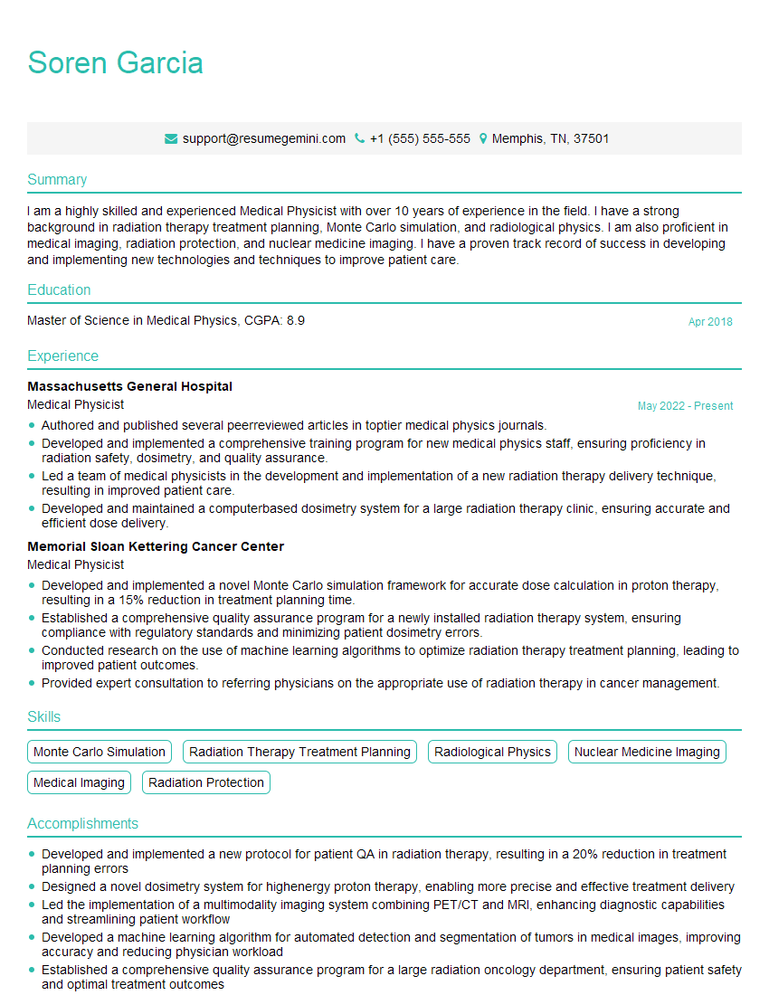
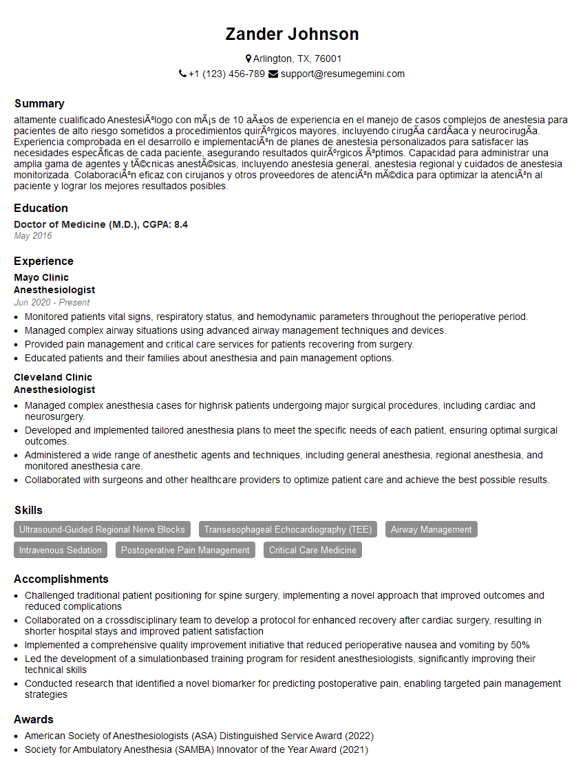
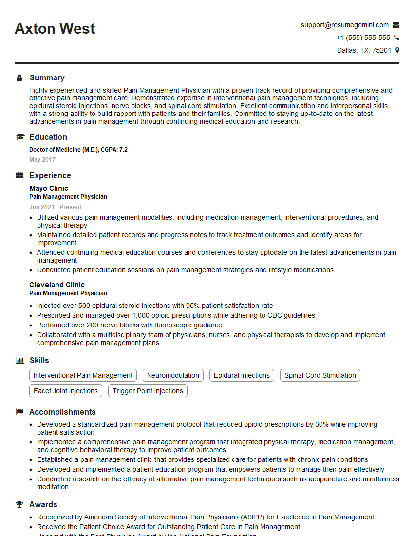
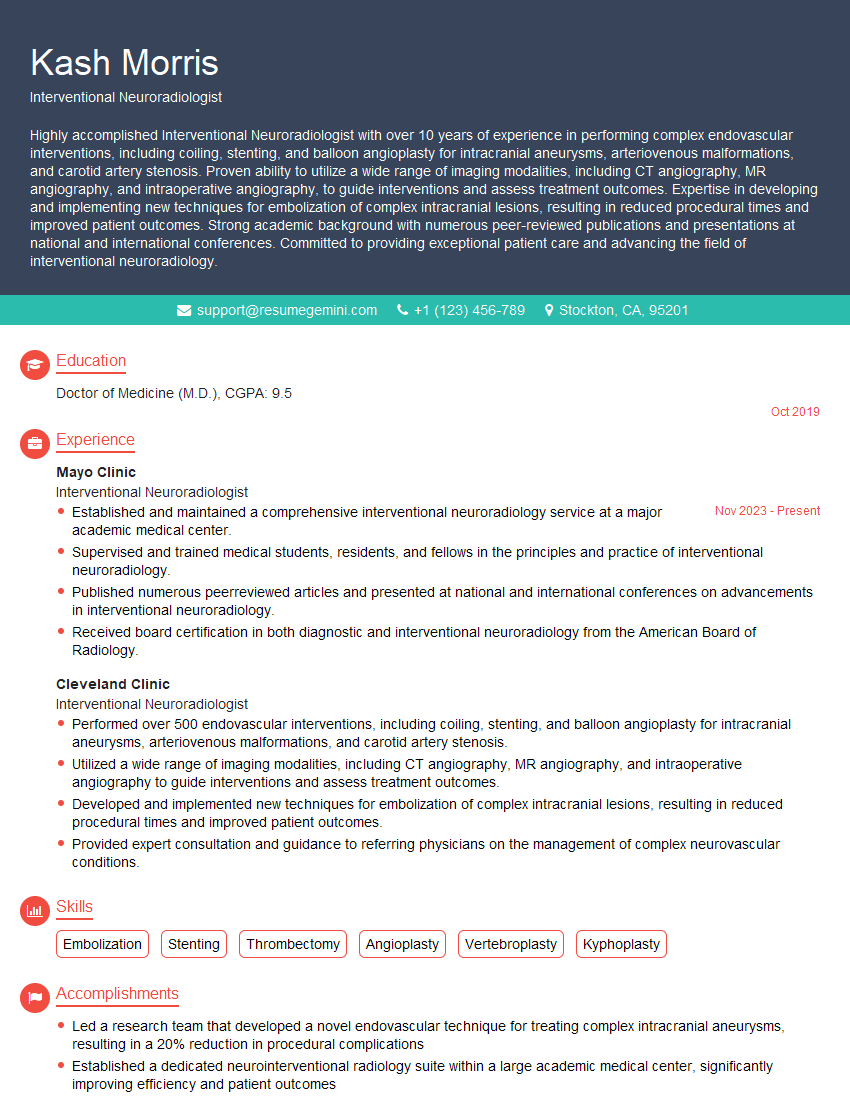
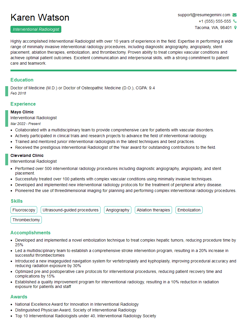
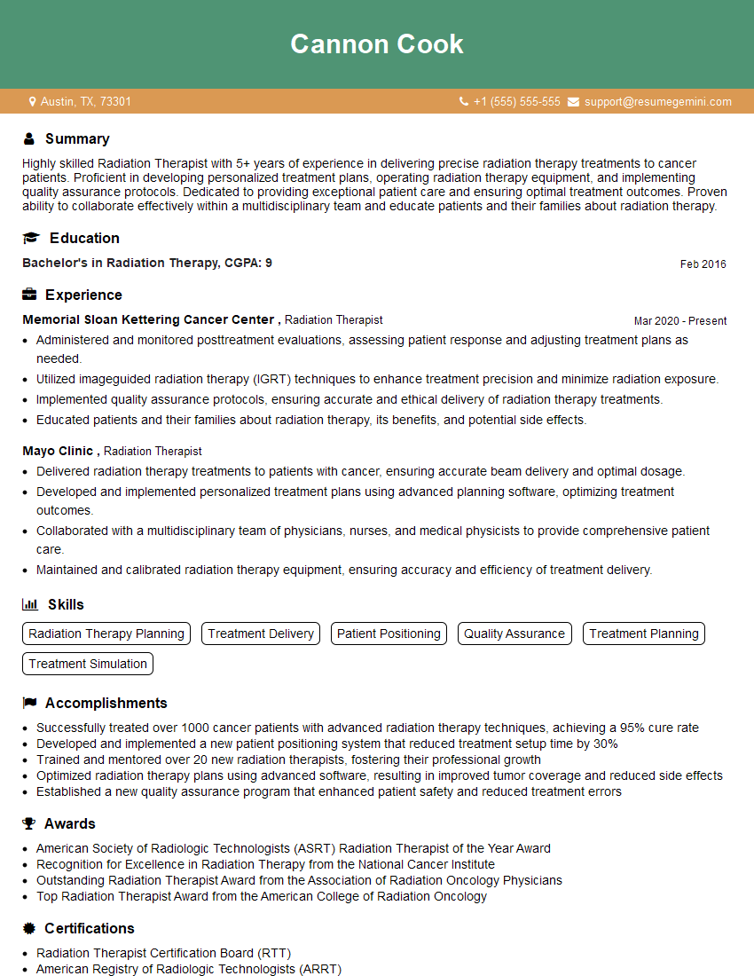
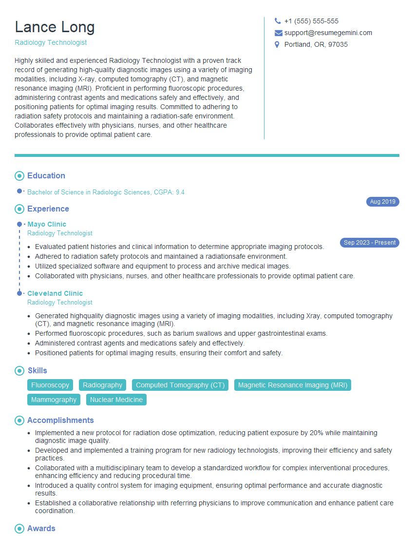
Mastering CT-Guided Injections opens doors to rewarding and specialized roles within the medical field, offering significant career advancement opportunities. A strong resume is crucial to showcasing your skills and experience to potential employers. Creating an ATS-friendly resume is essential for getting your application noticed. ResumeGemini is a trusted resource to help you build a professional and effective resume that highlights your qualifications for CT-Guided Injections positions. We provide examples of resumes tailored to this specialty to help you craft a compelling application. Invest in your future – build a standout resume today.
Explore more articles
Users Rating of Our Blogs
Share Your Experience
We value your feedback! Please rate our content and share your thoughts (optional).
What Readers Say About Our Blog
To the interviewgemini.com Webmaster.
Very helpful and content specific questions to help prepare me for my interview!
Thank you
To the interviewgemini.com Webmaster.
This was kind of a unique content I found around the specialized skills. Very helpful questions and good detailed answers.
Very Helpful blog, thank you Interviewgemini team.