Feeling uncertain about what to expect in your upcoming interview? We’ve got you covered! This blog highlights the most important Cubital Tunnel Release interview questions and provides actionable advice to help you stand out as the ideal candidate. Let’s pave the way for your success.
Questions Asked in Cubital Tunnel Release Interview
Q 1. Describe the surgical anatomy relevant to a Cubital Tunnel Release.
Understanding the surgical anatomy of the cubital tunnel is crucial for a successful Cubital Tunnel Release (CTR). The cubital tunnel is formed by the medial epicondyle of the humerus superiorly, the olecranon process posteriorly, and the arcuate ligament and fascia inferiorly. The ulnar nerve passes through this tunnel, which is a relatively fixed space. Key anatomical structures include:
- Ulnar Nerve: The main focus of the procedure. Understanding its branching patterns, particularly its relationship to the medial epicondyle and the articular branches, is paramount. Damage to these branches can lead to complications.
- Arcuate Ligament: A fibrous band that often contributes to ulnar nerve compression. The surgeon must carefully identify and release this ligament.
- Medial Epicondyle: A bony prominence that can contribute to nerve compression, especially in cases of osteophytes (bony spurs).
- Flexor Carpi Ulnaris (FCU) Muscle: This muscle’s relationship to the ulnar nerve must be carefully considered, as it’s involved in some surgical approaches.
- Ulnar Collateral Ligament: It’s vital to avoid injury to this ligament during the procedure.
Visualizing these structures pre-operatively through imaging techniques and meticulous intraoperative identification are key to minimizing complications.
Q 2. Explain the indications for Cubital Tunnel Release.
Indications for CTR are based on clinical findings consistent with ulnar nerve entrapment at the elbow. These include:
- Symptoms of ulnar neuropathy: This includes numbness, tingling, and weakness in the ulnar nerve distribution (little finger and ulnar half of the ring finger). These symptoms often worsen with elbow flexion.
- Positive clinical tests: Tinel’s sign (tingling with tapping over the ulnar nerve at the elbow) and Phalen’s sign (numbness and tingling with sustained elbow flexion) can be helpful but aren’t always definitive.
- Electrodiagnostic studies (NCS/EMG): Nerve conduction studies (NCS) and electromyography (EMG) are often used to confirm the diagnosis and rule out other causes of ulnar nerve symptoms. These tests can show slowed conduction across the cubital tunnel.
- Failure of conservative management: Conservative treatments, such as splinting, activity modification, and anti-inflammatory medications, should be tried initially. CTR is generally considered after conservative treatments fail to provide relief.
A patient presenting with persistent, progressive ulnar nerve symptoms despite conservative management is a clear indication for surgical intervention.
Q 3. What are the contraindications for Cubital Tunnel Release?
Contraindications to CTR are relatively few, but careful consideration is needed. These include:
- Active infection at the surgical site: Infection can significantly increase the risk of surgical site complications.
- Poor patient health: Patients with uncontrolled medical conditions such as diabetes, severe cardiovascular disease, or bleeding disorders may not be suitable candidates for surgery.
- Unrealistic patient expectations: Patients need to understand that surgery does not guarantee complete resolution of symptoms and that some degree of residual sensory or motor deficit may remain.
- Presence of other pathologies: Other conditions compressing the ulnar nerve, such as a mass or fracture, might require different approaches before considering CTR.
A thorough preoperative evaluation, including a discussion of the potential risks and benefits, is essential before proceeding with CTR.
Q 4. Detail the different surgical approaches for Cubital Tunnel Release.
Several surgical approaches exist for CTR, each with advantages and disadvantages. The most common approaches include:
- In situ decompression: This involves releasing the constricting elements of the cubital tunnel, such as the arcuate ligament and any osteophytes.
- Submuscular transposition: This technique involves moving the ulnar nerve anteriorly, either in front of the medial epicondyle or into the brachialis muscle. This approach is considered when in-situ decompression is insufficient or when there’s significant compression from the medial epicondyle.
- Medial epicondylectomy: In cases of significant bony compression, removing a portion of the medial epicondyle might be necessary to relieve pressure on the ulnar nerve. However, this is a more radical approach and is reserved for cases resistant to other techniques.
The choice of surgical approach is highly individualized and depends on the specific anatomy, the severity of the compression, and surgeon preference.
Q 5. Compare and contrast in-situ decompression versus transposition in Cubital Tunnel Release.
In-situ decompression and transposition represent different strategies for managing ulnar nerve compression at the elbow. Both aim to relieve pressure on the nerve, but they differ in their approach:
- In-situ decompression: This is a less invasive procedure, leaving the ulnar nerve in its anatomical location. It involves releasing the tight structures around the nerve, primarily the arcuate ligament and any osteophytes. It’s suitable for cases where nerve compression is mild to moderate and there isn’t significant extrinsic pressure from the medial epicondyle.
- Transposition: This is a more extensive procedure, relocating the ulnar nerve to a different location, usually anterior to the medial epicondyle or within the brachialis muscle. This provides more space for the nerve and is preferred when in-situ decompression is insufficient, or when there is significant bony compression. However, it may be associated with higher rates of certain complications.
The choice depends on the severity and nature of the compression. In-situ decompression is simpler, faster, and associated with fewer complications, but transposition offers greater decompression for severe cases. Ultimately, the surgeon carefully evaluates the patient’s specific anatomy and clinical presentation to decide the best approach.
Q 6. What are the potential complications of Cubital Tunnel Release?
Potential complications of CTR, though generally uncommon with experienced surgeons, include:
- Recurrence of ulnar nerve symptoms: This might be due to incomplete decompression or scar tissue formation.
- Ulnar nerve palsy: This is a rare but serious complication, resulting in weakness or paralysis of the ulnar nerve’s distribution.
- Infection: Post-operative infection is a risk of any surgery.
- Neuroma formation: Scar tissue forming around the ulnar nerve can cause pain and dysfunction.
- Pain and paresthesia: Persistent pain or numbness in the affected area is possible.
- Wound dehiscence: The incision might open.
- Damage to surrounding structures: Injury to the ulnar artery or collateral ligament is possible but less frequent.
Careful surgical technique and meticulous postoperative care significantly reduce the chances of these complications.
Q 7. How do you manage postoperative pain after Cubital Tunnel Release?
Postoperative pain management following CTR is crucial for patient comfort and successful rehabilitation. A multi-modal approach is generally employed:
- Analgesics: Nonsteroidal anti-inflammatory drugs (NSAIDs) are commonly prescribed to manage pain and inflammation. Opioids may be used for severe pain, but generally, the goal is to minimize opioid use due to potential side effects.
- Nerve blocks: Regional nerve blocks can provide excellent pain control in the immediate postoperative period.
- Ice therapy: Applying ice packs to the surgical site can reduce swelling and inflammation.
- Elevation: Elevating the arm can also minimize swelling.
- Early mobilization: Gradual range-of-motion exercises are important to prevent stiffness and promote healing.
- Patient education: Educating the patient on pain management strategies and the expected recovery process is essential.
Regular follow-up appointments allow for monitoring of pain levels and adjustment of the pain management plan as needed. A tailored approach to pain management ensures optimal patient comfort and a smooth recovery.
Q 8. Describe your preferred technique for Cubital Tunnel Release.
My preferred technique for Cubital Tunnel Release is a minimally invasive approach, utilizing a small incision just medial to the elbow. This allows for excellent visualization of the ulnar nerve without extensive dissection. I prioritize preserving the integrity of the surrounding tissues, minimizing post-operative scarring and complications. In cases of significant fibrosis or scarring around the nerve, I may incorporate a more extensive release, but this is approached cautiously to prevent iatrogenic injury.
I avoid overly aggressive surgical techniques that could potentially damage the nerve or surrounding vascular structures. The goal is to decompress the nerve, providing adequate space for it to function normally, not to drastically alter the anatomical landscape.
Q 9. What are the key steps involved in performing a Cubital Tunnel Release?
The key steps in performing a Cubital Tunnel Release are:
- Incision and Exposure: A small, longitudinal incision is made over the cubital tunnel, typically about 3-4 cm long. Careful dissection through the subcutaneous tissue exposes the ulnar nerve as it passes through the cubital tunnel.
- Nerve Identification and Mobilization: The ulnar nerve is gently identified and mobilized from its surrounding structures, including the medial epicondyle, flexor carpi ulnaris muscle, and overlying fascia. Meticulous dissection is crucial to avoid injury.
- Release of the Cubital Tunnel: The fibrous bands and thickened tissues constricting the ulnar nerve are carefully released using sharp dissection, freeing the nerve from any compression. In cases of significant ligamentous thickening, I may employ surgical instruments to carefully divide or incise the structures.
- Exploration and Assessment: A careful exploration of the cubital tunnel is performed to ensure complete decompression of the nerve. Any additional compression points or adhesions are addressed.
- Closure: The wound is thoroughly irrigated and closed in layers with absorbable sutures. A meticulous closure helps to minimize the risk of infection and hematoma formation.
Q 10. How do you assess the success of a Cubital Tunnel Release?
Assessing the success of a Cubital Tunnel Release involves a multi-faceted approach:
- Symptom Resolution: The primary measure of success is the reduction or elimination of the patient’s pre-operative symptoms, such as numbness, tingling, pain, and weakness in the hand and forearm. This is assessed through regular clinical examinations and patient-reported outcome measures.
- Electrodiagnostic Studies: Nerve conduction studies (NCS) and electromyography (EMG) are often repeated post-operatively to assess nerve function and confirm decompression. Improvements in nerve conduction velocities and reduction in denervation potentials indicate successful nerve decompression.
- Physical Examination: A thorough physical examination including assessment of sensory function, motor strength, and range of motion is vital. Improvements in muscle strength and sensory perception indicate the success of the surgery.
- Patient Satisfaction: Ultimately, the patient’s satisfaction with the outcome of the surgery is a key indicator of success. Improved quality of life and ability to perform daily activities are significant measures of the overall success.
Q 11. What are the common postoperative rehabilitation protocols for Cubital Tunnel Release?
Postoperative rehabilitation protocols following a Cubital Tunnel Release typically involve:
- Early Motion: Gentle range-of-motion exercises are encouraged immediately after surgery to prevent stiffness and promote healing. This typically involves simple exercises like wrist flexion and extension, finger movements, and elbow range of motion within pain tolerance.
- Splinting: A lightweight splint or brace may be used initially to provide support and protect the surgical site, typically for a few weeks. The duration of splinting is determined based on the patient’s progress and the surgeon’s assessment.
- Physical Therapy: Referral to a physical therapist is generally recommended to guide the patient through a structured program of exercises focused on strengthening, improving range of motion, and reducing any residual symptoms. The therapist will tailor the program to meet the individual patient’s needs.
- Gradual Return to Activity: Patients are encouraged to gradually return to their normal activities, avoiding strenuous activities or repetitive movements that might strain the ulnar nerve until sufficient healing has occurred. This process is carefully monitored and tailored to the individual patient.
Q 12. How do you counsel patients about the expected recovery after Cubital Tunnel Release?
Counseling patients regarding recovery expectations after Cubital Tunnel Release involves realistic and comprehensive communication:
I explain that recovery is gradual and can take several weeks or even months, depending on the severity of the nerve compression and individual healing capacity. I provide a timeline, which may include initial pain relief within days, improved sensation in weeks, and return to full function over months. I emphasize the importance of adherence to the prescribed rehabilitation program to optimize recovery. The need for patience and understanding is paramount. I also prepare patients for potential scenarios including the possibility of some persistent numbness or tingling, explaining that while most patients experience significant improvement, complete resolution isn’t always guaranteed.
I encourage open communication throughout the process, reassuring patients that any concerns or questions can be addressed promptly. I also discuss potential complications, however unlikely, so they feel completely informed.
Q 13. What are the signs and symptoms of ulnar nerve entrapment at the elbow?
Ulnar nerve entrapment at the elbow, also known as Cubital Tunnel Syndrome, presents with a variety of signs and symptoms, which often progress gradually. These include:
- Numbness and Tingling: The most common symptom is numbness and tingling in the little finger and ring finger, often extending to the ulnar aspect of the hand.
- Pain: Pain can be felt in the elbow, forearm, and hand, often exacerbated by prolonged elbow flexion or activities that put pressure on the elbow.
- Weakness: Weakness in the hand muscles innervated by the ulnar nerve may lead to difficulty with fine motor skills like buttoning clothes or gripping objects.
- Muscle Atrophy: In chronic cases, wasting or atrophy of the intrinsic hand muscles (those in the hand itself) may be observed, particularly in the hypothenar eminence (the fleshy area on the ulnar side of the palm).
- Clumsiness: Patients may report a decreased sense of coordination or clumsiness in the affected hand.
- Sensory Changes: The sensation of touch, temperature, and pain might be altered in the affected area.
It’s important to note that the severity of symptoms can vary greatly, ranging from mild intermittent discomfort to severe, debilitating pain and dysfunction.
Q 14. How do you differentiate Cubital Tunnel Syndrome from other conditions with similar symptoms?
Differentiating Cubital Tunnel Syndrome from other conditions with similar symptoms requires a thorough clinical evaluation:
- Thorough History and Physical Examination: A detailed patient history, including the location, onset, duration, and aggravating factors of symptoms, is essential. The physical examination, including assessment of sensory and motor function, helps to localize the area of nerve compression. Specific tests, such as Tinel’s sign (tapping over the ulnar nerve at the elbow causing paresthesia) and elbow flexion test (prolonged elbow flexion reproducing symptoms), are helpful in diagnosing Cubital Tunnel Syndrome.
- Electrodiagnostic Studies: Nerve conduction studies (NCS) and electromyography (EMG) are crucial for confirming the diagnosis and identifying the level of nerve involvement. NCS measures nerve conduction velocity, while EMG assesses muscle function. These studies can help to differentiate between Cubital Tunnel Syndrome and other conditions causing similar symptoms.
- Imaging Studies: While usually not necessary, imaging studies, like MRI or ultrasound, may be used to rule out other underlying conditions, such as bone spurs, tumors, or other anatomical abnormalities that can cause compression.
- Cervical Radiculopathy: This condition involves nerve root compression in the neck and may cause symptoms resembling Cubital Tunnel Syndrome. However, cervical radiculopathy often involves more widespread symptoms, including neck pain and different patterns of sensory and motor deficits.
- Carpal Tunnel Syndrome: This condition involves compression of the median nerve at the wrist and typically causes numbness and tingling in the thumb, index, middle, and radial half of the ring finger, not the ulnar aspect of the hand as seen in Cubital Tunnel Syndrome.
By combining a comprehensive patient history, physical examination, and appropriate diagnostic testing, a clear diagnosis can be established, differentiating Cubital Tunnel Syndrome from other similar conditions.
Q 15. What are the diagnostic tests used to confirm Cubital Tunnel Syndrome?
Diagnosing Cubital Tunnel Syndrome (CTS) relies on a combination of a thorough clinical examination and specific diagnostic tests. The clinical exam focuses on identifying symptoms like numbness, tingling, and weakness in the ulnar-innervated hand (the little finger and the ring finger’s ulnar half). We look for signs of ulnar nerve compression at the elbow, such as positive Tinel’s sign (tingling with tapping over the nerve) and positive elbow flexion test (reproducing symptoms with elbow flexion).
Beyond the physical examination, electromyography (EMG) and nerve conduction studies (NCS) are crucial. These electrodiagnostic tests objectively assess the function and conduction velocity of the ulnar nerve. Slowed conduction velocity across the elbow, particularly with prolonged latency, indicates nerve compression.
- EMG measures the electrical activity of muscles. In CTS, it may show denervation or reduced muscle activity in the ulnar-innervated hand muscles if the compression is severe and chronic.
- NCS measure the speed of nerve signal transmission. A significantly slower speed at the elbow compared to other sites strongly suggests Cubital Tunnel Syndrome.
Sometimes, imaging studies like ultrasound or magnetic resonance imaging (MRI) may be used to visualize the ulnar nerve and surrounding anatomy, especially to rule out other causes of symptoms or to identify anatomical variations that may be contributing to compression. However, these are generally supplementary to the clinical and electrodiagnostic findings.
Career Expert Tips:
- Ace those interviews! Prepare effectively by reviewing the Top 50 Most Common Interview Questions on ResumeGemini.
- Navigate your job search with confidence! Explore a wide range of Career Tips on ResumeGemini. Learn about common challenges and recommendations to overcome them.
- Craft the perfect resume! Master the Art of Resume Writing with ResumeGemini’s guide. Showcase your unique qualifications and achievements effectively.
- Don’t miss out on holiday savings! Build your dream resume with ResumeGemini’s ATS optimized templates.
Q 16. Explain the role of electrodiagnostic studies in Cubital Tunnel Syndrome.
Electrodiagnostic studies, specifically EMG and NCS, play a pivotal role in confirming the diagnosis of Cubital Tunnel Syndrome and differentiating it from other conditions presenting with similar symptoms. They provide objective evidence of ulnar nerve dysfunction.
NCS are particularly important because they assess the conduction velocity and latency of the ulnar nerve as it passes through the cubital tunnel. A significant slowing of conduction velocity at this specific point is diagnostic. The severity of the slowing can often correlate with the duration and severity of the symptoms.
EMG complements NCS by assessing the muscles innervated by the ulnar nerve. In advanced cases of Cubital Tunnel Syndrome, EMG may reveal denervation changes, indicating muscle damage secondary to prolonged nerve compression. This helps determine the extent of nerve damage and guide treatment decisions. For example, a more severe EMG finding might suggest a need for earlier surgical intervention.
The combination of NCS and EMG allows us to make a precise diagnosis, determine the severity of nerve involvement, and often predict the potential for recovery with and without surgery. It’s important to note that a normal electrodiagnostic study doesn’t always rule out Cubital Tunnel Syndrome, especially in early-stage cases where nerve damage may not be fully evident.
Q 17. How do you manage recurrent Cubital Tunnel Syndrome?
Managing recurrent Cubital Tunnel Syndrome requires a careful and thorough approach, focusing on identifying the reason for recurrence. Recurrence is frustrating for both the patient and surgeon. We typically begin with a detailed history and physical examination to assess the patient’s symptoms and identify potential contributing factors such as anatomical variations, poor surgical technique (in previous surgeries), or inadequate decompression.
Imaging such as ultrasound or MRI is often used to visualize the nerve and assess the surgical site, helping to identify potential areas of compression. This can show scar tissue formation or persistent compression points.
Surgical revision is frequently necessary in cases of recurrent CTS. The strategy varies depending on the cause. Sometimes a simple release of residual scar tissue is sufficient. Other times, a more extensive surgical technique, potentially involving different anatomical approaches, might be needed. The goal is to achieve complete decompression of the ulnar nerve and create sufficient space for it to glide freely.
In some cases, non-surgical management is attempted if the recurrence is mild. This might include splinting, avoiding prolonged elbow flexion, and anti-inflammatory medications. However, this is often less successful in recurrent cases.
Post-operative rehabilitation plays a crucial role in preventing recurrence. A tailored rehabilitation program, including range-of-motion exercises, strengthening, and nerve gliding exercises, will promote optimal recovery and reduce the risk of re-compression.
Q 18. Describe your experience with minimally invasive techniques for Cubital Tunnel Release.
Minimally invasive techniques for Cubital Tunnel Release are gaining popularity due to their potential advantages, including smaller incisions, less tissue trauma, reduced scarring, and potentially faster recovery. My experience encompasses both open and minimally invasive approaches, and I tailor the technique to the individual patient’s anatomy and clinical presentation.
One example of a minimally invasive technique is endoscopic cubital tunnel release. In this approach, small incisions are made, and an endoscope (a small camera) is used to visualize the ulnar nerve and surrounding structures. Specialized instruments are then used to release the nerve from any compressive structures. This approach allows for precise dissection with minimal tissue disruption.
I find that minimally invasive techniques are particularly well-suited for patients with mild-to-moderate Cubital Tunnel Syndrome, or for those who want to minimize the risk of complications and scarring associated with a more extensive open surgical release. However, it’s crucial to understand the limitations. More complex cases, such as those with significant anatomical variations or previous surgeries, may still require an open approach to ensure complete decompression.
The decision to use a minimally invasive approach versus an open approach is always patient-specific. I carefully consider the patient’s history, physical exam findings, electrodiagnostic results, and imaging studies before making a recommendation.
Q 19. What are the potential long-term complications of Cubital Tunnel Release?
While Cubital Tunnel Release is generally a successful procedure, potential long-term complications can occur. It’s crucial to understand these risks to manage patient expectations.
- Recurrence of symptoms: As discussed earlier, this can occur due to inadequate decompression, scar tissue formation, or anatomical variations.
- Ulnar nerve injury: While rare, inadvertent injury to the ulnar nerve during surgery is possible. This can result in persistent numbness, weakness, or other neurological deficits.
- Infection: As with any surgical procedure, infection is a possibility, though usually minimized with proper sterile techniques.
- Persistent paresthesia: Some patients may experience persistent numbness or tingling even after successful decompression. This is often less severe than the pre-operative symptoms, and usually improves over time.
- Scar tissue formation: Scar tissue can form around the ulnar nerve after surgery, potentially leading to recurrent compression. Proper surgical technique and post-operative care can help minimize this.
- Pain at the surgical site: Post-surgical pain is common but usually resolves over several weeks. However, persistent pain in the surgical area is a potential long-term complication.
It’s important to have an open discussion with patients about these potential complications before surgery so they can make an informed decision. We strive to minimize these risks through careful surgical planning, meticulous surgical technique, and post-operative care.
Q 20. How do you manage patients with comorbid conditions undergoing Cubital Tunnel Release?
Managing patients with comorbid conditions undergoing Cubital Tunnel Release necessitates a multidisciplinary approach. The presence of other medical conditions, such as diabetes, peripheral vascular disease, or chronic kidney disease, can significantly impact the surgical plan and post-operative course. Careful pre-operative assessment and optimization of these conditions are critical for minimizing surgical risk and improving patient outcomes.
For example, patients with diabetes may have impaired wound healing, increasing the risk of infection. Therefore, meticulous blood sugar control is essential both before and after surgery. Similarly, patients with peripheral vascular disease may have compromised blood flow to the surgical site, hindering healing. We may need to consult with a vascular surgeon to evaluate the vascular status and potentially improve blood flow before surgery.
Anesthesiologists play a crucial role in managing patients with comorbid conditions. Careful anesthetic management is needed to minimize the risks associated with surgery in patients with heart, lung, or kidney disease. Post-operative pain management must be tailored to the patient’s overall health status, considering potential drug interactions and side effects.
Close collaboration with other medical specialists, including diabetes educators, cardiologists, and nephrologists, is essential to provide comprehensive care and maximize the chance of a successful outcome. This teamwork ensures optimal pre-operative optimization and helps manage any complications that may arise during or after the procedure.
Q 21. Describe your experience with different types of surgical instruments used in Cubital Tunnel Release.
My experience with surgical instruments used in Cubital Tunnel Release includes a range of both standard and specialized tools, chosen based on the surgical approach and individual patient needs. The selection depends heavily on whether we are performing an open or minimally invasive procedure.
In open release, standard surgical instruments are used, including scalpels, scissors, forceps, retractors, and nerve hooks. Microsurgical instruments might be used in cases requiring more precise dissection. These allow for better visualization and manipulation of delicate neural structures.
For minimally invasive techniques, specialized instruments are necessary. These may include endoscopes with high-resolution cameras, small-diameter retractors, and specialized micro-scissors and forceps that can be manipulated through small incisions. These instruments allow for precise dissection and visualization within the confines of a small incision.
Regardless of the approach, careful use of instruments is crucial to avoid injury to the ulnar nerve and surrounding structures. The choice of instruments is often a matter of personal preference and surgeon experience. However, the principle of minimizing tissue trauma and maximizing precision remains consistent.
Beyond the basic tools, the use of surgical loupes or operating microscopes enhances precision and visualization, particularly in challenging cases. These enable better identification of the ulnar nerve and its branches, minimizing the risk of iatrogenic injury.
Q 22. Explain your approach to patient selection for Cubital Tunnel Release.
Patient selection for cubital tunnel release is crucial for optimizing outcomes. My approach involves a thorough evaluation focusing on three key areas: clinical presentation, electrodiagnostic studies, and patient expectations.
- Clinical Presentation: This includes a detailed history of symptoms such as numbness, tingling, weakness, or pain in the forearm and hand, particularly affecting the ulnar nerve distribution (ring and little fingers). I assess the severity, duration, and patterns of symptoms, including any nocturnal worsening or relief with elbow flexion or extension. I carefully examine the elbow for any anatomical abnormalities, such as an abnormally positioned ulnar nerve or evidence of prior trauma.
- Electrodiagnostic Studies (EDS): Nerve conduction studies (NCS) and electromyography (EMG) are vital in confirming the diagnosis and ruling out other conditions. These tests objectively assess the degree of ulnar nerve compression at the elbow. Specific findings like slowed nerve conduction velocity across the cubital tunnel support the diagnosis.
- Patient Expectations: Open and honest communication is key. I discuss the potential benefits and risks of surgery, including the possibility of incomplete recovery or complications. Realistic expectations are crucial for patient satisfaction. I may decline surgery for patients who have unrealistic expectations or whose symptoms do not significantly correlate with objective findings. For example, I may postpone surgery for a patient with mild symptoms that are well-managed conservatively, opting instead for a trial of conservative management including bracing and physical therapy.
Ultimately, I select patients for surgery who have significant, persistent symptoms despite conservative management and whose EDS confirm ulnar nerve compression at the elbow. The decision is always made collaboratively with the patient to ensure they are fully informed and comfortable with the chosen path.
Q 23. How do you address patient anxieties and concerns before surgery?
Addressing patient anxieties before cubital tunnel release is paramount. My approach focuses on building trust and providing comprehensive information.
- Pre-operative Education: I provide detailed explanations of the procedure, including the surgical technique, potential risks and complications (e.g., infection, nerve damage, scarring), and expected recovery timeline. I use clear, non-medical jargon to ensure understanding. Visual aids like anatomical diagrams or videos can be incredibly helpful.
- Open Communication: I actively encourage patients to ask questions and address their concerns. I create a safe space for them to voice their anxieties, whether they are related to pain, anesthesia, recovery time, or potential complications. Listening empathetically is crucial.
- Realistic Expectations: I emphasize that while surgery aims to alleviate symptoms, complete resolution isn’t always guaranteed. Setting realistic expectations helps manage post-operative disappointment and promotes a positive outlook.
- Meeting the Support System: I often include the patient’s family or caregiver in the pre-operative discussion, answering any questions they may have and ensuring a shared understanding of the surgical plan and post-operative care.
By taking a patient-centered approach, I aim to empower patients with knowledge and alleviate their fears, fostering a sense of confidence and control over their surgical journey.
Q 24. What are the key performance indicators you track for Cubital Tunnel Release procedures?
Key performance indicators (KPIs) for cubital tunnel release procedures are essential for evaluating surgical success and guiding improvements. I track several parameters:
- Patient-reported outcome measures (PROMs): These include validated questionnaires like the Patient-Rated Tennis Elbow Evaluation (PRTEE) or the Disabilities of the Arm, Shoulder, and Hand (DASH) questionnaire, administered pre-operatively and at regular intervals post-operatively. These tools assess symptom severity, functional limitations, and overall patient satisfaction.
- Electrodiagnostic study (EDS) improvements: Post-operative EDS are compared to pre-operative results to assess changes in nerve conduction velocities and demonstrate the effectiveness of decompression.
- Surgical complications: I meticulously document any complications, such as infection, hematoma formation, nerve injury, or persistent paresthesia. Tracking these helps identify areas for improvement in surgical technique or post-operative care.
- Return to work/activity: I monitor the time it takes for patients to return to their normal activities, including work and recreational pursuits. This provides a measure of functional recovery.
- Patient satisfaction scores: Post-operative surveys assessing overall patient satisfaction with the procedure, recovery, and communication with the surgical team provide crucial feedback.
Regularly reviewing these KPIs allows me to identify trends, improve surgical techniques, refine post-operative care protocols, and optimize patient outcomes.
Q 25. Describe a challenging case of Cubital Tunnel Release you encountered and how you managed it.
A particularly challenging case involved a patient with a recurrent cubital tunnel syndrome following a previous surgical release at another institution. The patient presented with persistent ulnar nerve symptoms despite prior surgery.
During the initial examination, I discovered significant scar tissue and an unusual anatomical variation in the cubital tunnel’s architecture. Standard surgical techniques were not sufficient to adequately decompress the nerve. I employed intraoperative nerve monitoring to carefully dissect the scar tissue, meticulously identify the ulnar nerve, and perform a more extensive release than in a typical case. This included addressing the underlying anatomical variation that had not been addressed in the prior procedure.
Post-operatively, the patient required a more extensive physical therapy program to regain full function. While the outcome wasn’t a complete resolution of symptoms (some mild residual paresthesia remained), the patient experienced significant improvement in their strength and functionality, achieving a considerable increase in their quality of life. This case highlighted the importance of thorough pre-operative evaluation, intra-operative nerve monitoring, flexible surgical strategies and tailored post-operative rehabilitation to manage complex cases.
Q 26. How do you stay updated with the latest advancements in Cubital Tunnel Release techniques?
Staying current with advancements in cubital tunnel release is crucial. My strategies include:
- Continuing Medical Education (CME): I actively participate in relevant conferences, workshops, and online courses focusing on hand surgery and peripheral nerve surgery. I attend national and international meetings to stay abreast of the latest research and techniques.
- Professional Societies: I am a member of professional societies such as the American Society for Surgery of the Hand (ASSH), which provides access to journals, publications, and educational resources.
- Peer-reviewed literature: I regularly review peer-reviewed journals, such as the Journal of Hand Surgery and the Journal of Bone and Joint Surgery, focusing on articles related to cubital tunnel syndrome and surgical techniques.
- Collaboration with colleagues: I maintain a strong network of colleagues specializing in hand surgery and share knowledge and experience through case discussions and collaborations.
- Mentorship: I engage in mentorship with other surgeons, allowing for ongoing learning and knowledge exchange.
This multi-pronged approach allows me to stay informed about emerging techniques, new technologies, and evidence-based best practices in cubital tunnel release.
Q 27. Discuss the role of evidence-based medicine in your practice related to Cubital Tunnel Release.
Evidence-based medicine is the cornerstone of my practice related to cubital tunnel release. I strive to incorporate the best available research into my clinical decision-making process.
This begins with a thorough review of the literature to assess the efficacy and safety of various surgical techniques, for instance, comparing the outcomes of open release versus endoscopic techniques. I consider factors like recurrence rates, functional recovery times, and complication profiles when selecting a surgical approach. I also utilize evidence-based guidelines for patient selection, pre-operative assessment, post-operative care, and rehabilitation protocols.
For example, I incorporate the use of validated patient-reported outcome measures (PROMs) as described previously to objectively assess the impact of surgery and track improvements. When choosing between different surgical techniques, I look for studies comparing their outcomes to determine which provides the best balance of efficacy, safety, and cost-effectiveness. This ensures that my surgical decisions are based on scientific evidence, leading to optimized patient outcomes.
Q 28. What are your strategies for improving patient outcomes in Cubital Tunnel Release?
Improving patient outcomes in cubital tunnel release involves a multi-faceted approach encompassing all aspects of care:
- Minimally invasive techniques: Employing minimally invasive techniques such as endoscopic release when appropriate can result in reduced scarring, quicker recovery times, and potentially less post-operative pain.
- Precise surgical technique: Meticulous surgical technique, including careful dissection, thorough decompression, and precise nerve handling, reduces the risk of complications and improves nerve recovery.
- Personalized rehabilitation: Tailoring the post-operative rehabilitation program to the individual needs of each patient, considering factors such as age, activity levels, and co-morbidities, ensures optimal recovery.
- Early mobilization: Encouraging early, supervised mobilization post-operatively helps reduce stiffness, improve range of motion, and accelerate functional recovery.
- Patient education and communication: Continuous patient education and open communication help alleviate anxiety, manage expectations, and promote active participation in the recovery process.
- Multidisciplinary approach: Collaboration with physical and occupational therapists, pain management specialists, and other healthcare professionals can provide comprehensive care and optimized outcomes.
By focusing on each of these strategies, I aim to provide excellent care and achieve the best possible results for my patients undergoing cubital tunnel release.
Key Topics to Learn for Cubital Tunnel Release Interview
- Anatomy and Physiology: Thorough understanding of the ulnar nerve’s anatomy, its course through the cubital tunnel, and the structures contributing to potential compression.
- Pathophysiology of Cubital Tunnel Syndrome: Mastering the mechanisms leading to ulnar nerve compression, including repetitive movements, anatomical variations, and underlying conditions.
- Clinical Presentation: Fluency in recognizing and differentiating the symptoms of cubital tunnel syndrome, from subtle paresthesias to significant motor deficits. Consider variations based on the severity of compression.
- Diagnostic Techniques: Familiarity with physical examination maneuvers, nerve conduction studies (NCS), and electromyography (EMG) in diagnosing cubital tunnel syndrome and differentiating it from other conditions.
- Surgical Techniques: Understanding the various surgical approaches to cubital tunnel release, including in situ decompression, transposition, and their respective indications and contraindications.
- Post-operative Management: Knowledge of post-operative care, including pain management, rehabilitation protocols, and potential complications.
- Complications and Management: Awareness of potential complications such as persistent paresthesia, recurrence, infection, and strategies for their prevention and management.
- Non-Surgical Management: Understanding conservative treatment options like splinting, activity modification, and medication, and when to consider surgical intervention.
- Case Studies and Problem Solving: Ability to analyze patient presentations, interpret diagnostic findings, and formulate appropriate treatment plans, considering various clinical scenarios.
Next Steps
Mastering Cubital Tunnel Release is crucial for career advancement in orthopedics, hand surgery, and related fields. Demonstrating a comprehensive understanding of this procedure will significantly enhance your candidacy for desirable positions. To maximize your job prospects, create a compelling and ATS-friendly resume that effectively highlights your skills and experience. ResumeGemini is a trusted resource to help you build a professional and impactful resume. Examples of resumes tailored to Cubital Tunnel Release are available to guide you in crafting a document that showcases your expertise and secures your next career opportunity.
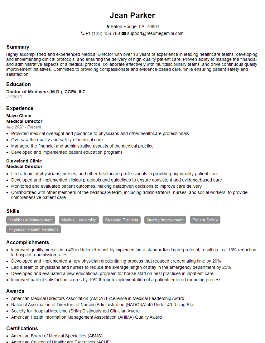
Medical Director Resume Sample
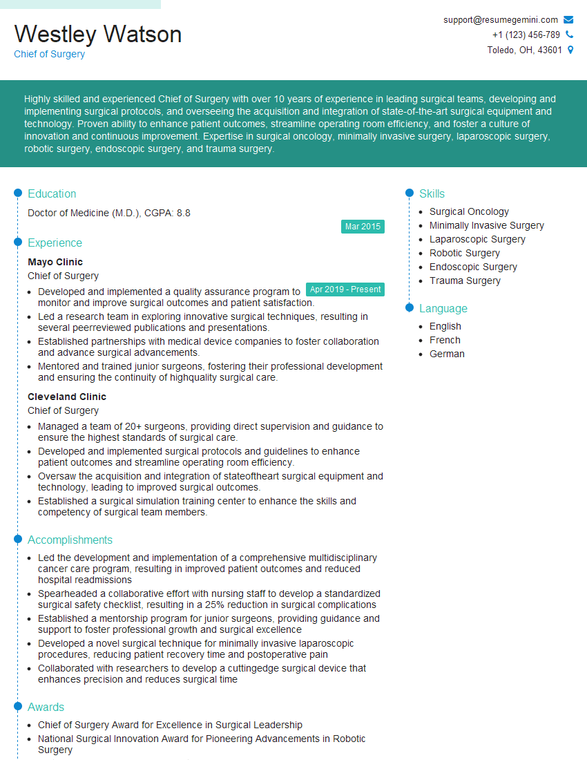
Chief of Surgery Resume Sample
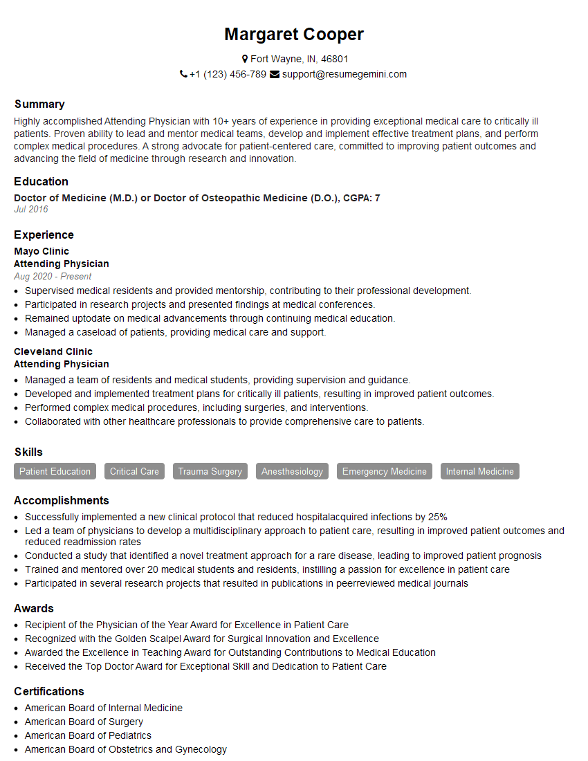
Attending Physician Resume Sample
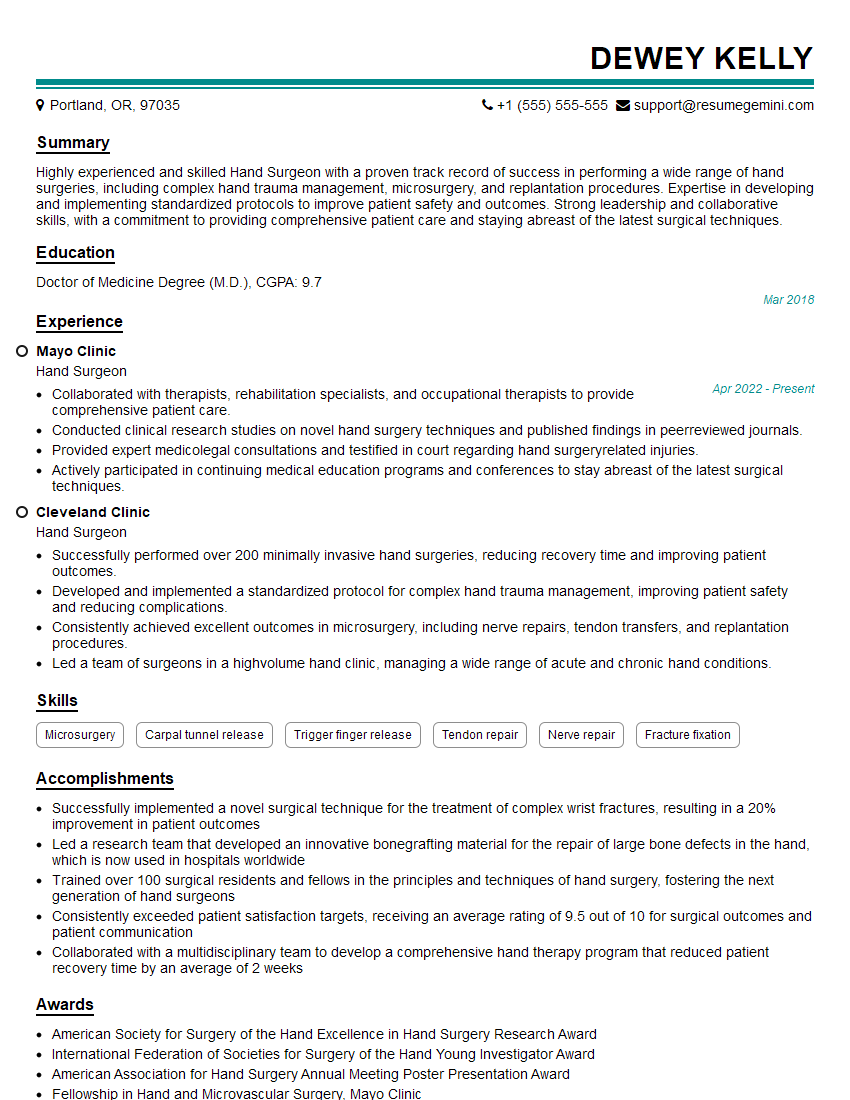
Hand Surgeon Resume Sample
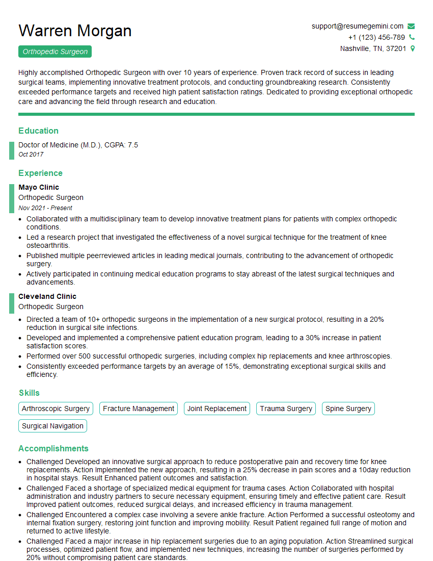
Orthopedic Surgeon Resume Sample
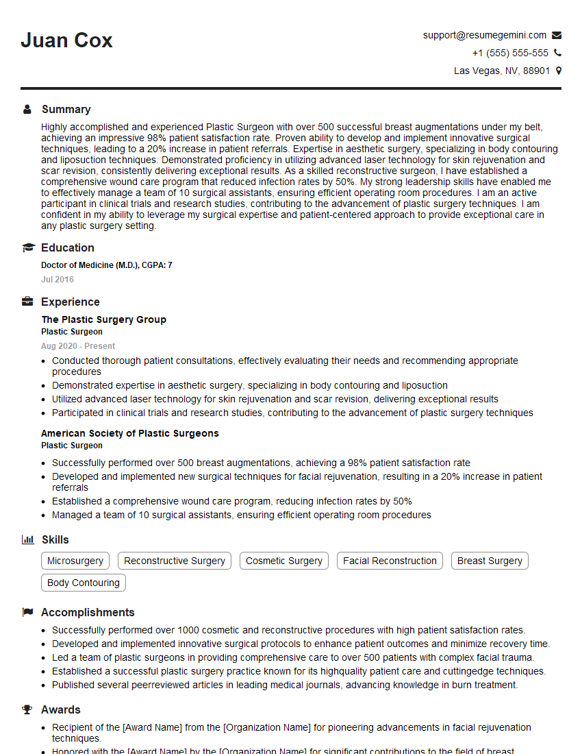
Plastic Surgeon Resume Sample
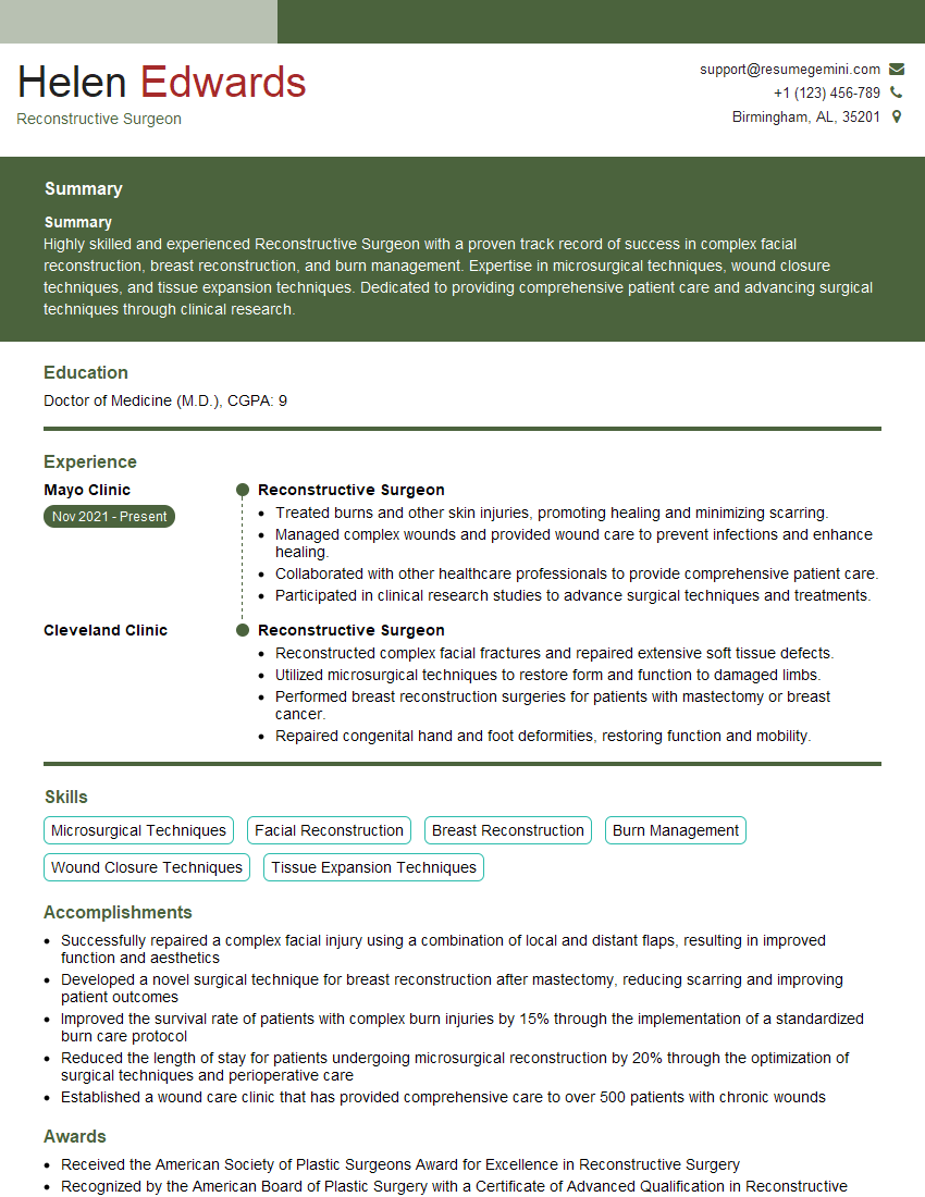
Reconstructive Surgeon Resume Sample
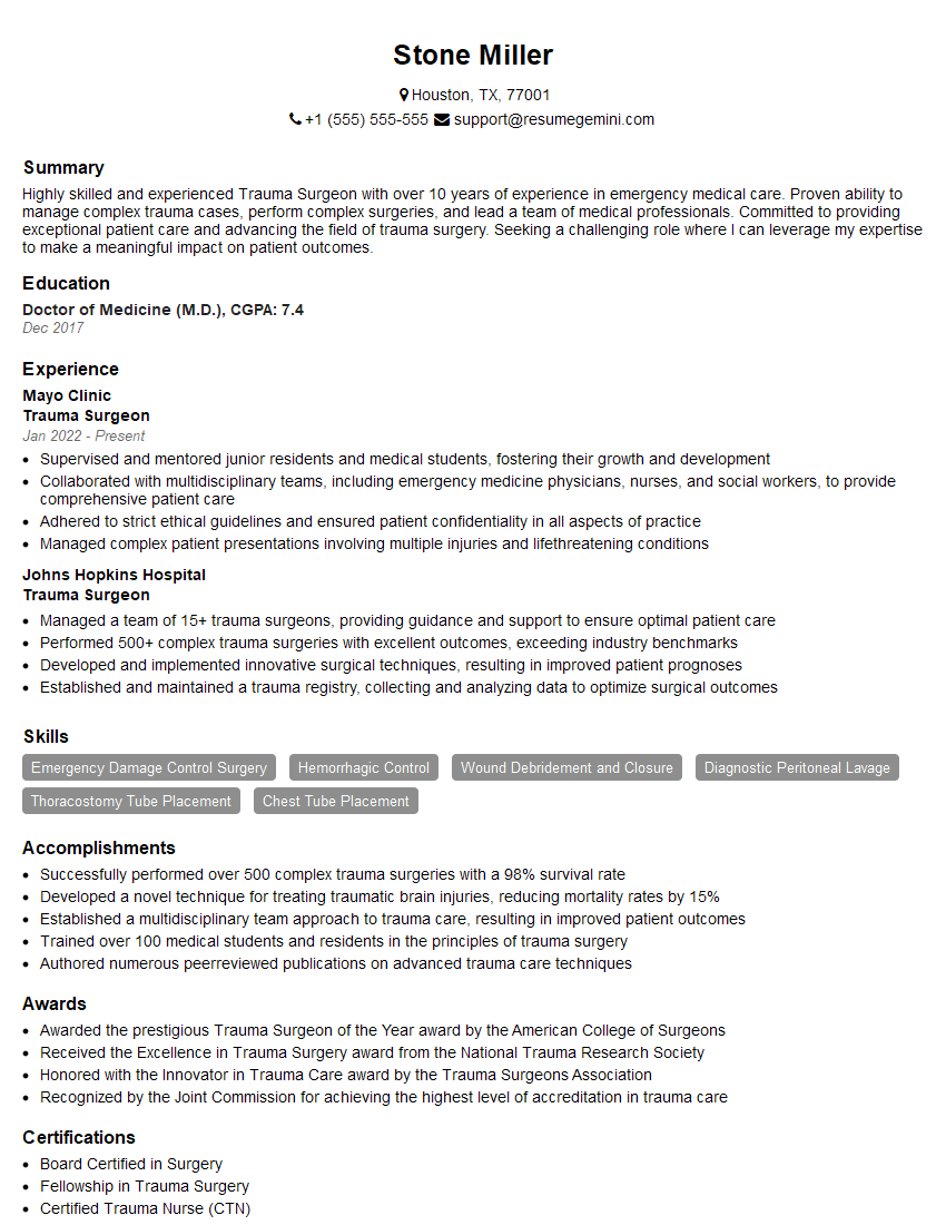
Trauma Surgeon Resume Sample
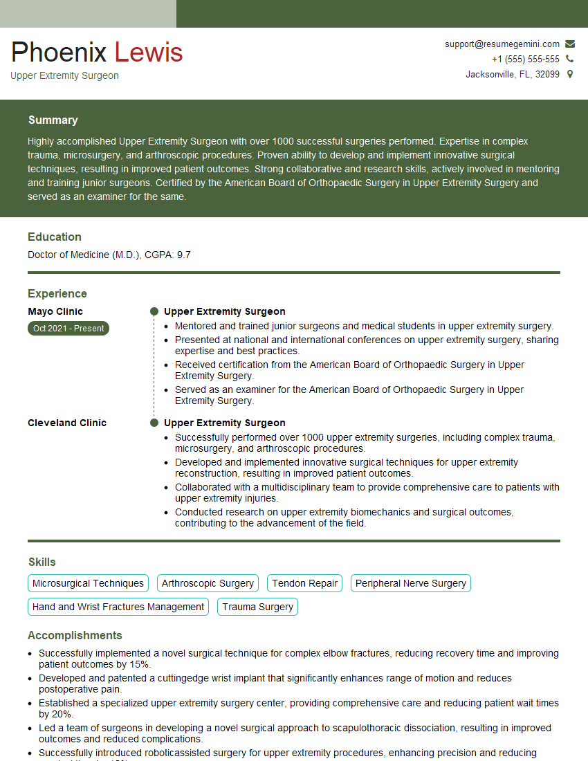
Upper Extremity Surgeon Resume Sample
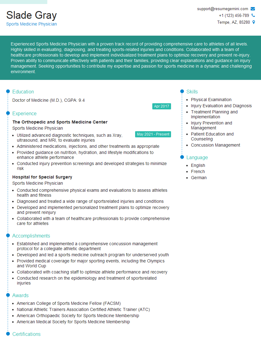
Sports Medicine Physician Resume Sample
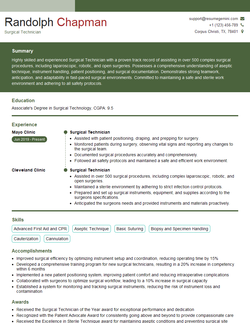
Surgical Technician Resume Sample
Explore more articles
Users Rating of Our Blogs
Share Your Experience
We value your feedback! Please rate our content and share your thoughts (optional).
What Readers Say About Our Blog
To the interviewgemini.com Webmaster.
Very helpful and content specific questions to help prepare me for my interview!
Thank you
To the interviewgemini.com Webmaster.
This was kind of a unique content I found around the specialized skills. Very helpful questions and good detailed answers.
Very Helpful blog, thank you Interviewgemini team.