The right preparation can turn an interview into an opportunity to showcase your expertise. This guide to Cytopathology interpretation interview questions is your ultimate resource, providing key insights and tips to help you ace your responses and stand out as a top candidate.
Questions Asked in Cytopathology interpretation Interview
Q 1. Describe the process of Pap smear preparation and interpretation.
The Pap smear, or cervical cytology, is a crucial screening test for cervical cancer. It involves collecting cells from the cervix using a spatula and a brush, then spreading them thinly onto a glass slide. The slide is then fixed and stained, typically using the Papanicolaou stain (Pap stain), which allows for clear visualization of the cellular details under a microscope. Interpretation involves a systematic examination of the cellular morphology – the size, shape, nuclear characteristics, and cytoplasmic features of individual cells and their arrangement. The cytologist looks for abnormalities that might suggest infection, inflammation, precancerous changes, or cancer.
For example, a normal Pap smear will show predominantly mature squamous cells with a low nuclear-to-cytoplasmic ratio and no significant abnormalities in nuclear structure. In contrast, a smear suggestive of precancerous changes might exhibit cells with larger, darker nuclei and altered cytoplasmic features.
The entire process, from sample collection to microscopic examination and interpretation, requires meticulous attention to detail to ensure accurate diagnosis and appropriate patient management.
Q 2. Explain the Bethesda System for reporting cervical cytology.
The Bethesda System is a standardized reporting system for cervical cytology results. Its primary goal is to provide clear, consistent, and reproducible reporting across different laboratories, thus improving communication between cytologists, clinicians, and patients. The system organizes findings into specific categories, avoiding ambiguous terminology.
Key components of the Bethesda System include:
- Specimen adequacy: Indicates whether enough cells were collected for accurate interpretation.
- General categorization: Describes the overall cellular components of the smear (e.g., presence of inflammation, infection).
- Squamous cell abnormalities: Classifies abnormalities of squamous cells, ranging from atypical squamous cells of undetermined significance (ASCUS) to low-grade squamous intraepithelial lesion (LSIL) and high-grade squamous intraepithelial lesion (HSIL).
- Glandular cell abnormalities: Classifies abnormalities of glandular cells, including atypical glandular cells (AGC) and adenocarcinoma.
- Other findings: Includes notes on any other significant observations, such as infections (e.g., HPV, Trichomonas).
This standardized approach helps clinicians make informed decisions about further management, such as follow-up testing or colposcopy (direct visualization of the cervix).
Q 3. Differentiate between reactive and neoplastic changes in cervical cytology.
Differentiating between reactive and neoplastic changes in cervical cytology is crucial for accurate diagnosis and management. Reactive changes are benign alterations in the cells caused by inflammation, infection, or irritation. Neoplastic changes, on the other hand, represent abnormal cell growth that can be precancerous or cancerous.
Reactive changes are characterized by features like:
- Increased cellularity and inflammation
- Nuclear enlargement, but usually with regular chromatin pattern
- Abundant cytoplasm
- Absence of significant cytologic atypia
Neoplastic changes display more concerning features like:
- Increased nuclear-to-cytoplasmic ratio
- Nuclear hyperchromasia (darkly stained nucleus)
- Irregular nuclear membranes
- Increased mitotic activity (cell division)
- Loss of cell polarity and cohesiveness
It’s important to note that some reactive changes can mimic neoplastic ones, hence the need for experienced cytologists and often the utilization of ancillary tests like HPV testing to make a definitive diagnosis.
Q 4. How do you interpret a Pap smear showing atypical squamous cells of undetermined significance (ASCUS)?
ASCUS (atypical squamous cells of undetermined significance) is an indeterminate cytological finding that indicates the presence of abnormal squamous cells whose nature cannot be definitively classified as either reactive or neoplastic. It’s essentially a ‘gray area’ diagnosis requiring further investigation.
Interpreting an ASCUS result involves careful consideration of the patient’s age, risk factors (e.g., smoking, multiple sexual partners), and the extent of cellular atypia. Further evaluation is almost always recommended, commonly involving HPV testing. If the HPV test is negative, the likelihood of a significant abnormality is low, and the patient may undergo repeat cytology in a specified time frame. A positive HPV test, however, suggests a higher risk of precancerous lesions and often necessitates colposcopy for direct visualization and potential biopsy.
The management of ASCUS illustrates the importance of a collaborative approach between the cytologist, clinician, and the patient, ensuring appropriate follow-up and timely intervention when necessary.
Q 5. Describe the criteria for diagnosing low-grade squamous intraepithelial lesion (LSIL) and high-grade squamous intraepithelial lesion (HSIL).
LSIL (low-grade squamous intraepithelial lesion) and HSIL (high-grade squamous intraepithelial lesion) represent precancerous changes in the squamous epithelium of the cervix. The distinction is crucial because HSIL has a much greater potential to progress to cancer.
LSIL is characterized by:
- Mild nuclear atypia
- Koilocytosis (perinuclear clearing and cytoplasmic vacuolation, often associated with HPV infection)
- Relatively well-preserved cell architecture
HSIL shows more significant abnormalities:
- Marked nuclear atypia (pleomorphism, hyperchromasia)
- Increased nuclear-to-cytoplasmic ratio
- Disrupted cell architecture with cellular maturation abnormalities
- Cells may show features consistent with carcinoma in situ
LSIL often regresses spontaneously, while HSIL requires close monitoring and often colposcopic evaluation with directed biopsy to assess for the presence of cervical intraepithelial neoplasia (CIN) of varying grades.
Q 6. What are the cytological features of endometrial adenocarcinoma?
Endometrial adenocarcinoma is a cancer of the uterine lining. Cytologically, it manifests with a variety of appearances, depending on the differentiation grade of the tumor.
Common cytological features include:
- Individual malignant glandular cells: These cells are typically enlarged with a high nuclear-to-cytoplasmic ratio, prominent nucleoli, irregular nuclear membranes, and hyperchromatic nuclei. They often exhibit nuclear pleomorphism (variation in size and shape).
- Glandular clusters: Malignant glandular cells may form clusters with varying degrees of cohesiveness. Loss of normal glandular architecture is a significant indicator of malignancy.
- Nuclear atypia: The most striking feature is the significant variation in nuclear size and shape. Chromatin pattern is often coarsely clumped and irregular.
- Intracytoplasmic vacuoles: In some cases, intracytoplasmic vacuoles (clear spaces within the cytoplasm) may be observed.
The diagnosis of endometrial adenocarcinoma from cytology specimens often requires considerable experience and careful correlation with clinical information. The sensitivity and specificity of cytology in detecting endometrial cancer are lower compared to other cytological diagnoses, often necessitating further investigation such as endometrial biopsy.
Q 7. How do you differentiate between benign and malignant effusions?
Differentiating between benign and malignant effusions (fluid accumulations in body cavities) relies on a thorough cytological examination. Benign effusions typically result from inflammation or congestive heart failure, while malignant effusions are often associated with cancers spreading to the serosal surfaces (e.g., pleura, peritoneum).
Benign effusions typically show:
- Predominantly reactive mesothelial cells (lining cells of body cavities)
- Few inflammatory cells (lymphocytes, macrophages)
- Absence of malignant cells
Malignant effusions often demonstrate:
- Presence of malignant cells (e.g., adenocarcinoma, mesothelioma)
- Abundant atypical cells with features described earlier (nuclear pleomorphism, high nuclear-to-cytoplasmic ratio)
- Cell clusters and papillae, indicative of malignancy
- Increased cellularity overall
However, it’s essential to remember that some malignant effusions may have low cellularity, making diagnosis challenging. Immunocytochemistry (staining for specific cellular markers) can be extremely helpful in identifying the origin and nature of malignant cells. The interpretation of effusion cytology requires considerable experience and is often combined with clinical information and imaging studies to reach a definitive conclusion.
Q 8. Describe the cytological features of thyroid cancer.
Thyroid cancer cytology shows a wide range of appearances depending on the specific type. Papillary thyroid carcinoma (PTC), the most common type, often presents with characteristic nuclear features: nuclear grooves (like a ‘coffee bean’ shape), psammoma bodies (concentrically layered calcifications), and nuclear enlargement with finely dispersed chromatin (giving a translucent, ‘Orphan Annie eye’ appearance). Follicular thyroid carcinomas may display more uniform cells arranged in follicles or sheets, often with a distinct nuclear enlargement and sometimes with invasion into the surrounding stroma. Medullary thyroid carcinoma, arising from parafollicular C cells, shows nests or sheets of polygonal cells with abundant cytoplasm, often with amyloid stroma visible on special stains. Anaplastic thyroid carcinoma, the most aggressive type, demonstrates bizarre, pleomorphic cells with giant nuclei and marked atypia, often making diagnosis challenging based on cytology alone. Accurate diagnosis requires careful evaluation of nuclear features, cellular arrangement, and the presence of any specific features like psammoma bodies or amyloid.
Q 9. Explain the significance of finding suspicious cells in a fine-needle aspiration (FNA) of a breast mass.
Finding suspicious cells in a breast FNA is a critical finding that mandates careful interpretation and often requires correlation with clinical and radiological data. Suspicious features include nuclear pleomorphism (variation in nuclear size and shape), high nuclear-to-cytoplasmic ratio (meaning the nucleus is large compared to the cell body), prominent nucleoli (dark spots inside the nucleus), loss of cell polarity (disorganized cellular arrangement), and evidence of mitosis (cell division). These features can suggest malignancy, but it’s crucial to remember that benign conditions can sometimes mimic these findings. For example, reactive atypia, often seen in inflammatory or fibrocystic changes, can cause cellular changes that overlap with those of cancer. The cytologist will consider the overall context, the number of suspicious cells, and the presence of any other features to render a diagnosis, which may range from benign to suspicious for malignancy to definitively malignant, often necessitating further investigation like core biopsy or surgical excision.
Q 10. What are the cytological features of lymphoma?
Lymphoma cytology reveals a heterogeneous population of lymphocytes, usually showing a significant increase in the number of lymphocytes compared to a normal blood smear. The specific cytological features depend on the type of lymphoma. In Hodgkin lymphoma, Reed-Sternberg cells are diagnostic; these are large, binucleate or multinucleate cells with prominent nucleoli. Non-Hodgkin lymphomas display a broader range of morphologies, often categorized by the size and shape of the lymphocytes, the presence of cleaved or non-cleaved nuclei, and the amount of cytoplasm. Some non-Hodgkin lymphomas show a monotonous population of small, mature lymphocytes, while others exhibit larger, less differentiated cells. Immunocytochemistry is crucial for classifying lymphoma subtypes, distinguishing between B-cell and T-cell lymphomas, and guiding treatment decisions. It’s important to note that a reactive lymphoid hyperplasia (a benign increase in lymphocytes) can sometimes mimic low-grade lymphomas, so correlation with clinical information is essential for accurate diagnosis.
Q 11. Describe the different types of lung cancer and their cytological features.
Lung cancer encompasses several subtypes, each with distinct cytological characteristics. Adenocarcinoma, the most common type, often displays glandular formations or individual cells with abundant cytoplasm and mucin. Squamous cell carcinoma typically shows cells with abundant, keratinized cytoplasm, often showing ‘intercellular bridges’ connecting adjacent cells. Small cell carcinoma is characterized by small, intensely dark nuclei and scant cytoplasm; these cells often appear in clusters or sheets. Large cell carcinoma is a heterogeneous group with a variable appearance that lacks the defining features of the other types and is often diagnosed through exclusion. Cytologic examination can be helpful in identifying the type of lung cancer, but it’s usually not sufficient for precise sub-classification. Molecular testing on tissue samples is often necessary for targeted therapy selection.
Q 12. How do you approach the interpretation of a poorly cellular specimen?
Interpreting a poorly cellular specimen is challenging because it limits the amount of diagnostic material available. The approach involves meticulously examining every cell present. This requires careful scanning of the entire slide at low power magnification to ensure no cellular clusters are missed. Then, higher magnification is used to carefully evaluate the morphology of individual cells and any cellular groupings present. The clinical history and radiological findings become even more crucial in guiding the interpretation. The cytologist must weigh the possibility of a truly non-diagnostic specimen against the chance of a low-cellularity lesion that still contains diagnostic features. Even a single suspicious cell in a scanty background can have significant diagnostic implications, necessitating further investigation. Reporting should carefully reflect the limitations imposed by the poor cellularity.
Q 13. What are the limitations of cytology in diagnosis?
Cytology has limitations, primarily related to its inherent nature as a sampling technique. The sample obtained may not be representative of the entire lesion. This is especially true for heterogeneous lesions, where only a small portion is sampled. The quality of the specimen significantly impacts diagnostic accuracy; poorly cellular specimens or those with artifacts can be difficult to interpret. Furthermore, some lesions may have subtle cytological features that are difficult to distinguish from benign processes. Overlapping cytological features between benign and malignant conditions can lead to diagnostic uncertainty. Cytology should be considered a component of a broader diagnostic workup, not a standalone test, often needing correlation with clinical data, imaging, and other tests like immunocytochemistry and molecular diagnostics.
Q 14. Discuss the role of ancillary techniques in cytopathology (e.g., immunocytochemistry, flow cytometry).
Ancillary techniques significantly enhance the diagnostic capabilities of cytopathology. Immunocytochemistry uses antibodies to identify specific proteins within cells, allowing for precise classification of various cell types, especially crucial in lymphomas and other tumors. For example, identifying CD20 in a lymphoma helps to confirm its B-cell origin. Flow cytometry allows for rapid and quantitative analysis of cellular populations, useful in assessing the proportion of different cell types and identifying unique markers, invaluable for diagnosing hematologic malignancies. Other techniques include special stains (e.g., for amyloid in medullary thyroid cancer) and molecular tests (like PCR for specific gene mutations or rearrangements), which add layers of information for more definitive diagnoses and personalized treatment strategies. These ancillary techniques bridge the gap between morphology and molecular information, ultimately improving diagnostic accuracy and patient management.
Q 15. How do you manage discrepancies between cytology and histology results?
Discrepancies between cytology and histology results are unfortunately common and necessitate a careful, multi-step approach. The key is to understand that these methods sample tissue differently; cytology examines individual cells, while histology assesses tissue architecture. A discrepancy doesn’t automatically invalidate one result; instead, it often highlights the limitations of each technique.
My approach involves several steps:
- Reviewing both reports meticulously: I carefully examine the cytology smears and the histology slides, looking for potential explanations such as sampling error (different areas of the lesion sampled), cellular changes missed on the smaller cytology sample, or the presence of artifacts in either method.
- Clinical correlation: I reassess the patient’s clinical history, imaging findings, and other relevant data to see if any information can reconcile the findings. For instance, a small, poorly cellular lesion may yield inadequate cytological material but show more extensive disease on histological examination.
- Considering diagnostic possibilities: Sometimes, both results are partially correct but reflect different facets of the disease. For example, cytology might reveal atypia, while histology reveals a low-grade malignancy. This requires careful interpretation to arrive at an accurate overall diagnosis.
- Consultation with colleagues: If the discrepancy remains unresolved, I consult with experienced colleagues, pathologists, and clinicians involved in the patient’s care to reach a consensus on management. This ensures a multidisciplinary approach and avoids potential misinterpretations.
- Additional testing if needed: In some cases, further investigations like immunohistochemistry or molecular studies on the histology specimen may be necessary to clarify the discrepancy.
For example, I once encountered a case where fine-needle aspiration cytology (FNAC) suggested a benign thyroid nodule, while subsequent surgical histology revealed a follicular adenoma (a benign tumor). The discrepancy was likely due to the limited sampling provided by FNAC. In such cases, careful clinical correlation, along with detailed evaluation of both reports, is critical to providing appropriate patient care.
Career Expert Tips:
- Ace those interviews! Prepare effectively by reviewing the Top 50 Most Common Interview Questions on ResumeGemini.
- Navigate your job search with confidence! Explore a wide range of Career Tips on ResumeGemini. Learn about common challenges and recommendations to overcome them.
- Craft the perfect resume! Master the Art of Resume Writing with ResumeGemini’s guide. Showcase your unique qualifications and achievements effectively.
- Don’t miss out on holiday savings! Build your dream resume with ResumeGemini’s ATS optimized templates.
Q 16. What are the common artifacts that can be seen in cytology specimens?
Artifacts in cytology specimens are non-diagnostic structures that can mimic true cellular abnormalities, leading to misinterpretations if not carefully recognized. These artifacts stem from various sources during specimen collection, processing, or staining.
Some common artifacts include:
- Air drying artifacts: These appear as cellular shrinkage and nuclear pyknosis (small, condensed nuclei), often resulting from inadequate or slow fixation.
- Crush artifacts: Result from excessive pressure during slide preparation, causing overlapping cells and distorted morphology.
- Red blood cell aggregates: These can obscure cellular details and, if numerous, might mimic cell clusters.
- Fixative artifacts: Improper fixation can lead to altered cell morphology, including nuclear changes and cytoplasmic artifacts.
- Stain artifacts: Uneven staining, precipitation of stain, and background staining can interfere with cellular interpretation.
- Degeneration artifacts: Cell death and autolysis can lead to altered cellular features, potentially mistaken for malignancy.
Recognizing these artifacts is crucial for accurate diagnosis. Experience plays a vital role in differentiating true pathological changes from artifacts. For instance, while nuclear pyknosis can mimic malignancy, the presence of widespread and uniform pyknosis in a background of drying artifact usually points towards a technical issue, not a malignant process.
Q 17. Discuss your experience with quality control and quality assurance procedures in a cytopathology laboratory.
Quality control (QC) and quality assurance (QA) are paramount in a cytopathology laboratory to ensure the accuracy and reliability of diagnoses. In my experience, these processes encompass several key areas:
- Specimen handling and processing: Strict protocols are followed for specimen accessioning, fixation, and slide preparation to minimize artifacts and maintain specimen integrity. Regular checks on fixative quality and staining reagents are essential.
- Microscopy and slide review: Regular calibration of microscopes and maintenance of equipment are critical. We conduct internal quality control checks, involving review of slides by multiple pathologists, often utilizing peer review and proficiency testing programs to maintain consistent diagnostic accuracy.
- Proficiency testing: Participation in external proficiency testing programs helps assess our laboratory’s performance relative to other facilities. This ongoing evaluation ensures we meet established standards and identify areas for improvement.
- Record keeping and reporting: Detailed record keeping of all cases, including patient information, laboratory procedures, and diagnostic reports, is vital. Regular review of these records helps identify any patterns or issues that could affect the quality of our work. Electronic health records (EHRs) and laboratory information systems (LISs) significantly streamline this process.
- Continuing education and training: Ongoing training for technicians and pathologists on best practices, new technologies, and emerging diagnostic challenges is essential. Attendance at conferences, participation in workshops, and review of latest research articles maintain competency and improve accuracy.
For instance, during my tenure at a previous lab, we implemented a new digital slide scanning system. This required extensive training for the staff and the development of new quality control procedures to ensure image quality and data security. This meticulous approach to QC and QA ensures reliable and accurate results which are vital for optimal patient care.
Q 18. Describe your experience with different types of staining methods used in cytology.
Various staining methods are employed in cytology, each offering unique advantages for highlighting specific cellular components and features. My experience includes the routine use of several techniques:
- Papanicolaou (Pap) stain: This is the most common stain used in gynecological cytology and other exfoliative cytology specimens. It utilizes a combination of dyes (hematoxylin, eosin, orange G, and others) to highlight nuclear and cytoplasmic details, facilitating assessment of cellular morphology and differentiation.
- Hematoxylin and eosin (H&E) stain: While used primarily in histology, H&E is also helpful in cytology, particularly in fine-needle aspiration (FNA) specimens. It provides good overall nuclear and cytoplasmic staining, revealing basic architectural features, if present in the sample.
- Romanowsky stains (e.g., Giemsa, Wright-Giemsa): These stains are particularly useful for hematological specimens and can be valuable in assessing inflammatory cells and certain microorganisms in cytological samples. These are especially helpful when dealing with infectious entities within cells.
- Special stains: Various special stains are used to highlight specific cellular components, including periodic acid-Schiff (PAS) for glycogen detection, immunohistochemical (IHC) stains for specific markers (e.g., cytokeratins for epithelial cells, CD markers for lymphocytes), and other stains like Prussian blue for hemosiderin, to refine diagnosis. Examples of their use include diagnosing fungal infections using PAS and distinguishing between different types of lymphomas using IHC.
The choice of staining method depends on the type of specimen and the clinical questions being addressed. For instance, Pap stain is optimal for evaluating cervical smears, while H&E is better suited for FNA samples from solid organs. The judicious use of special stains significantly increases diagnostic accuracy.
Q 19. Explain your understanding of the legal and ethical aspects of cytopathology.
Cytopathology involves significant legal and ethical responsibilities. Accuracy in diagnosis is paramount; an incorrect diagnosis can lead to delayed or inappropriate treatment, causing significant harm to the patient. Ethical considerations encompass several key areas:
- Maintaining confidentiality: Patient information must be protected and handled in accordance with relevant privacy laws (e.g., HIPAA). This includes careful storage and transmission of patient data, as well as the appropriate access and disclosure of diagnostic information to authorized personnel only.
- Ensuring diagnostic accuracy: This involves following established protocols, participating in quality assurance programs, and seeking consultation when needed. This ensures responsible interpretation of cytological findings.
- Professional competence: Continuing education and staying abreast of the latest advancements in the field are vital for maintaining diagnostic accuracy. Failure to do so is a breach of ethical and professional standards.
- Informed consent: Patients must be adequately informed about the procedure (e.g., FNA), potential risks and benefits, and the limitations of cytological assessment. This should be part of the patient’s overall treatment plan.
- Legal implications: Cytology reports are often pivotal in clinical decision-making; thus, careful wording and clear communication are essential to avoid ambiguity and potential legal liability. Accurate reporting is crucial to avoid misleading medical information.
In my experience, adhering to these principles necessitates a conscientious approach to every aspect of the diagnostic process. Maintaining a culture of quality assurance and ongoing learning ensures ethical and legal compliance.
Q 20. Describe your experience with image analysis software in cytopathology.
Image analysis software is increasingly important in cytopathology, offering tools that enhance diagnostic accuracy and efficiency. My experience encompasses the use of several systems for various purposes:
- Digital slide scanning and viewing: Whole slide imaging (WSI) systems allow for digital scanning of glass slides, facilitating remote viewing, teleconsultation, and improved workflow. This greatly improves the workflow compared to the traditional microscope.
- Computer-aided diagnosis (CAD) software: Certain systems provide algorithms that can assist in identifying potentially abnormal cells, although these tools are not intended to replace human interpretation. These programs can detect certain cellular features that may help with early detection.
- Quantitative image analysis: Some software allows for quantification of cellular features (e.g., nuclear size, nuclear-to-cytoplasmic ratio), which can be helpful in certain diagnostic situations. These objective measurements can help with standardized diagnostic reporting.
- Automated cell counting and classification: Specialized image analysis systems are being developed to automate the counting and classification of cells in cytology preparations, but such technology is still in an early stage of development for routine use in cytopathology.
While these tools enhance workflow and assist in analysis, they do not replace the expertise of a trained cytopathologist. Human review and interpretation remain crucial for accurate diagnosis.
Q 21. How do you stay up-to-date with the latest advancements in cytopathology?
Keeping abreast of the latest advancements in cytopathology is crucial for maintaining competence and providing optimal patient care. My approach to continuous learning involves several strategies:
- Regular review of scientific literature: I regularly read peer-reviewed journals, such as the American Journal of Clinical Pathology and the Diagnostic Cytopathology, to stay updated on new research findings and diagnostic techniques.
- Attendance at professional meetings and conferences: Participation in professional meetings like those held by the American Society of Cytopathology (ASC) provides opportunities to learn from experts, network with colleagues, and participate in continuing medical education (CME) activities.
- Participation in online courses and webinars: Various online platforms offer CME courses and webinars on current topics in cytopathology, providing a convenient way to update knowledge and earn CME credits.
- Collaboration with colleagues: Discussions with colleagues in cytopathology and other related fields often spark new ideas and highlight recent advancements.
- Utilizing online resources: Numerous reputable online resources, such as medical databases (PubMed, Medline), and professional society websites, offer access to updated guidelines, best practices, and other relevant information.
This multi-faceted approach ensures I remain knowledgeable about new diagnostic techniques, treatment strategies, and emerging challenges in the field, thereby directly benefiting my diagnostic capabilities and patient care.
Q 22. Discuss your experience with teaching or mentoring in cytopathology.
Throughout my career, I’ve had extensive experience teaching and mentoring in cytopathology. This has ranged from formal lectures and workshops for medical students and residents to one-on-one guidance for fellows and junior colleagues. My approach focuses on a practical, case-based learning style. For instance, I often present complex cases with challenging diagnostic features, encouraging collaborative problem-solving amongst my mentees. We discuss differential diagnoses, analyze imaging and clinical correlations, and critically review the microscopic features. This interactive methodology promotes critical thinking and strengthens their ability to interpret cytological findings independently. I also actively participate in the development and delivery of continuing medical education (CME) programs, keeping my colleagues abreast of the latest advancements and best practices in cytopathology.
For example, during a recent fellowship teaching session, we reviewed a case of a fine needle aspiration (FNA) of a thyroid nodule displaying atypia. I guided the fellows through a step-by-step analysis, highlighting the subtle nuclear features indicative of malignancy versus benign processes. This hands-on experience significantly enhanced their diagnostic abilities.
Q 23. How would you approach a case with unusual or unexpected cytological findings?
Encountering unusual or unexpected cytological findings requires a systematic and methodical approach. My first step involves a thorough review of the clinical history, correlating it with the cytological features. This includes assessing the patient’s age, sex, medical history, risk factors, and the nature of the lesion sampled. Next, I carefully re-examine the slides at low and high magnification, focusing on identifying any additional clues that might have been missed in the initial review.
If the initial assessment remains unclear, I employ several strategies. This might involve utilizing special stains such as immunocytochemistry (ICC) and flow cytometry to further characterize the cellular population. For example, in cases of uncertain malignant potential, ICC for keratins or other markers can be crucial for differentiation. If necessary, consultation with colleagues in other specialties (e.g., surgical pathology, hematopathology) is invaluable for reaching a consensus and arriving at the most accurate diagnosis. In challenging situations, I also consider obtaining additional specimens – perhaps a repeat FNA or surgical excision biopsy – for better clarification. Thorough documentation of the findings and the diagnostic reasoning is crucial for transparency and potential peer review.
Q 24. Describe your experience with different types of cytological specimens (e.g., body fluids, smears, imprints).
My experience encompasses a broad range of cytological specimens, including body fluids (serous effusions, cerebrospinal fluid, urine), smears (cervical, gynecological, non-gynecological), and imprints (fine-needle aspiration biopsies, surgical biopsies). Each specimen type presents unique challenges and requires specific interpretive skills.
For example, analyzing serous effusions requires expertise in differentiating between reactive mesothelial cells and malignant cells, often requiring detailed assessment of nuclear morphology and cellular arrangement. Processing and interpretation of fine-needle aspiration (FNA) biopsies involve understanding the sampling artifacts and recognizing the limitations of this technique. Cervical smears, on the other hand, require proficiency in identifying precancerous and cancerous lesions within the context of the Bethesda classification system. I am adept at handling and interpreting the nuances of each specimen type, always mindful of the limitations of each technique and integrating the cytological findings with clinical data for accurate diagnosis.
Q 25. What are your strengths and weaknesses in cytopathology?
My strengths lie in my meticulous attention to detail, strong diagnostic skills, and ability to correlate cytological findings with clinical information. I am adept at handling complex and challenging cases, and I possess excellent communication skills, enabling me to effectively convey information to colleagues, patients, and referring physicians. I thrive in collaborative environments and am always eager to learn and incorporate new technologies and techniques into my practice.
One area I’m actively working on is expanding my proficiency in advanced molecular techniques, such as next-generation sequencing (NGS), to further enhance my diagnostic capabilities. While I am familiar with the principles of these techniques, hands-on experience with NGS would further strengthen my skills in this rapidly evolving field.
Q 26. Why are you interested in this specific cytopathology position?
I am deeply interested in this cytopathology position because it aligns perfectly with my career goals and expertise. Your institution’s reputation for excellence in cytopathology, along with its commitment to cutting-edge research and innovative diagnostic techniques, is particularly appealing. The opportunity to collaborate with experienced professionals and contribute to a dynamic team is highly motivating. I am particularly drawn to [mention a specific program, research area, or aspect of the position that excites you]. The potential to contribute to both patient care and advancements in the field is an exciting prospect.
Q 27. What are your salary expectations?
My salary expectations are commensurate with my experience, qualifications, and the responsibilities of this position. I am open to discussing this further based on the specifics of the compensation package and benefits offered.
Key Topics to Learn for Cytopathology Interpretation Interview
- Specimen Adequacy Assessment: Understanding criteria for evaluating specimen quality, including cellularity, preservation, and artifacts. Practical application involves analyzing Pap smears and fine needle aspirates to determine suitability for diagnosis.
- Cellular Morphology: Mastering the identification and differentiation of normal and abnormal cells based on nuclear and cytoplasmic features. This includes recognizing reactive changes, inflammatory processes, and neoplastic transformations. Problem-solving involves distinguishing benign from malignant cellular atypia.
- Inflammatory and Reactive Processes: Differentiating various inflammatory conditions and reactive changes from neoplastic processes. Practical application includes identifying infectious agents and evaluating the context of inflammation in the specimen.
- Neoplastic Diagnosis: Accurately identifying and classifying different types of cancers based on cytological features. This includes understanding grading systems and prognostic implications. Problem-solving involves differentiating subtle cytological features between different tumor types and grades.
- Benign vs. Malignant Differentiation: Developing expertise in distinguishing benign from malignant cellular changes, a crucial skill for accurate diagnosis and patient management. Practical application includes interpreting Pap smears for cervical cancer screening and analyzing fine needle aspirates of suspicious masses.
- Quality Assurance and Control: Understanding the importance of quality control measures in cytopathology. This includes familiarity with proficiency testing and internal quality assurance programs.
- Reporting and Communication: Effectively communicating cytological findings in clear and concise reports, tailored to the needs of the clinicians involved. This involves understanding appropriate terminology and communicating diagnostic uncertainty when needed.
Next Steps
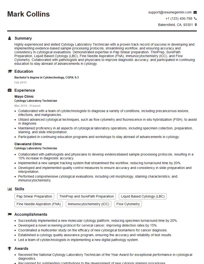
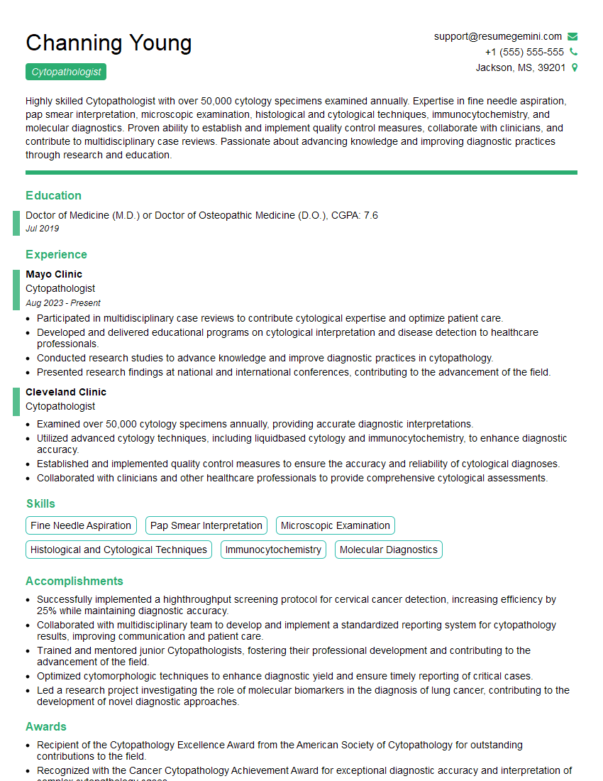
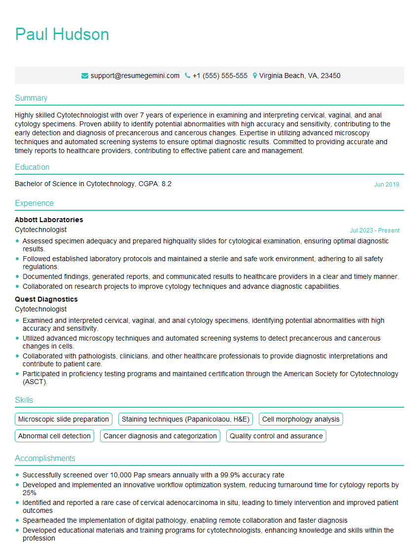
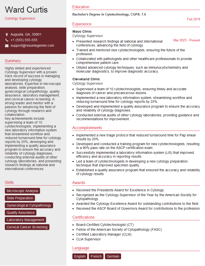
Mastering cytopathology interpretation is crucial for advancing your career in this specialized field. It opens doors to leadership roles, research opportunities, and increased professional recognition. To maximize your job prospects, crafting an ATS-friendly resume is essential. ResumeGemini is a trusted resource that can significantly enhance your resume-building experience, helping you showcase your skills and experience effectively. ResumeGemini provides examples of resumes tailored to cytopathology interpretation to guide you in creating a compelling application. Invest time in building a strong resume – it’s your first impression to potential employers.
Explore more articles
Users Rating of Our Blogs
Share Your Experience
We value your feedback! Please rate our content and share your thoughts (optional).
What Readers Say About Our Blog
To the interviewgemini.com Webmaster.
Very helpful and content specific questions to help prepare me for my interview!
Thank you
To the interviewgemini.com Webmaster.
This was kind of a unique content I found around the specialized skills. Very helpful questions and good detailed answers.
Very Helpful blog, thank you Interviewgemini team.