Every successful interview starts with knowing what to expect. In this blog, we’ll take you through the top Fluoroscopic-Guided Procedures interview questions, breaking them down with expert tips to help you deliver impactful answers. Step into your next interview fully prepared and ready to succeed.
Questions Asked in Fluoroscopic-Guided Procedures Interview
Q 1. Explain the principles of fluoroscopy.
Fluoroscopy is a dynamic imaging technique that uses X-rays to create real-time moving images of the internal structures of the body. Imagine it like a continuous X-ray movie, allowing physicians to visualize the movement of instruments, contrast agents, or organs during a procedure. The principle rests on the ability of X-rays to penetrate soft tissues and be differentially absorbed by denser materials like bone. This difference in absorption creates variations in image brightness, allowing us to differentiate different structures.
A fluoroscopic system consists of an X-ray source, an image intensifier, and a monitor displaying the live images. The X-ray beam passes through the patient and interacts with the tissues, causing some X-rays to be absorbed and others to pass through. The remaining X-rays are detected by the image intensifier, which converts them into a visible image. This image is then displayed on the monitor, allowing the physician to guide instruments in real-time.
Q 2. Describe the different types of fluoroscopic equipment and their applications.
Fluoroscopic equipment varies in its design and capabilities, catering to different procedures and needs. There are portable units, ideal for bedside procedures and operating rooms with limited space, and stationary units found in dedicated fluoroscopy suites. These stationary units often provide enhanced image quality and functionalities. Mobile C-arms are particularly versatile, allowing for imaging from various angles and positions around the patient.
- Mobile C-arms: These are widely used in operating rooms for orthopedic surgeries, vascular interventions, and other procedures requiring precise instrument placement.
- Fixed fluoroscopy units: Found in dedicated cardiac catheterization labs or interventional radiology suites, these units offer superior image quality and advanced features such as digital subtraction angiography (DSA).
- Portable fluoroscopy units: These are smaller, lightweight systems often used for procedures outside of a dedicated suite, providing flexibility but potentially with slightly lower image quality.
The choice of equipment depends on the specific procedure, the patient’s condition, and the resources available. For example, a minimally invasive procedure might utilize a mobile C-arm, whereas a complex cardiac intervention would demand a dedicated fixed fluoroscopy unit with advanced capabilities.
Q 3. What are the safety precautions associated with fluoroscopy?
Fluoroscopy involves ionizing radiation, posing potential risks to both patients and healthcare staff. Strict adherence to safety protocols is paramount. These precautions include:
- Minimizing exposure time: The principle of ALARA (As Low As Reasonably Achievable) should always be followed. Procedures should be planned meticulously and executed efficiently to reduce the time spent under fluoroscopy.
- Optimizing beam collimation: The X-ray beam should be carefully collimated (restricted) to encompass only the area of interest, minimizing unnecessary radiation to surrounding tissues.
- Using appropriate shielding: Lead aprons, thyroid collars, and other protective shielding should be used by both the patient and staff to reduce radiation exposure.
- Distance optimization: Maintaining a safe distance from the X-ray source is crucial. The inverse square law dictates that radiation intensity decreases with the square of the distance.
- Proper training and education: All personnel involved in fluoroscopy procedures must receive comprehensive training on radiation safety and proper equipment operation.
Q 4. How do you minimize radiation exposure to both the patient and the staff?
Minimizing radiation exposure requires a multifaceted approach. For patients, it involves:
- Pulse fluoroscopy: This technique delivers radiation in short bursts instead of continuous exposure, significantly reducing the total dose.
- Last image hold (LIH): This allows the operator to review the last acquired image without continuous radiation exposure.
- Roadmapping: This technique overlays a fluoroscopic image onto a subsequent angiographic image to guide procedures.
For staff, radiation safety relies on:
- Lead shielding: Consistent use of lead aprons, thyroid shields, and other personal protective equipment.
- Distance and time: Staying as far as possible from the radiation source and minimizing exposure time.
- Rotation of staff: Rotating personnel involved in fluoroscopy procedures helps distribute radiation exposure.
- Monitoring devices: Using dosimeters to track individual radiation exposure levels.
Regular monitoring and adherence to established protocols are essential for ensuring safety.
Q 5. Explain the concept of image intensification.
Image intensification is a crucial element of fluoroscopy that enhances the brightness and visibility of the X-ray image. Imagine trying to watch a faint movie; image intensification is like turning up the brightness dramatically. It involves converting the weak X-ray signal into a much brighter visible light image. This is achieved using a complex chain of components within the image intensifier tube.
The process begins with the X-rays striking an input phosphor screen. This screen converts the X-rays into visible light. This light is then amplified by a photocathode, which converts the light into electrons. These electrons are accelerated and focused onto an output phosphor screen. This final screen converts the amplified electron signal back into visible light, resulting in a significantly brighter image than the original X-ray signal. This intensified image can then be viewed directly or captured digitally.
Q 6. Describe the process of image acquisition and processing in fluoroscopy.
Image acquisition in fluoroscopy involves the generation of X-rays, their interaction with the patient’s body, and their detection by the image intensifier. The X-ray source emits a continuous beam of X-rays, which pass through the patient. The image intensifier converts the transmitted X-rays into a visible light image, which is then captured by a camera and displayed on a monitor.
Image processing can range from basic brightness and contrast adjustments to more advanced techniques like digital subtraction angiography (DSA). DSA involves subtracting a pre-contrast image from a post-contrast image, effectively removing overlying structures and highlighting the blood vessels filled with contrast agent. This allows for clearer visualization of the vascular system during procedures like angiograms. Modern systems also often allow for image manipulation, such as zooming, rotating, and measuring distances.
Q 7. What are the common artifacts seen in fluoroscopic images and how are they addressed?
Several artifacts can affect the quality of fluoroscopic images, potentially leading to misinterpretations. These include:
- Scatter radiation: X-rays scattered within the patient’s body can reduce image contrast and sharpness. This can be minimized through careful collimation and the use of anti-scatter grids.
- Motion blur: Patient movement during the procedure can lead to blurred images. This can be reduced by patient cooperation and the use of immobilization devices.
- Quantum mottle: This granular appearance of the image is due to statistical fluctuations in the number of X-rays detected. It can be reduced by increasing the exposure time or X-ray intensity (while remaining within ALARA principles).
- Beam hardening: The differential absorption of lower energy X-rays by the patient can lead to image artifacts. This can be mitigated by using beam filtration.
Addressing these artifacts often involves a combination of technical adjustments, careful procedural technique, and an understanding of the underlying causes. For instance, if motion blur is an issue, better patient immobilization should be sought; if scatter radiation is prominent, improved collimation is often needed.
Q 8. Discuss the role of contrast agents in fluoroscopy-guided procedures.
Contrast agents are crucial in fluoroscopy-guided procedures because they allow us to visualize structures within the body that are otherwise invisible on X-ray imaging. They are substances that absorb X-rays differently than the surrounding tissues, creating a clear distinction between the area of interest and the surrounding anatomy. This allows the physician to accurately guide instruments, catheters, or needles to the target site during a procedure.
Think of it like highlighting a specific paragraph in a book. The contrast agent is like the highlighter, making the target area stand out so we can easily see it on the fluoroscopy screen.
Q 9. Explain the different types of contrast agents and their properties.
Contrast agents are broadly categorized into two main types: iodinated contrast media and barium sulfate.
- Iodinated Contrast Media: These are commonly used for vascular studies (angiograms), and are injected intravenously, intra-arterially, or directly into a specific organ. They contain iodine, which is a highly effective X-ray absorber. They are available in different osmolarities (concentration of solute), affecting their potential for side effects. Low-osmolality contrast media (LOCM) are generally preferred due to a lower risk of adverse reactions.
- Barium Sulfate: This is a non-iodinated contrast agent primarily used for imaging the gastrointestinal tract. It’s ingested or administered rectally and is relatively safe, though rare complications can still occur. It is not used for vascular studies as it’s not soluble in blood.
The choice depends heavily on the specific organ or vascular system being imaged and the route of administration.
Q 10. How do you select the appropriate contrast agent for a specific procedure?
Selecting the appropriate contrast agent involves considering several factors:
- The target anatomy: Barium sulfate is ideal for the GI tract, while iodinated contrast is necessary for vascular imaging.
- Route of administration: Intravenous, intra-arterial, or oral/rectal routes necessitate different contrast agents and concentrations.
- Patient factors: Patient allergies, renal function (crucial for iodinated contrast), and other medical conditions significantly impact the choice. For patients with impaired renal function, we might use lower-osmolarity contrast or even consider alternative imaging modalities.
- Procedure specifics: The type of procedure, such as a coronary angiogram versus a peripheral angiogram, guides the selection of the contrast agent and the technique of administration.
For instance, in a coronary angiogram, we’d use a high-quality iodinated LOCM injected directly into the coronary arteries via a catheter.
Q 11. Describe the complications associated with contrast agents.
Complications associated with contrast agents, though generally rare, can be serious. The most common adverse reactions are related to iodinated contrast media and include:
- Mild Reactions: These are the most frequent and usually include nausea, vomiting, flushing, and itching. These are generally managed with supportive care.
- Moderate Reactions: These include hives, bronchospasm, and hypotension (low blood pressure). They require prompt medical intervention.
- Severe Reactions: These are life-threatening and involve severe hypotension, cardiac arrhythmias, seizures, and anaphylaxis. Immediate resuscitation and supportive care are critical.
- Nephrotoxicity: Iodinated contrast can be harmful to the kidneys, especially in patients with pre-existing renal impairment. Careful assessment of renal function before and after the procedure is essential.
We always take a thorough patient history, including allergies and renal function, to minimize the risk of complications and have appropriate management plans in place.
Q 12. What are the different types of fluoroscopy-guided procedures?
Fluoroscopy-guided procedures encompass a wide range of interventions. Some common examples include:
- Angiography: Visualization and intervention of blood vessels (e.g., coronary angiography, peripheral angiography).
- Interventional Radiology: Procedures like thrombolysis (clot removal), angioplasty (widening narrowed vessels), stent placement, and embolotherapy (blocking blood vessels).
- Gastrointestinal Procedures: Fluoroscopy guides placement of tubes (e.g., nasogastric tubes, feeding tubes), biopsies, and other interventions.
- Orthopedic Procedures: Fluoroscopy helps in fracture reduction, joint injections, and placement of implants.
- Urological Procedures: Fluoroscopy guides placement of catheters and stents in the urinary tract.
The applications are diverse and constantly evolving with advancements in technology.
Q 13. Explain the steps involved in performing a specific fluoroscopy-guided procedure (e.g., angiogram).
Let’s outline the steps involved in a coronary angiogram, a common fluoroscopy-guided procedure:
- Patient Preparation: Includes obtaining informed consent, assessing the patient’s medical history, and confirming renal function.
- Vascular Access: A catheter is inserted into a suitable artery (usually the femoral artery) under local anesthesia.
- Catheter Advancement: Using fluoroscopy, the catheter is carefully advanced into the coronary arteries.
- Contrast Injection: Iodinated contrast media is injected, and fluoroscopy provides real-time imaging of the coronary arteries, revealing any blockages or narrowing.
- Image Acquisition: Digital subtraction angiography (DSA) creates clear images of the coronary arteries by subtracting background images.
- Intervention (if necessary): If blockages are found, interventional procedures such as angioplasty or stent placement can be performed under fluoroscopic guidance.
- Catheter Removal: Once the procedure is complete, the catheter is removed, and hemostasis (stopping bleeding) is ensured.
- Post-Procedure Care: The patient is monitored for complications and receives appropriate post-procedure instructions.
The entire process relies heavily on the real-time visualization provided by fluoroscopy.
Q 14. Describe your experience with different fluoroscopy-guided procedures.
Throughout my career, I’ve been involved in a wide variety of fluoroscopy-guided procedures, including coronary angiograms, peripheral angiograms, uterine fibroid embolization, vertebroplasties, and the placement of various catheters and drains. I have experience with both diagnostic and interventional procedures. One particularly memorable case involved a complex peripheral angiogram where we successfully treated a critical limb ischemia using a combination of angioplasty and stent placement, significantly improving the patient’s prognosis. Working with different modalities and techniques continually challenges me to stay at the forefront of medical advancements in interventional radiology.
Q 15. How do you ensure accurate image interpretation during fluoroscopy?
Accurate image interpretation during fluoroscopy is paramount for procedure success and patient safety. It relies on a combination of technical skill, anatomical knowledge, and meticulous attention to detail. I ensure accuracy through several key strategies:
Optimal Image Acquisition: I carefully select the appropriate fluoroscopic settings (kVp, mA, pulse rate) to minimize radiation exposure while maximizing image quality. This includes considering the patient’s size and the anatomical area being visualized. For instance, a denser region like the pelvis might require higher kVp than a less dense region like the wrist.
Systematic Image Review: I employ a systematic approach to image review, starting with a broad overview to establish anatomical landmarks and then focusing on specific areas of interest. This methodical approach minimizes the risk of overlooking crucial details.
Image Manipulation Techniques: Fluoroscopy allows for real-time image manipulation, including magnification, image subtraction, and the use of different image intensifier modes. Mastering these techniques allows for better visualization of subtle anatomical structures and the differentiation of tissues. For example, image subtraction can help to better visualize a stent within a vessel by removing overlying bone structures from the image.
Cross-referencing with other imaging modalities: Whenever available, I cross-reference fluoroscopic images with pre-procedural imaging such as CT scans or MRIs for precise anatomical localization and planning. This helps contextualize the fluoroscopic images and reduces the risk of errors.
Continuous Learning and Quality Assurance: Regular participation in continuing medical education programs keeps me up-to-date on the latest techniques and technologies. Internal quality assurance protocols, including case reviews, help identify and address any areas for improvement in interpretation techniques.
Career Expert Tips:
- Ace those interviews! Prepare effectively by reviewing the Top 50 Most Common Interview Questions on ResumeGemini.
- Navigate your job search with confidence! Explore a wide range of Career Tips on ResumeGemini. Learn about common challenges and recommendations to overcome them.
- Craft the perfect resume! Master the Art of Resume Writing with ResumeGemini’s guide. Showcase your unique qualifications and achievements effectively.
- Don’t miss out on holiday savings! Build your dream resume with ResumeGemini’s ATS optimized templates.
Q 16. Explain the importance of proper patient positioning during fluoroscopy.
Proper patient positioning is critical for successful fluoroscopy-guided procedures. Incorrect positioning can lead to inaccurate image interpretation, increased radiation exposure, and even complications during the procedure. Key aspects include:
Anatomical Alignment: The patient must be positioned so that the target anatomical structure is optimally aligned with the x-ray beam. This often involves careful consideration of the angle of approach and the use of appropriate immobilization devices to prevent movement during the procedure.
Minimizing Radiation Exposure: Positioning should minimize radiation exposure to both the patient and the operator. This may involve using shielding devices and optimizing the fluoroscopy beam to cover only the area of interest. For example, using lead aprons and thyroid shields on patients and staff.
Patient Comfort: Although accuracy is paramount, patient comfort is also crucial. Positioning should be as comfortable as possible for the patient while still ensuring accurate image acquisition. Pain and discomfort can lead to patient movement, which can compromise the procedure. For example, using appropriate padding and supports.
Collaboration with the Procedural Team: Effective communication with the other members of the procedural team (e.g., the surgeon, anesthesiologist) is essential for achieving optimal patient positioning. It often involves a collaborative effort between the fluoroscopy technician, radiologist and other team members to ensure the patient is appropriately aligned and stabilized.
Q 17. Describe your experience with troubleshooting equipment malfunctions.
Troubleshooting equipment malfunctions is a regular part of my work. My approach is systematic and prioritizes patient safety:
Safety First: If the malfunction poses an immediate safety risk (e.g., electrical hazard), I immediately cease the procedure and follow established emergency protocols. I contact biomedical engineering immediately.
Systematic Troubleshooting: I utilize a structured approach to troubleshoot the problem: I start by reviewing basic system checks (power supply, connections, etc.) before moving onto more complex issues. I check logs for error messages. If the issue persists, I escalate to the hospital’s biomedical engineering department who has specialized training to repair equipment
Documentation: Detailed documentation of the malfunction, troubleshooting steps, and any actions taken is crucial for both quality assurance and legal purposes.
Example: During a recent procedure, the image intensifier experienced a sudden drop in brightness. After initial checks confirmed no obvious power issues, I contacted biomedical engineering, who traced the problem to a failing component within the image intensifier. The procedure was temporarily halted, and the patient’s safety maintained before resuming the procedure after repair.
Q 18. How do you handle emergency situations during a fluoroscopy-guided procedure?
Handling emergencies during a fluoroscopy-guided procedure requires swift, decisive action and a calm demeanor. My response is guided by established protocols and prioritizes patient safety:
Immediate Assessment: The first step is to rapidly assess the nature and severity of the emergency. This involves identifying the immediate threat and the actions needed.
Activation of Emergency Response System: I immediately initiate the hospital’s emergency response system, notifying relevant personnel (e.g., the surgical team, anesthesiologist, emergency department). This ensures a coordinated response and access to appropriate resources.
Stabilization of the Patient: Appropriate steps are taken to stabilize the patient’s condition, such as administering oxygen, managing bleeding, or addressing any other life-threatening concerns. This often includes working with the other procedural team members.
Post-Emergency Documentation: Thorough documentation of the emergency, actions taken, and the outcome is vital for quality assurance, patient care, and legal purposes.
Example: If the patient experiences a sudden drop in blood pressure during a procedure, I would immediately cease the procedure, communicate the emergency with the procedural team, administer oxygen, initiate IV fluids, and call for rapid response assistance.
Q 19. Explain your knowledge of radiation protection regulations.
My understanding of radiation protection regulations is extensive, and it governs every aspect of my work. Key aspects include:
ALARA Principle: I meticulously adhere to the ALARA principle (As Low As Reasonably Achievable) to minimize radiation exposure to patients and personnel. This includes optimizing fluoroscopy settings, using shielding devices, and minimizing exposure time.
Radiation Safety Training: I have undergone regular radiation safety training and maintain relevant certifications to ensure competency in radiation safety practices.
Radiation Safety Protocols: I am thoroughly familiar with and strictly follow all hospital protocols related to radiation safety, including those relating to radiation badges, radiation safety officers and the use of protective equipment. This includes ensuring proper use and maintenance of shielding equipment.
Patient Safety: I emphasize patient safety by explaining the procedure, risks, and radiation exposure in a clear and understandable manner. This ensures informed consent and reduces patient anxiety. I also optimize for the least amount of radiation to achieve the desired diagnostic goal.
Regulatory Compliance: I am aware of the legal and regulatory requirements governing the use of ionizing radiation in medical procedures and ensure full compliance with these regulations.
Q 20. Describe your experience with image-guided navigation systems.
I have significant experience with image-guided navigation systems, which significantly enhance the precision and safety of many fluoroscopy-guided procedures. My experience includes:
System Operation: I am proficient in operating various image-guided navigation systems, including those using CT, MRI, or fluoroscopy as the primary imaging modality. This includes knowledge of calibration and registration techniques.
Data Integration: I am experienced in integrating data from different imaging modalities (e.g., pre-operative CT scans) into the navigation system to create a precise three-dimensional model for guidance. This allows for more accurate placement of instruments.
Procedural Planning: Navigation systems aid in procedural planning by allowing for virtual simulation of the procedure, improving the accuracy of instrument placement and reducing procedure time. This reduces the need for repeat fluoroscopic exposures.
Minimally Invasive Procedures: Image-guided navigation systems are especially useful in minimally invasive procedures, allowing for precise targeting of structures and reducing trauma to surrounding tissues. This leads to better patient outcomes.
Example: In spinal procedures, image-guided navigation allows for precise placement of pedicle screws, reducing the risk of neurological complications. The navigation system overlays the planned trajectory onto the live fluoroscopic image, guiding the surgeon to the precise location.
Q 21. How do you maintain sterility during fluoroscopy-guided procedures?
Maintaining sterility during fluoroscopy-guided procedures is crucial to prevent infection. My approach combines rigorous adherence to established protocols and a meticulous attention to detail:
Sterile Field Preparation: I ensure the sterile field is meticulously prepared before the procedure begins. This includes the appropriate draping of the patient, the use of sterile gloves, gowns, and instruments, and the maintenance of a sterile environment.
Aseptic Technique: I rigorously follow aseptic techniques throughout the procedure to prevent contamination. This includes careful handling of sterile instruments and avoiding unnecessary touching of sterile surfaces.
Sterile Drapes and Barriers: I use sterile drapes and barriers to protect the sterile field from contamination. Proper draping techniques are crucial to minimize exposure of the sterile field.
Equipment Sterilization: Any equipment that comes into contact with the sterile field is appropriately sterilized or disinfected before and after the procedure. Fluoroscopy equipment itself is routinely cleaned and disinfected, though it doesn’t directly enter the sterile field.
Post-Procedure Cleanup: Following the procedure, I ensure the proper disposal of contaminated materials and the thorough cleaning and disinfection of the area to prevent the spread of infection.
Q 22. What is your approach to managing complications during a procedure?
Managing complications during a fluoroscopy-guided procedure requires a calm, systematic approach prioritizing patient safety. My first step is always to immediately assess the patient’s vital signs and the nature of the complication. This could range from bleeding at the puncture site to a perforation of an organ. Then, I systematically address the immediate threat. This may involve stopping the procedure, applying pressure to control bleeding, administering medications (e.g., fluids, vasopressors), or summoning additional support such as interventional radiology colleagues, anesthesia, or surgery.
After stabilizing the patient, I conduct a thorough reassessment, possibly involving further imaging (e.g., CT scan) to fully understand the extent of the complication. Depending on the severity and type of complication, I’d determine the most appropriate course of action, which could range from conservative management and observation to emergency surgery. Comprehensive documentation of the event, including the steps taken to manage it, is essential for both patient care and quality improvement purposes.
For instance, during a transjugular liver biopsy, if I encountered significant bleeding, I would immediately withdraw the needle, apply direct pressure, and potentially insert a compression device. Concurrently, I’d closely monitor the patient’s blood pressure and heart rate, administering intravenous fluids as needed. A follow-up CT scan might then be required to assess the extent of bleeding and the need for further intervention.
Q 23. Explain your understanding of image quality parameters such as resolution and contrast.
Image quality in fluoroscopy is paramount. It directly impacts the accuracy and safety of the procedure. Two crucial parameters are resolution and contrast. Resolution refers to the sharpness and detail of the image. High resolution allows clear visualization of small structures, such as the tip of a catheter or a subtle vessel. Poor resolution, on the other hand, can lead to misinterpretations and potentially unsafe maneuvers.
Contrast, in this context, describes the difference in brightness between different structures in the image. Good contrast enhances the visibility of structures of interest, especially against a background of similar density. For instance, differentiating a blood vessel from surrounding tissue depends heavily on sufficient contrast. Factors influencing image quality include kilovoltage (kVp), milliamperage (mA), pulse rate, image intensifier size, and the patient’s body habitus.
Imagine trying to find a specific small detail on a blurry photograph (low resolution) versus a sharp, well-lit image (high resolution and contrast). The latter makes the task significantly easier and reduces the risk of error. Adjusting kVp and mA is like adjusting the brightness and contrast on your television. We carefully select these parameters to optimize the image for the specific anatomy and procedure.
Q 24. How do you document procedures performed using fluoroscopy?
Documentation of fluoroscopy-guided procedures is meticulously detailed and follows standardized protocols. The documentation must be comprehensive and accurate, reflecting a clear chronological sequence of events and incorporating specific details about equipment, patient status, and any complications. I use a structured reporting system that includes the following:
- Patient demographics and medical history: Essential to contextualize the procedure.
- Indication for the procedure: Why was the procedure performed?
- Procedure details: Step-by-step description of the intervention.
- Equipment used: Including catheter sizes, guidewire specifications, and fluoroscopy settings.
- Imaging findings: Clear description of the fluoroscopic images, noting any relevant anatomical variations.
- Fluoroscopy time: Total duration of fluoroscopic exposure for ALARA compliance tracking.
- Dose Report: Quantified radiation dose delivered to the patient.
- Complications: Any adverse events during or after the procedure.
- Post-procedure instructions: Discharge instructions and follow-up plans.
- Digital Images: Storing relevant fluoroscopic images digitally as part of the electronic health record.
This detailed approach ensures clarity, accountability, and the preservation of information for future reference and quality assurance review. I make sure the digital images are correctly labeled with patient identifiers, date and time, and procedure type.
Q 25. Describe your experience with different types of catheters and guidewires.
My experience encompasses a wide range of catheters and guidewires, tailored to specific procedures and anatomical locations. Catheter selection depends on factors like vessel size, target location, and the nature of the procedure (e.g., diagnostic or therapeutic). For example, a smaller-diameter catheter would be used for coronary angiography, while a larger-diameter catheter might be needed for angioplasty. Similarly, guidewires come in various compositions (e.g., hydrophilic, stiff, floppy), tip shapes (e.g., J-tip, angled), and lengths, each suited for different challenges during navigation.
I’m proficient with various types, including diagnostic catheters (for angiography), therapeutic catheters (for interventions like angioplasty or stent placement), and specialized catheters for specific areas such as the heart (e.g., coronary catheters), brain (e.g., neurovascular catheters), and peripheral vasculature. I’m also experienced in using guidewires to negotiate tortuous vessels and access challenging locations. My selection is based on a careful assessment of the patient’s anatomy and the procedural needs. For instance, in a complex peripheral vascular intervention, I might choose a hydrophilic guidewire to help navigate a tortuous artery, whereas a stiffer guidewire might be used for support during stent deployment.
Q 26. What is your understanding of the ALARA principle in fluoroscopy?
The ALARA principle, which stands for “As Low As Reasonably Achievable,” is fundamental to radiation safety in fluoroscopy. It guides us to minimize radiation exposure to both patients and healthcare professionals. We achieve this through several strategies:
- Optimizing Fluoroscopic Settings: Carefully adjusting kVp and mA to obtain optimal image quality with the lowest possible radiation dose. Using pulsed fluoroscopy instead of continuous fluoroscopy helps significantly reduce radiation exposure.
- Using appropriate shielding: Employing lead aprons, thyroid shields, and other protective gear for both patients and staff.
- Minimizing fluoroscopy time: Using intermittent fluoroscopy and completing the procedure efficiently to reduce cumulative dose.
- Image Intensifier Selection: Choosing the appropriate size of the image intensifier to reduce radiation dose. Smaller image intensifiers offer better magnification with reduced radiation dose.
- Distance Optimization: Maintaining a safe distance from the radiation source to minimize exposure.
The importance of ALARA cannot be overstated. Every effort to minimize radiation exposure is vital for reducing the long-term health risks associated with ionizing radiation.
Q 27. How do you adapt your technique for patients with different anatomical variations?
Adapting fluoroscopy techniques to individual anatomical variations is crucial for accurate and safe procedures. Patients exhibit significant anatomical differences in size, shape, and positioning of organs and vessels. Ignoring these variations can lead to complications like vessel perforation or suboptimal results. My approach involves a combination of pre-procedural planning and intra-procedural adjustments.
Pre-procedural planning often includes reviewing imaging studies such as CT scans and MRIs to assess individual anatomy. This helps anticipate potential challenges and allows me to plan the approach accordingly. During the procedure, I use fluoroscopy to continually assess anatomical landmarks and adjust the catheter trajectory as needed. I employ techniques like using angled catheters, altering guidewire manipulation, or selecting different access sites to navigate anatomical variations. For instance, in a patient with severe scoliosis, I would carefully adjust the fluoroscopic angles to better visualize the target vessels, ensuring a safe and effective procedure. In a patient with an aberrant vascular anatomy, I would use my knowledge of anatomical variations to adapt my technique accordingly and potentially avoid complications.
Key Topics to Learn for Fluoroscopic-Guided Procedures Interview
- Image Acquisition and Interpretation: Understanding different fluoroscopy techniques, image quality optimization, and recognizing anatomical landmarks. Consider the impact of various factors on image quality.
- Radiation Safety Protocols: Demonstrate knowledge of ALARA principles (As Low As Reasonably Achievable), radiation protection measures, and patient safety guidelines specific to fluoroscopy. Be prepared to discuss practical applications in reducing radiation exposure.
- Procedure-Specific Knowledge: Thoroughly review the specific fluoroscopic-guided procedures you’ve performed or are familiar with. Focus on the steps, potential complications, and post-procedure care for each. Examples include biopsies, catheterizations, and injections.
- Equipment Operation and Troubleshooting: Showcase your understanding of fluoroscopy equipment, including C-arms, image intensifiers, and associated technologies. Be ready to discuss common malfunctions and troubleshooting strategies.
- Patient Positioning and Comfort: Explain how optimal patient positioning affects image quality and procedural success. Discuss techniques for ensuring patient comfort and minimizing discomfort during procedures.
- Anatomy and Physiology: Solid knowledge of relevant anatomy and physiology is crucial. Be prepared to discuss how this knowledge informs your procedural decisions and interpretation of fluoroscopic images.
- Sterile Technique and Infection Control: Highlight your understanding and adherence to strict sterile techniques and infection control protocols relevant to fluoroscopy-guided procedures.
- Emergency Response and Crisis Management: Discuss your approach to handling potential complications or emergencies during fluoroscopy-guided procedures. Show your understanding of appropriate response protocols.
Next Steps
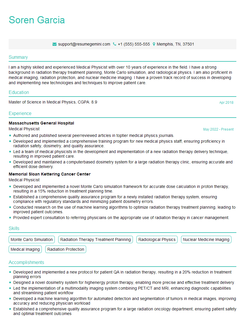
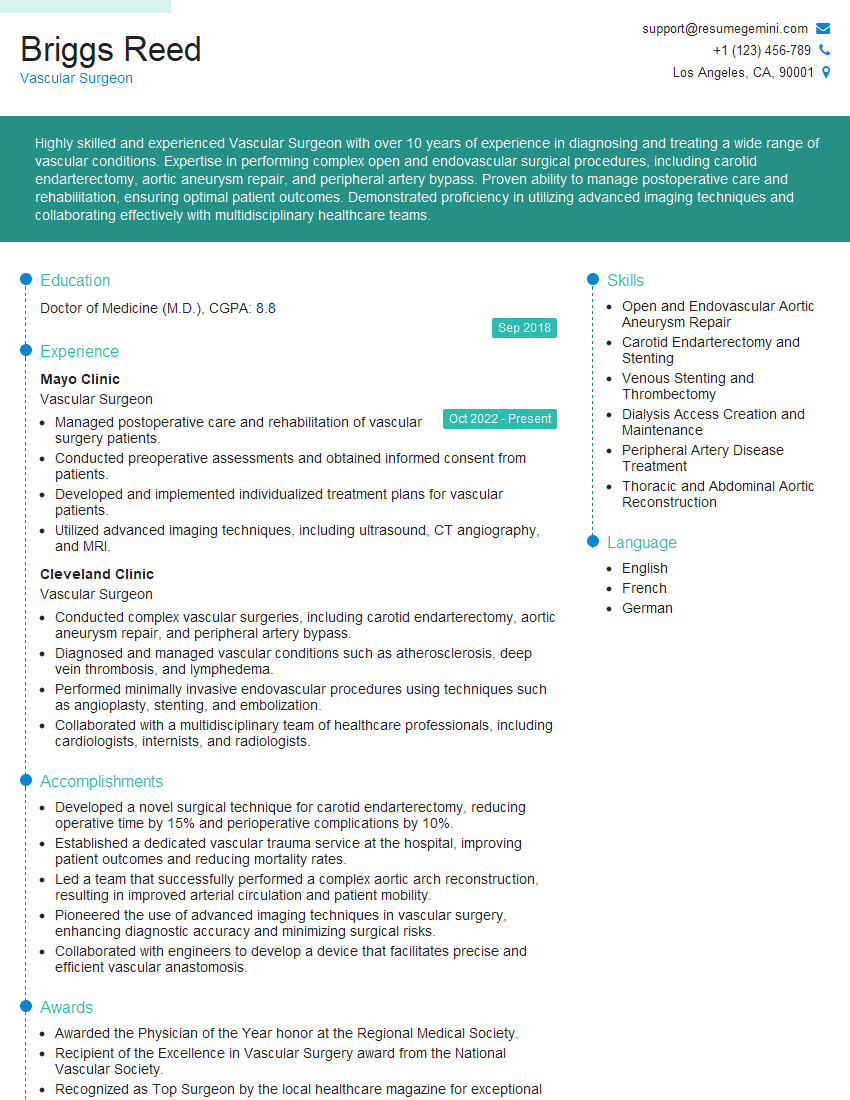
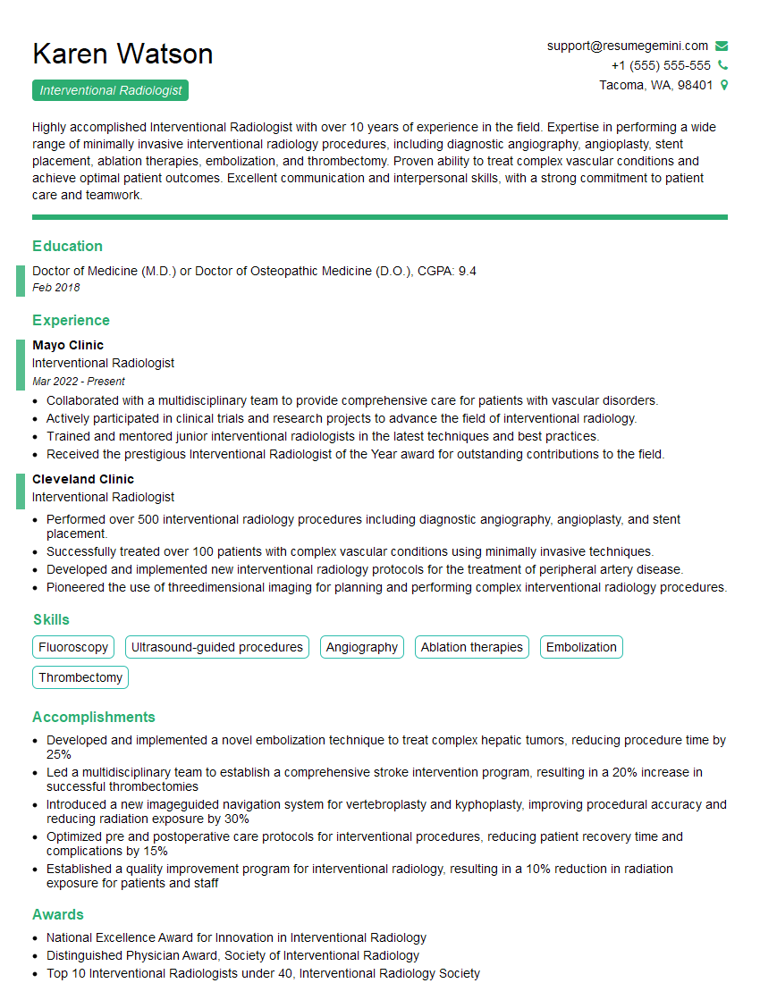
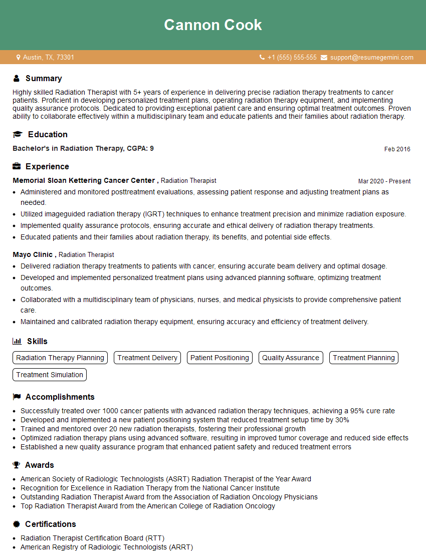
Mastering Fluoroscopic-Guided Procedures is crucial for career advancement in the medical imaging field, opening doors to specialized roles and increased responsibility. To maximize your job prospects, crafting a strong, ATS-friendly resume is essential. ResumeGemini is a trusted resource to help you build a professional and impactful resume that highlights your skills and experience effectively. Examples of resumes tailored to Fluoroscopic-Guided Procedures are available to help guide your process.
Explore more articles
Users Rating of Our Blogs
Share Your Experience
We value your feedback! Please rate our content and share your thoughts (optional).
What Readers Say About Our Blog
To the interviewgemini.com Webmaster.
Very helpful and content specific questions to help prepare me for my interview!
Thank you
To the interviewgemini.com Webmaster.
This was kind of a unique content I found around the specialized skills. Very helpful questions and good detailed answers.
Very Helpful blog, thank you Interviewgemini team.