The thought of an interview can be nerve-wracking, but the right preparation can make all the difference. Explore this comprehensive guide to Fluoroscopy-Guided Injections interview questions and gain the confidence you need to showcase your abilities and secure the role.
Questions Asked in Fluoroscopy-Guided Injections Interview
Q 1. Describe the principles of fluoroscopy-guided injections.
Fluoroscopy-guided injections utilize real-time X-ray imaging to visualize the injection process, ensuring accurate placement of the needle into the target area. Imagine it like having a live X-ray movie playing as you inject – this allows for precise targeting of structures, minimizing the risk of complications. The principles revolve around using the fluoroscopic image to guide the needle’s trajectory to the desired location, constantly verifying its position to avoid damaging surrounding tissues. This is crucial for procedures requiring pinpoint accuracy, such as epidural injections or placing catheters within joints.
Q 2. Explain the different types of imaging modalities used in fluoroscopy-guided injections.
While fluoroscopy is the primary modality, other imaging techniques can be integrated or used in conjunction. The most common is conventional fluoroscopy, using continuous X-ray imaging to guide the needle. Digital subtraction angiography (DSA) enhances visualization by subtracting background images, resulting in clearer views of blood vessels. Some procedures may incorporate computed tomography (CT) fluoroscopy, offering cross-sectional images which provide additional spatial information. The choice depends on the specific procedure and the complexity of the target anatomy. For example, DSA might be used during arterial injections, while CT fluoroscopy may be preferred for complex spinal injections.
Q 3. What are the safety precautions to be taken during fluoroscopy-guided injections?
Safety is paramount. Strict adherence to sterile technique is vital to prevent infection. This involves proper hand hygiene, sterile draping, and using sterile needles, gloves, and other equipment. A comprehensive patient history, including allergies and coagulation status, is crucial. The radiation exposure needs to be minimized (discussed in the next question). Continuous monitoring of the patient’s vital signs is essential to detect any adverse reactions, such as allergic responses or sudden changes in blood pressure. We always ensure we have emergency resuscitation equipment readily available.
- Sterile technique
- Patient history and allergy checks
- Radiation safety measures
- Vital signs monitoring
- Emergency equipment readily available
Q 4. How do you minimize radiation exposure to both the patient and the healthcare professional?
Minimizing radiation exposure is critical for both patient and healthcare professional. Techniques like pulse fluoroscopy (using intermittent rather than continuous X-ray pulses), low-dose fluoroscopy settings, and lead shielding for the patient and operator significantly reduce exposure. Using image intensifiers with high sensitivity and employing optimal imaging techniques also helps. Furthermore, the principle of ALARA (As Low As Reasonably Achievable) should be followed meticulously. This involves optimizing the fluoroscopy parameters to achieve the necessary image quality with the minimum radiation dose. For example, we might use a higher mA setting for a shorter period rather than a lower mA setting for a longer one.
Q 5. Explain the procedure for selecting the appropriate needle size and type for a fluoroscopy-guided injection.
Needle selection depends on the target anatomy, the viscosity of the injected substance, and the depth of the injection site. For superficial injections, a smaller gauge needle (e.g., 25-gauge) might be sufficient, whereas deeper injections often require larger bore needles (e.g., 18-gauge) to facilitate smooth injection of viscous fluids. The needle type is also important: beveled needles are standard for most procedures. Longer needles may be required for deeper injections. Careful consideration of the needle’s length and gauge is essential to prevent tissue damage and ensure accurate placement. For example, a longer, smaller gauge needle might be chosen for a facet joint injection to avoid injuring the spinal cord. Always consider patient factors, such as body habitus and the location of relevant vascular structures.
Q 6. Describe the process of identifying the target anatomy using fluoroscopy.
Identifying the target anatomy starts with reviewing the patient’s imaging studies (MRI, CT) prior to the procedure to define the target’s location and surrounding anatomy. Fluoroscopy is then used to confirm the target in real-time. Different views (AP, lateral, oblique) are often employed. The contrast medium (if used) helps define the target structure’s boundaries and relationships to surrounding structures. For instance, in a spinal injection, the fluoroscopic image allows us to visualize the intervertebral space and the relationship of the needle to the spinal cord and nerve roots. We use the images to carefully guide the needle towards the target, making small adjustments as needed based on the ongoing fluoroscopic feedback.
Q 7. How do you manage complications such as extravasation or bleeding during a fluoroscopy-guided injection?
Extravasation (leakage of injected fluid into surrounding tissues) is managed by immediately stopping the injection. If the contrast medium is extravasated, the affected area may be observed for swelling or discomfort. Cold compresses may be applied to reduce swelling. Significant extravasation may require additional management, such as aspiration of the extravasated fluid or potentially surgical intervention in severe cases. Bleeding is rare but may be managed by applying pressure to the injection site, using appropriate hemostatic agents if needed, and monitoring the patient’s vital signs. In cases of significant bleeding, surgical intervention may be necessary. Documentation of any complications and their management is essential.
Q 8. What are the common indications for fluoroscopy-guided injections?
Fluoroscopy-guided injections are used to precisely deliver medication or contrast agents to specific anatomical locations within the body. This is crucial when accuracy is paramount to avoid damaging surrounding tissues. Common indications include:
- Pain management: Targeting nerve roots (e.g., epidural steroid injections for back pain), facet joints (for facet joint arthritis), or other pain generators.
- Diagnostic procedures: Assessing the extent of a lesion, such as a tumor or abscess, by injecting contrast dye and visualizing the spread under fluoroscopy.
- Therapeutic interventions: Administering medication directly to a joint affected by arthritis (e.g., intra-articular injections), or injecting medication into a cyst.
- Abscess drainage: Guiding a needle to drain an abscess under fluoroscopic visualization to ensure complete drainage.
- Vertebroplasty/Kyphoplasty: Injecting bone cement to stabilize vertebral fractures caused by osteoporosis or trauma. Fluoroscopy ensures accurate placement of the cement.
Essentially, any situation where precise, targeted delivery of medication or contrast agent is necessary and real-time imaging is beneficial can be a suitable indication for a fluoroscopy-guided injection.
Q 9. What are the contraindications for fluoroscopy-guided injections?
Contraindications for fluoroscopy-guided injections are situations where the procedure poses a significant risk to the patient or where the benefits don’t outweigh the risks. These include:
- Patient refusal or lack of informed consent: The procedure should only proceed with the patient’s full understanding and agreement.
- Active infection at the injection site: Infection at the site could be exacerbated by the procedure, leading to complications.
- Severe coagulopathy: Patients with bleeding disorders are at increased risk of hematoma formation.
- Allergy to contrast media: Contrast agents can cause allergic reactions, including anaphylaxis. A thorough allergy history is crucial.
- Uncontrolled hypertension: Elevated blood pressure can increase the risk of complications.
- Lack of appropriate anatomical landmarks: Sometimes, significant anatomical variations make needle placement extremely difficult and potentially unsafe.
Careful patient assessment and a thorough review of the patient’s medical history are crucial to identifying any contraindications before proceeding with the injection.
Q 10. Explain the process of post-procedural care following a fluoroscopy-guided injection.
Post-procedural care following a fluoroscopy-guided injection focuses on monitoring the patient for potential complications and ensuring their comfort. This typically involves:
- Monitoring vital signs: Closely observing blood pressure, heart rate, and respiratory rate for any significant changes.
- Assessing injection site: Checking for signs of bleeding, swelling, or infection.
- Pain management: Administering analgesics as needed to control pain.
- Patient education: Instructing the patient on potential side effects, activity restrictions, and follow-up care.
- Neurological assessment: In procedures near the spinal cord, careful neurological examination is essential to detect any signs of nerve damage.
The duration and intensity of post-procedural care will vary depending on the type of injection performed and the patient’s individual needs. For example, a simple joint injection requires less intensive monitoring compared to a complex epidural injection.
Q 11. How do you interpret fluoroscopic images to guide needle placement?
Interpreting fluoroscopic images to guide needle placement requires a detailed understanding of anatomy and experience in image interpretation. We visualize the anatomy in real-time, making adjustments as needed to reach the target. Here’s a breakdown of the process:
- Identifying anatomical landmarks: First, we identify bony structures, soft tissues, and other anatomical markers visible on the fluoroscopic image.
- Assessing needle position: We constantly observe the needle’s trajectory on the fluoroscopic screen, ensuring it’s advancing towards the target site.
- Adjusting needle position: Based on the image, we make small adjustments to the needle’s angle and depth to achieve optimal placement.
- Using contrast media (if necessary): Sometimes, injection of a small amount of contrast medium is useful to visualize the spread and confirm accurate placement. This helps prevent accidental injection into undesirable areas.
- Evaluating the injection spread: Once the injection is completed, we reassess the fluoroscopic images to ensure the medication/contrast has spread as expected within the target area.
Imagine it like using a GPS to reach a specific destination; the fluoroscopic image is our real-time map, and the needle is our vehicle. We use the image to navigate and ensure we reach the correct location precisely.
Q 12. Describe your experience with different types of fluoroscopy equipment.
My experience encompasses various fluoroscopy equipment, from older analog systems to modern digital systems. Digital fluoroscopy offers significant advantages, including:
- Improved image quality: Digital systems provide higher resolution and better contrast, leading to more accurate needle placement.
- Image post-processing: Digital images can be processed to enhance visibility and reduce noise.
- Reduced radiation exposure: Advanced digital systems offer features to minimize radiation dose to both patients and staff.
- Image storage and retrieval: Digital images are easily stored and retrieved for review, facilitating quality assurance and patient record-keeping.
I’m comfortable working with different manufacturers’ equipment and adapting to different imaging techniques and software interfaces. Adaptability is key in this field, as new technologies are constantly being developed.
Q 13. How do you assess the patient’s anatomical landmarks before starting the procedure?
Assessing anatomical landmarks is the foundational step for successful fluoroscopy-guided injections. This involves a combination of:
- Palpation: Feeling the patient’s skin and underlying bony structures to locate anatomical landmarks. This gives us a three-dimensional sense of the area.
- Surface anatomy: Visual inspection of the patient’s body surface to identify skin markings and other external clues to guide needle placement.
- Fluoroscopic imaging: Using fluoroscopy to visualize the internal anatomy and confirm the location of the target site in real-time. We use this to correlate our palpation findings with the actual anatomy.
- Patient history and medical imaging: Prior imaging studies (like CT or MRI scans) can provide additional information about the anatomy and can inform our pre-procedural planning.
For example, in a lumbar epidural injection, identifying the spinous processes, intervertebral spaces, and sacrum is crucial for accurate needle placement. Each procedure has its specific set of critical anatomical landmarks that need to be carefully identified before proceeding.
Q 14. Explain your understanding of different injection techniques (e.g., interlaminar, transforaminal).
Fluoroscopy-guided injections employ various techniques depending on the target location. Examples include:
- Interlaminar injection: This technique involves inserting the needle between the lamina of adjacent vertebrae to reach the epidural space. It’s used to treat pain in the spine.
- Transforaminal injection: The needle is inserted through the intervertebral foramen (the opening between vertebrae) to target a specific nerve root. This approach is particularly beneficial when targeting specific nerve roots responsible for radiculopathy.
- Intra-articular injection: The needle is inserted directly into a joint cavity to deliver medication. This is commonly done for joint pain relief in conditions like osteoarthritis.
- Facet joint injection: The needle is guided to inject medication into the facet joints, which are small synovial joints in the spine that can be a source of back pain.
Choosing the appropriate injection technique requires a comprehensive understanding of anatomy, pathology, and the patient’s specific clinical presentation. Each technique carries its own set of potential risks and benefits, and proper technique is essential for minimizing complications and maximizing efficacy.
Q 15. What are the different types of contrast media used in fluoroscopy-guided injections?
Fluoroscopy-guided injections utilize contrast media to visualize the target area and guide the needle placement. The choice of contrast agent depends heavily on the specific procedure and patient factors. Common types include:
- Iodinated contrast media: These are the most frequently used, offering excellent visualization due to their high X-ray attenuation. They come in ionic and non-ionic forms, with non-ionic agents generally preferred due to a lower incidence of adverse reactions. Examples include iohexol and iopamidol.
- Barium sulfate: This is primarily used for gastrointestinal studies, not commonly in other fluoroscopy-guided injections. It’s not absorbed systemically and is therefore generally safer, although it can cause bowel obstruction if extravasation occurs.
- Air or gas: Sometimes, air or carbon dioxide may be used as a contrast agent, particularly in procedures involving the spinal column or joints. This is less common than iodinated contrast.
The selection of the contrast agent is a crucial step, considering factors like patient allergy history, the specific anatomical location being targeted, and the desired image characteristics. Always consider the potential risks and benefits of each type.
Career Expert Tips:
- Ace those interviews! Prepare effectively by reviewing the Top 50 Most Common Interview Questions on ResumeGemini.
- Navigate your job search with confidence! Explore a wide range of Career Tips on ResumeGemini. Learn about common challenges and recommendations to overcome them.
- Craft the perfect resume! Master the Art of Resume Writing with ResumeGemini’s guide. Showcase your unique qualifications and achievements effectively.
- Don’t miss out on holiday savings! Build your dream resume with ResumeGemini’s ATS optimized templates.
Q 16. How do you manage a patient with an allergy to contrast media?
Managing a patient with a contrast media allergy requires careful planning and a multi-faceted approach. The severity of the previous reaction dictates the management strategy. A detailed history is crucial – including the type of contrast used previously, the symptoms experienced, and the treatment received.
- Mild reactions (e.g., hives, itching): Pre-medication with antihistamines (like diphenhydramine) and corticosteroids (like methylprednisolone) can significantly reduce the risk of a recurrence.
- Moderate to severe reactions (e.g., anaphylaxis): These patients require very careful monitoring and may need more intensive pre-medication including broader antihistamines and potentially even a short hospital stay for observation. The use of non-ionic contrast media can also significantly reduce the chance of a reaction.
- Alternative imaging modalities: In cases of severe allergy or previous anaphylaxis, alternative imaging techniques like ultrasound or MRI might be considered if they are appropriate for the clinical situation. However, this is often dependent on the anatomical location and the clinical question being addressed.
Collaboration with an allergist or anesthesiologist may be necessary for high-risk patients. Having emergency medications and resuscitation equipment readily available is paramount.
Q 17. Describe your experience with different types of needles and catheters used in fluoroscopy-guided injections.
My experience encompasses a wide range of needles and catheters, chosen based on the specific injection site, target vessel or tissue, and the volume of contrast media required. For example:
- Needles: I use various sizes and types, from small gauge needles (22-27 gauge) for superficial injections to larger gauge needles (18-22 gauge) for deeper injections. The choice depends on the viscosity of the contrast and the size of the target vessel. I’ve experience with spinal needles (e.g., Quincke, Sprotte) for epidurals and other spinal injections.
- Catheters: These are often used for more complex procedures requiring precise placement and continuous injection. I have experience with various types including hydrophilic catheters for smooth insertion, microcatheters for navigating tortuous vessels, and specialized catheters designed for specific injections (e.g., vertebroplasty catheters). Choosing the right catheter length, gauge, and material is vital for effective and safe delivery.
Proper selection and handling of needles and catheters are crucial for minimizing patient discomfort, reducing the risk of complications like bleeding and nerve injury, and ensuring the accuracy of the injection.
Q 18. How do you troubleshoot common technical issues encountered during fluoroscopy-guided injections?
Troubleshooting technical issues is a common part of fluoroscopy-guided injections. Problems can arise from various sources, including equipment malfunction, patient movement, or difficulty in accessing the target area. Here’s a structured approach:
- Image quality issues: Poor image quality (blurriness, insufficient contrast) can be caused by incorrect settings on the fluoroscopy unit, patient movement, or inadequate contrast agent administration. Troubleshooting involves checking the equipment settings, stabilizing the patient, and adjusting the contrast media injection rate and volume.
- Needle placement issues: Difficulty in advancing the needle may be due to anatomical variations, unexpected tissue resistance, or incorrect needle angulation. Careful observation of fluoroscopic images, adjusting the needle angle and depth incrementally, and perhaps using image intensification with magnification, assists here.
- Equipment malfunction: Should the fluoroscopy unit malfunction (e.g., image freezing, poor resolution), immediate actions should be to check power connections, assess equipment settings, and escalate the issue to appropriate biomedical engineering staff.
- Extravasation: Extravasation (leakage of contrast agent outside of the intended area) requires immediate cessation of injection and implementation of supportive measures such as local compression to minimize swelling and discomfort.
A systematic approach, coupled with a calm and methodical demeanor, is essential for effective troubleshooting and patient safety. Thorough documentation of any issues, interventions, and outcomes is paramount.
Q 19. Explain your knowledge of radiation safety regulations and protocols.
Radiation safety is a top priority in fluoroscopy-guided injections. I adhere strictly to the ALARA principle (As Low As Reasonably Achievable) to minimize radiation exposure to both the patient and the healthcare team. This involves:
- Time optimization: Limiting the fluoroscopy time to the absolute minimum necessary for procedure completion. We use pulse fluoroscopy whenever possible to decrease radiation dose.
- Distance maximization: Maintaining the maximum possible distance from the radiation source, utilizing lead shielding where available.
- Shielding: Using lead aprons, thyroid shields, and other protective equipment consistently for both the patient and the medical team. I always advise pregnant or breastfeeding colleagues to avoid close proximity.
- Collimation: Precise collimation of the X-ray beam to encompass only the region of interest reduces unnecessary radiation exposure to surrounding tissues.
- Image intensification: Using the lowest acceptable fluoroscopic image intensifier brightness, which can also be helpful in minimizing radiation dose.
Regular training and adherence to institutional radiation safety protocols are essential for maintaining a safe environment. Documentation of radiation exposure is also kept according to hospital regulations.
Q 20. How do you ensure the sterility and asepsis of the procedure?
Maintaining sterility and asepsis is critical to prevent infection during fluoroscopy-guided injections. We follow a strict sterile technique, which includes:
- Hand hygiene: Thorough handwashing or use of an alcohol-based hand rub before and after the procedure.
- Sterile field preparation: Creating a sterile field using sterile drapes and appropriate antiseptic solutions (e.g., chlorhexidine or iodine) to disinfect the patient’s skin at the injection site.
- Sterile equipment: Using only sterile needles, catheters, syringes, and other equipment. Careful handling to prevent contamination is key.
- Gowning and gloving: Wearing sterile gowns and gloves to maintain a barrier between the healthcare worker and the sterile field.
- Environmental hygiene: Maintaining a clean and disinfected environment in the procedure room to minimize the risk of airborne contamination.
Regular monitoring of equipment sterility and adherence to infection control guidelines are essential components of our practice. Any deviation from strict aseptic technique can lead to serious complications, which is why it’s given absolute priority.
Q 21. What is your experience with documentation and reporting of fluoroscopy-guided injections?
Accurate and comprehensive documentation and reporting are crucial for patient safety, legal compliance, and quality assurance. My documentation includes:
- Patient demographics: Full patient identification, medical history, and relevant allergies.
- Procedure details: Precise description of the procedure performed, including the indication, type of contrast used, needle/catheter details, injection site, and the volume injected.
- Fluoroscopic images: Relevant fluoroscopic images are included in the patient’s record to document needle placement and procedure outcome.
- Complications and adverse events: Any complications or adverse events, including pain, bleeding, infection, extravasation are carefully documented along with the interventions undertaken.
- Radiation exposure: The radiation dose delivered to the patient is recorded.
- Post-procedure instructions: Clear instructions for post-procedure care, including activity restrictions and follow-up appointments.
This documentation ensures continuity of care, facilitates communication between healthcare professionals, and provides essential data for audit and quality improvement purposes. Electronic health records are utilized to ensure efficient and accessible documentation.
Q 22. How do you communicate with patients and their families before, during, and after the procedure?
Effective communication is paramount in fluoroscopy-guided injections. Before the procedure, I explain the process in clear, simple terms, addressing any patient concerns and answering questions patiently. I ensure they understand the procedure’s purpose, potential benefits, risks, and alternatives. I obtain informed consent, confirming their understanding and willingness to proceed. During the procedure, I maintain a calm and reassuring demeanor, providing real-time updates on the progress. I use simple language, avoiding medical jargon, and I encourage the patient to communicate any discomfort or concerns immediately. After the procedure, I provide detailed post-procedure instructions, including pain management, activity restrictions, and follow-up appointments. I answer any remaining questions and provide contact information for any emergencies. For example, I might explain a spinal injection by using an analogy of a targeted medication delivery system to address the patient’s pain rather than using complex anatomical terms.
With families, I tailor my communication to their understanding, ensuring they feel informed and involved, yet respecting the patient’s autonomy. I always encourage open communication and welcome questions from both the patient and their family members.
Q 23. Describe your experience working within a multidisciplinary team.
Fluoroscopy-guided injections require a highly coordinated multidisciplinary team. My experience involves seamless collaboration with radiologists, nurses, anesthesiologists (if needed), and other specialists. For instance, in a spinal injection, the radiologist provides expertise in image interpretation and needle guidance, while the nurse assists with patient positioning, monitoring vital signs, and preparing the equipment. The anesthesiologist may be involved in providing sedation or analgesia if required. Effective teamwork relies on clear communication, respect for each team member’s expertise, and a shared goal of patient safety and optimal procedural outcomes. I actively participate in team discussions, contributing my knowledge of anatomy, injection techniques, and potential complications to ensure the best possible approach for each patient. I believe that a collaborative environment leads to the best patient care; we all support each other to ensure the procedure is carried out successfully and safely. I actively participate in regular team meetings to ensure open communication and address any challenges that might arise.
Q 24. How do you handle unexpected situations or complications during a procedure?
Unexpected situations during fluoroscopy-guided injections require quick thinking and decisive action. My approach involves a structured, systematic response. First, I assess the situation carefully, identifying the nature of the complication. This could range from a patient experiencing unexpected pain or discomfort to accidental needle misplacement or bleeding. Second, I immediately inform the team, clearly articulating the problem and initiating appropriate countermeasures. These might involve adjusting the injection technique, administering medication to manage pain or bleeding, contacting additional specialists, and potentially stopping the procedure. Third, I continuously monitor the patient’s vital signs and overall condition. Fourth, documentation of the complication, the steps taken to address it, and the patient’s response is meticulously recorded. Finally, I debrief with the team after the procedure to analyze the event, learn from the experience, and implement preventative measures to reduce the likelihood of similar events in the future. For example, if the needle deviates from the planned trajectory, I would immediately stop the injection and re-evaluate the imaging to reposition the needle, ensuring the target area is accurately reached.
Q 25. Describe your understanding of the anatomy relevant to fluoroscopy-guided injections (e.g., spinal anatomy).
A thorough understanding of relevant anatomy is fundamental to safe and effective fluoroscopy-guided injections. For spinal injections, this includes detailed knowledge of the vertebral column, including the individual vertebrae, intervertebral discs, spinal cord, nerve roots, and surrounding ligaments and muscles. I have a comprehensive understanding of the various spinal levels (cervical, thoracic, lumbar, sacral), their anatomical variations, and their proximity to critical structures. This knowledge allows me to plan the injection trajectory precisely, minimizing the risk of nerve damage or other complications. For other procedures, such as joint injections, I possess similar in-depth knowledge of the specific joint anatomy, including ligaments, tendons, and cartilage, allowing me to accurately target the injection site. A strong foundation in anatomical knowledge, combined with the guidance of fluoroscopy imaging, enables the accurate and safe delivery of medication.
Q 26. What is your experience with image post-processing and analysis?
Image post-processing and analysis are critical aspects of fluoroscopy-guided injections. I am proficient in using various software programs to enhance image quality, including adjusting brightness, contrast, and magnification. This allows for clearer visualization of anatomical structures and the needle tip, improving accuracy during needle placement. I can also use post-processing techniques to measure distances, angles, and needle depth, confirming the injection site’s precision. Furthermore, I can analyze post-injection images to assess the distribution of contrast media or medication, ensuring the injection was successful and appropriately targeted. For example, we might use digital subtraction angiography to improve image clarity by removing static background noise, aiding us to accurately assess the placement of contrast media. Analysis of images can be crucial for confirming successful procedures as well as for identifying any complications.
Q 27. How do you stay updated with the latest advancements in fluoroscopy-guided injection techniques?
Staying current with the latest advancements in fluoroscopy-guided injection techniques is an ongoing process. I actively participate in professional conferences and workshops, attending presentations and engaging with colleagues to learn about new techniques, technologies, and best practices. I subscribe to relevant peer-reviewed journals and regularly read publications on advancements in fluoroscopy and injection techniques. I also participate in continuing medical education courses, which often incorporate hands-on training with new equipment and methods. Keeping abreast of the latest research ensures I can offer my patients the safest and most effective procedures available, maximizing their chances of positive outcomes and minimizing complications. Additionally, I am committed to lifelong learning in this rapidly developing area of medicine.
Key Topics to Learn for Fluoroscopy-Guided Injections Interview
- Fluoroscopic Anatomy & Imaging Principles: Understanding anatomical landmarks and their visualization under fluoroscopy is fundamental. Practice interpreting fluoroscopic images and identifying potential complications.
- Injection Techniques & Procedures: Master various injection techniques (e.g., epidural, facet joint, nerve root) including needle placement, contrast media administration, and patient positioning. Be prepared to discuss the rationale behind choosing specific techniques.
- Radiation Safety & ALARA Principle: Demonstrate a thorough understanding of radiation safety protocols, ALARA (As Low As Reasonably Achievable) principles, and patient shielding techniques. This is crucial for both your safety and the patient’s.
- Complications & Management: Be prepared to discuss potential complications (e.g., bleeding, infection, nerve damage) and the strategies for their prevention and management. Showcase your problem-solving skills in hypothetical scenarios.
- Equipment Operation & Troubleshooting: Familiarize yourself with the operation of fluoroscopy equipment, including image intensifiers, C-arms, and injection systems. Be ready to discuss basic troubleshooting and maintenance procedures.
- Patient Assessment & Communication: Highlight your ability to assess patient suitability for fluoroscopy-guided injections, explain procedures clearly, and manage patient anxieties. Effective communication is paramount.
- Legal and Ethical Considerations: Understand the legal and ethical implications of performing fluoroscopy-guided injections, including informed consent and documentation procedures.
Next Steps
Mastering Fluoroscopy-Guided Injections opens doors to exciting career opportunities and increased earning potential within the medical field. To significantly enhance your job prospects, creating a strong, ATS-friendly resume is crucial. ResumeGemini is a trusted resource that can help you craft a compelling resume tailored to highlight your skills and experience. Leverage ResumeGemini’s tools to build a professional resume that stands out to recruiters. Examples of resumes specifically tailored for Fluoroscopy-Guided Injections professionals are available to help guide your creation process.

Quality Assurance Coordinator Resume Sample
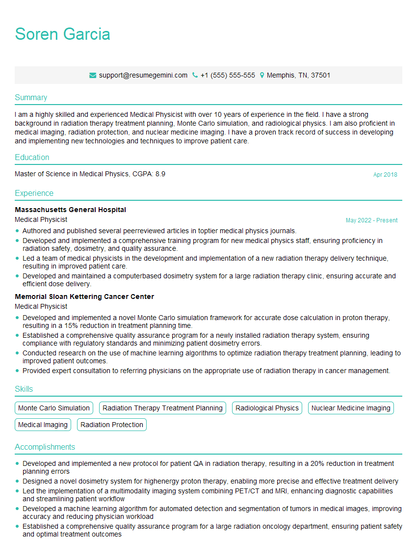
Medical Physicist Resume Sample

Clinical Instructor Resume Sample
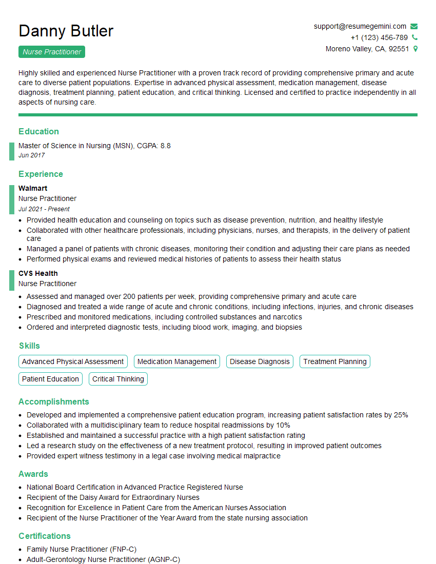
Nurse Practitioner Resume Sample
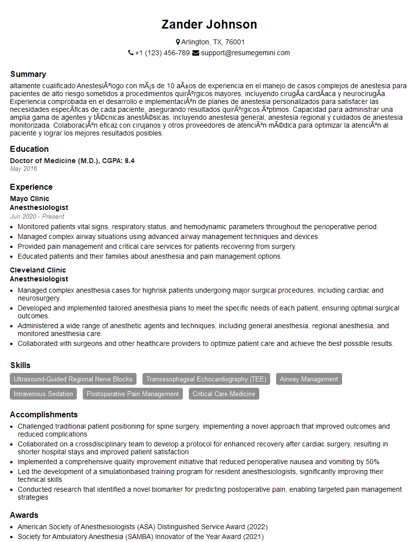
Anesthesiologist Resume Sample

Physician Assistant Resume Sample
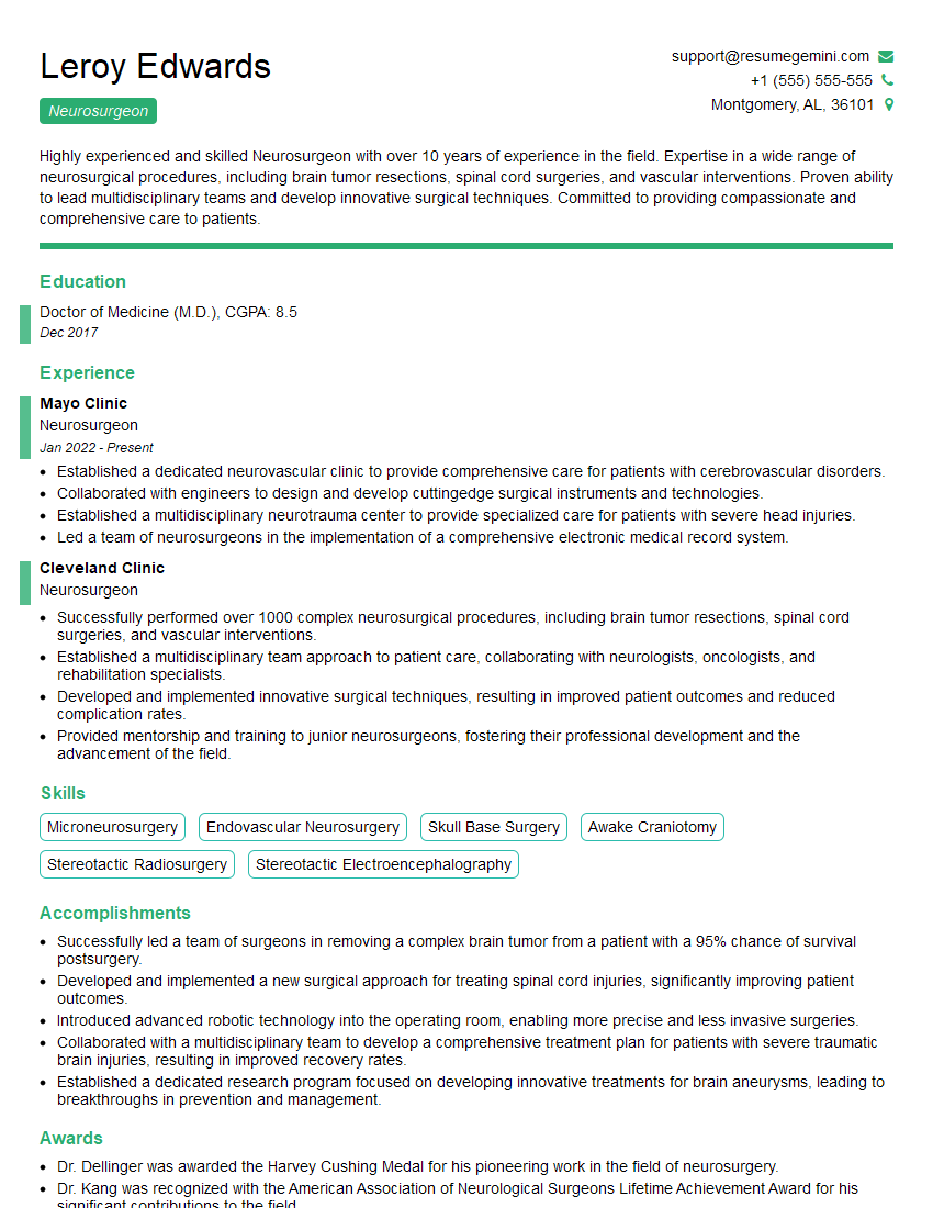
Neurosurgeon Resume Sample
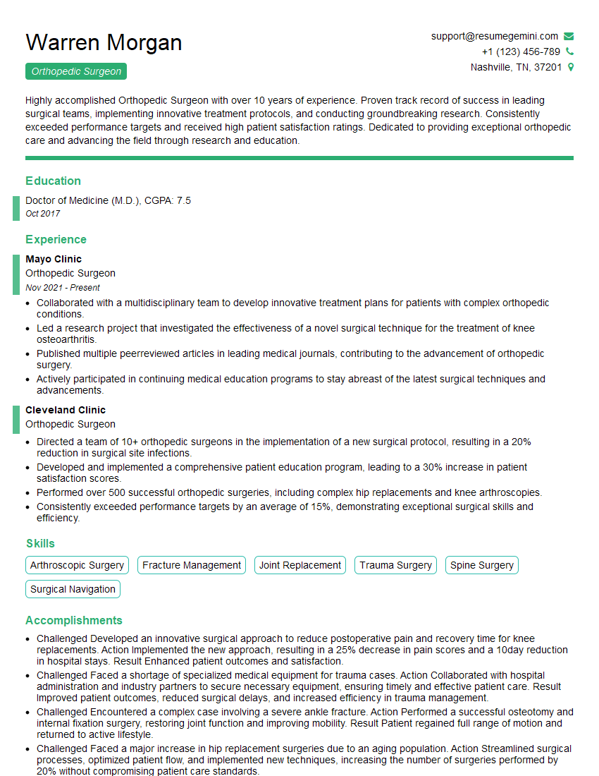
Orthopedic Surgeon Resume Sample
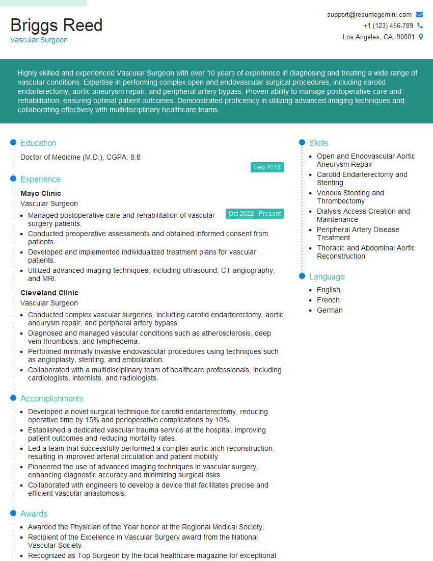
Vascular Surgeon Resume Sample
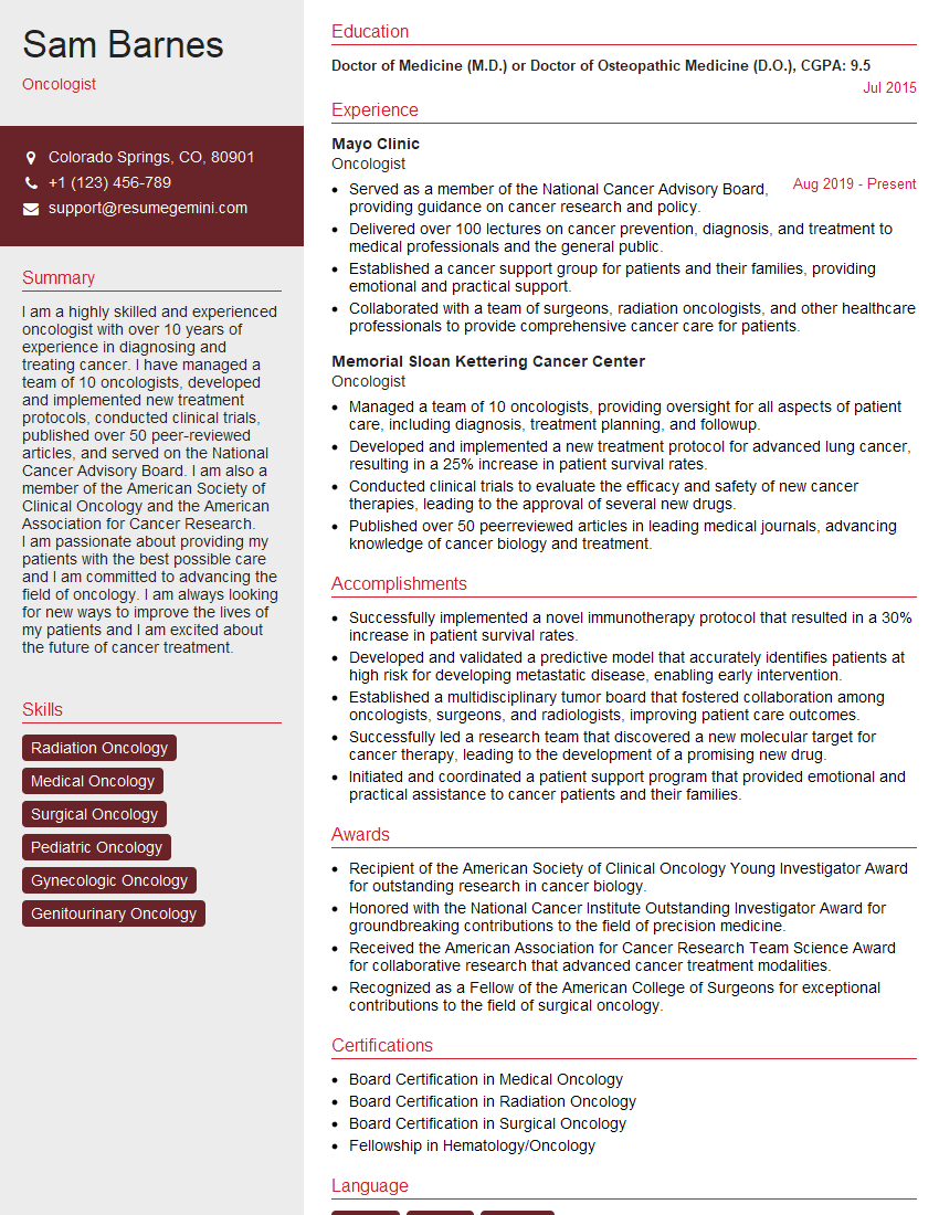
Oncologist Resume Sample
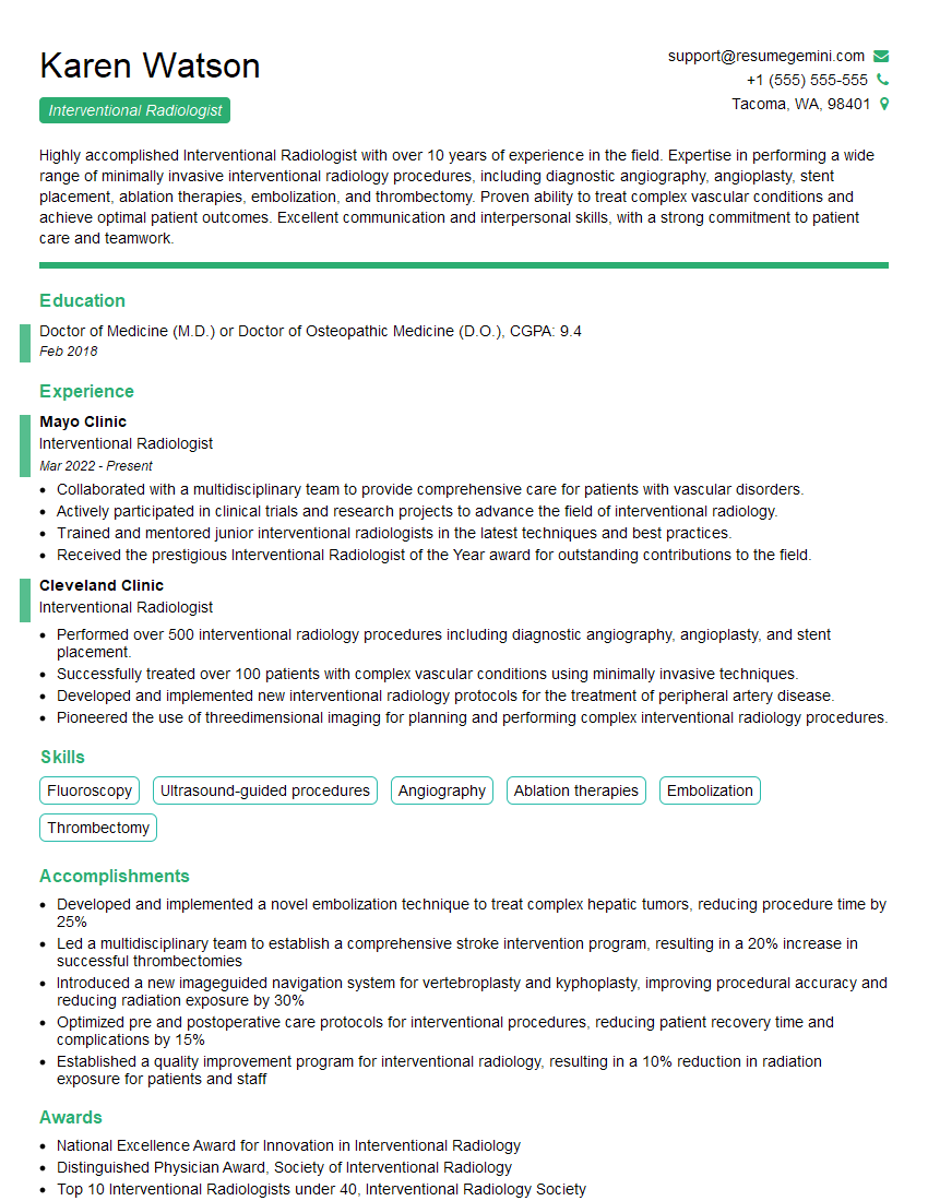
Interventional Radiologist Resume Sample
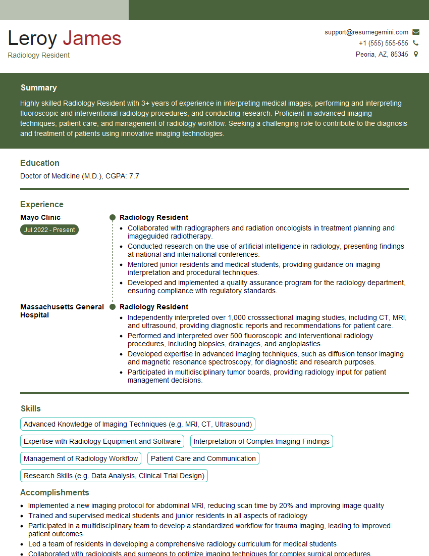
Radiology Resident Resume Sample
Explore more articles
Users Rating of Our Blogs
Share Your Experience
We value your feedback! Please rate our content and share your thoughts (optional).
What Readers Say About Our Blog
To the interviewgemini.com Webmaster.
Very helpful and content specific questions to help prepare me for my interview!
Thank you
To the interviewgemini.com Webmaster.
This was kind of a unique content I found around the specialized skills. Very helpful questions and good detailed answers.
Very Helpful blog, thank you Interviewgemini team.