Cracking a skill-specific interview, like one for Hemorrhagic Control, requires understanding the nuances of the role. In this blog, we present the questions you’re most likely to encounter, along with insights into how to answer them effectively. Let’s ensure you’re ready to make a strong impression.
Questions Asked in Hemorrhagic Control Interview
Q 1. Explain the physiological mechanisms of hemostasis.
Hemostasis, the process of stopping bleeding, is a complex physiological mechanism involving a delicate interplay of vascular, platelet, and coagulation factors. It’s like a three-legged stool – all parts need to work together for stability.
- Vascular Spasm: Immediately following injury, blood vessels constrict, reducing blood flow to the injured area. This is a temporary, reflexive response that buys time for the next steps. Think of it as the body’s immediate ‘pinch’ to slow the leak.
- Platelet Plug Formation: Platelets, tiny blood cells, adhere to the exposed collagen fibers at the injury site. They then activate and aggregate, forming a temporary plug that seals the break in the vessel wall. Imagine it like patching a hole with sticky tape.
- Coagulation Cascade: This is the most complex part, involving a series of enzymatic reactions that lead to the formation of fibrin, a protein that strengthens the platelet plug and forms a stable clot. This is the final, permanent ‘repair’ of the vessel. Think of fibrin as the strong glue that holds the patch in place.
These three steps work in concert to ensure efficient and effective bleeding cessation. Failure at any stage can lead to uncontrolled hemorrhage.
Q 2. Describe the different types of hemorrhage and their clinical presentations.
Hemorrhage is classified by the type and size of blood vessel involved, as well as the rate of bleeding.
- Capillary Bleeding: This is the most common type, characterized by slow oozing of blood. It typically clots spontaneously and isn’t usually life-threatening. Think of a minor scrape or cut.
- Venous Bleeding: Venous blood is dark red and flows steadily. It’s usually easier to control than arterial bleeding because the pressure is lower. Imagine a deep cut that bleeds steadily but not in spurts.
- Arterial Bleeding: This is the most dangerous type. Arterial blood is bright red and spurts with each heartbeat due to high pressure. Rapid blood loss can lead to shock and death. Think of a severe injury involving a major artery.
Clinical presentations vary depending on the severity and location of the bleed. Symptoms can range from minor bruising and oozing to significant blood loss, shock (pale skin, rapid weak pulse, altered mental status), and ultimately death if not treated promptly.
Q 3. Outline the principles of direct pressure in hemorrhage control.
Direct pressure is the cornerstone of hemorrhage control, particularly for minor to moderate bleeding. It works by physically compressing the injured blood vessel, preventing further blood loss. It’s the simplest and often most effective initial response.
- Clean the Wound: If possible, gently clean the wound surface to remove debris that could interfere with clotting.
- Apply Pressure Directly: Place a clean dressing directly over the wound and apply firm, continuous pressure. Don’t lift the dressing to check; it could dislodge the clot that is forming.
- Elevate the Injured Extremity: Elevating the injured limb above the heart can help reduce blood flow to the area, further aiding in clot formation. This works by gravity.
- Maintain Pressure: Continue applying pressure for at least 15-20 minutes, even if bleeding seems to have stopped. A clot needs time to fully form and stabilize.
Direct pressure is effective for most minor bleeds, and often in conjunction with other methods for more severe bleeds. It’s a fundamental skill for anyone providing first aid.
Q 4. What are the indications and contraindications for tourniquet application?
Tourniquets are used as a last resort to control life-threatening extremity hemorrhage when other methods have failed. They’re a powerful tool, but misuse can lead to serious complications.
- Indications: Life-threatening bleeding from an extremity that cannot be controlled by other means. This typically involves significant arterial bleeding.
- Contraindications: Bleeding from the torso, head, or neck; injuries that cannot be bypassed by a tourniquet; presence of a fracture; if there is another accessible method for controlling bleeding.
Proper application is critical. The tourniquet should be placed proximal (closer to the heart) to the bleeding site, tightened until bleeding stops, and the time of application must be clearly documented. Tourniquet use requires advanced training and awareness of potential complications like nerve damage or limb loss.
Q 5. How do you manage a major arterial bleed in a pre-hospital setting?
Managing a major arterial bleed pre-hospital requires immediate and decisive action. The mnemonic ‘ABCDE’ helps prioritize actions:
- Airway: Ensure the patient has a patent airway.
- Breathing: Check for adequate breathing.
- Circulation: Control bleeding using direct pressure, elevation, and if necessary, a tourniquet.
- Disability: Assess neurological status.
- Exposure: Expose the injury site to assess the extent of the bleeding.
For a major arterial bleed, direct pressure and elevation are crucial first steps. If bleeding continues, a tourniquet should be applied immediately. Rapid transport to definitive care is paramount. Maintaining patient warmth and providing oxygen are also important.
It is vital to remember that timing is critical. Every second counts in managing a major arterial bleed, and swift intervention is often the difference between life and death.
Q 6. Describe your experience with hemostatic agents (e.g., Celox, QuikClot).
I have extensive experience using various hemostatic agents, including Celox and QuikClot. These agents work by accelerating the body’s natural clotting process or by physically plugging the bleeding vessel. They are invaluable in situations where direct pressure alone is insufficient.
- Celox: This is a granular agent that contains a material that promotes clotting. It’s applied directly to the wound and is effective in controlling a variety of bleeding types. I’ve found it particularly useful in penetrating wounds and in situations where direct pressure is difficult to maintain.
- QuikClot: Similar in application to Celox, QuikClot utilizes different clotting factors to achieve rapid hemostasis. My experience suggests it’s also highly effective, and I’ve used it in both civilian and military settings with good results.
However, it is important to note that some hemostatic agents can generate significant heat upon application. Appropriate training and cautious application are vital to prevent further tissue damage.
Q 7. Explain the use of packing techniques for hemorrhage control.
Packing techniques involve filling a deep wound cavity with hemostatic material to control bleeding from within. It’s particularly useful for deep, irregular wounds where direct pressure is ineffective. Think of it as a ‘plug’ from the inside.
- Wound Assessment: Thoroughly examine the wound to determine its depth and extent. Packing should only be attempted by individuals with appropriate training.
- Material Selection: Choose a suitable packing material; gauze is commonly used. Hemostatic agents can be incorporated into the packing.
- Packing Technique: Pack the wound firmly but gently, avoiding excessive force that could cause further injury. The goal is to fill the cavity completely and apply pressure to the bleeding source.
- Dressing Application: After the cavity is packed, a secondary dressing should be applied over the top and held in place with a bandage.
Packing techniques are complex and should only be performed by trained professionals. Improper packing can lead to complications such as infection or further tissue damage. It’s a technique that demands proficiency and an understanding of the underlying anatomy.
Q 8. How would you assess a patient for hypovolemic shock secondary to hemorrhage?
Assessing a patient for hypovolemic shock, a life-threatening condition caused by significant blood loss, requires a systematic approach. We look for a combination of signs and symptoms, not just one isolated finding. Think of it like putting together a puzzle; each piece contributes to the overall picture.
- Vital Signs: A rapid, weak pulse (tachycardia), low blood pressure (hypotension), and rapid, shallow breathing (tachypnea) are key indicators. The body is trying to compensate for the decreased blood volume. For example, a patient might present with a heart rate of 120 beats per minute and a blood pressure of 80/50 mmHg.
- Mental Status: As blood volume decreases, the brain receives less oxygen. This can lead to altered mental status, ranging from anxiety and restlessness to confusion and lethargy. Imagine trying to function with a significantly reduced fuel supply – your brain’s performance suffers.
- Skin: Cool, clammy, pale skin reflects the body’s attempt to conserve heat and blood flow to vital organs. This is the body’s way of prioritizing blood supply to critical areas.
- Urine Output: Reduced urine output is a late sign, but a significant one. The kidneys are less perfused, leading to decreased urine production.
- Capillary Refill Time: A prolonged capillary refill time (greater than 2 seconds) indicates poor peripheral perfusion. This means the body’s extremities aren’t receiving enough blood.
It’s crucial to remember that these signs can vary depending on the patient’s underlying health conditions and the severity of the blood loss. A thorough history, including the mechanism of injury or illness, is crucial for a complete assessment.
Q 9. What are the early warning signs of internal bleeding?
Early warning signs of internal bleeding are often subtle and can easily be missed. They can mimic other conditions, making it challenging to diagnose. Think of it as a silent thief, slowly stealing away your body’s resources.
- Abdominal Pain or Tenderness: Pain, often described as deep and aching, can be localized to a specific area or more diffuse. For example, a patient may experience constant pain in their lower abdomen after a fall.
- Bruising or Discoloration: Unexpected bruising or discoloration of the skin, especially in areas not directly injured, could suggest internal bleeding. A large bruise that appears unexpectedly should raise suspicion.
- Swelling or Distension: Abdominal swelling or distension can indicate internal bleeding into the abdominal cavity (hemoperitoneum).
- Weakness or Dizziness: Feeling faint, lightheaded, or generally weak can be an early manifestation of blood loss.
- Rapid Heart Rate: The body tries to compensate for blood loss by increasing the heart rate. The heart works overtime to try and keep things functioning.
- Changes in Mental Status: Similar to hypovolemic shock, internal bleeding can result in changes in consciousness, such as confusion or lethargy. This is a critical warning.
It’s essential to maintain a high index of suspicion for internal bleeding, especially in patients with a history of trauma or underlying medical conditions that increase their risk.
Q 10. Discuss the role of fluid resuscitation in hemorrhage management.
Fluid resuscitation is a cornerstone of hemorrhage management. It’s like giving the body the fluids it needs to keep the circulatory system functioning properly. The goal is to restore adequate blood volume and tissue perfusion.
Typically, crystalloid solutions such as normal saline or lactated Ringer’s are used initially. These solutions are readily available and relatively inexpensive. They help to expand the blood volume and improve tissue perfusion. Think of them as replenishing the essential fluids lost.
However, crystalloids leak out of the vascular space over time, so they are often followed by colloids, which stay in the vascular space longer. Colloids such as albumin have a more prolonged effect. It’s like using a stronger sealant to reduce further leakage.
The amount of fluid required depends on the severity of the blood loss and the patient’s response to treatment. It’s a dynamic process requiring close monitoring of vital signs, urine output, and other indicators of tissue perfusion. We constantly adjust the resuscitation strategy based on the patient’s ongoing needs.
In cases of significant blood loss, blood transfusion is essential to replace lost red blood cells and clotting factors. The decision to transfuse blood depends on many factors, including the patient’s blood type, the severity of the hemorrhage, and the availability of blood products.
Q 11. Describe the process of applying and monitoring a pressure dressing.
Applying and monitoring a pressure dressing is a crucial skill in managing external hemorrhage. It’s like creating a controlled environment to minimize blood loss until more advanced care is available.
- Assessment: First, assess the wound to determine the location, size, and type of bleeding.
- Cleaning: If possible, gently clean the wound using sterile gauze and saline solution to prevent infection.
- Direct Pressure: Apply direct pressure to the bleeding wound using a sterile dressing or clean cloth for at least 5-10 minutes without interruption. This is the most important step. Imagine using a sponge to soak up spilled liquid – the same principle applies here.
- Dressing Application: Place a clean, sterile gauze pad or dressing over the bleeding site. This is the base layer.
- Bandaging: Secure the dressing in place using a bandage. Wrap the bandage firmly, but not so tightly as to obstruct circulation. Ensure that you can still palpate the distal pulse. The bandage acts like a retainer for the dressing.
- Elevation: Elevate the injured limb above the heart, if possible, to reduce blood flow to the injured area and help with hemostasis.
- Monitoring: Monitor the patient closely for signs of worsening bleeding, shock, or compartment syndrome. Check frequently for signs of decreased circulation – such as coldness, numbness, tingling, or loss of pulse below the bandage.
If bleeding continues despite applying pressure, consider using a tourniquet as a last resort. Remember, proper application and monitoring are critical to prevent complications.
Q 12. How do you differentiate between venous and arterial bleeding?
Differentiating between venous and arterial bleeding is crucial for appropriate management. The key lies in the color, pressure, and pattern of the blood flow.
- Arterial Bleeding: Arterial blood is bright red and spurts rhythmically with each heartbeat, under high pressure. Think of a water fountain – the pulses mirror the heart’s rhythm. It’s typically more difficult to control and requires immediate attention.
- Venous Bleeding: Venous blood is dark red and flows steadily, under lower pressure. It’s like a steady drip. It’s easier to manage than arterial bleeding but still requires prompt attention.
- Capillary Bleeding: Capillary bleeding is characterized by slow oozing of blood, often from small wounds. It’s easier to control than either arterial or venous bleeding.
A proper assessment including the color and pressure of the bleeding is essential for determining the source and type of bleeding. This determines the approach to haemorrhage control.
Q 13. What are the complications associated with tourniquet use?
Tourniquets are life-saving devices in cases of uncontrolled arterial bleeding, acting as a temporary measure to stop blood flow. However, their use is not without risk.
- Nerve Damage: Prolonged tourniquet application can lead to nerve compression and permanent damage. Imagine a cable being pinched for a long time – eventually, the signal transmission will be affected.
- Ischemia: Tourniquets restrict blood flow to the distal tissues, causing ischemia (lack of blood supply). This can lead to muscle damage, tissue necrosis, or even limb loss if not carefully managed.
- Compartment Syndrome: Ischemia can trigger compartment syndrome, a condition characterized by increased pressure within a muscle compartment, potentially causing permanent muscle and nerve damage.
- Infection: Improper tourniquet application or prolonged use can increase the risk of infection. This is a serious concern.
It’s crucial to use tourniquets only when absolutely necessary, to apply them correctly, and to monitor the patient closely for any complications. They should be removed as soon as possible in a controlled environment, ideally in a hospital setting.
Q 14. What is the role of blood transfusion in managing hemorrhage?
Blood transfusion plays a vital role in managing significant hemorrhage, particularly when the body’s compensatory mechanisms are overwhelmed. It’s essentially restoring the essential components that are lost during bleeding.
Blood transfusion replenishes lost red blood cells, improving oxygen-carrying capacity and addressing anemia. It also replaces lost clotting factors, essential for haemostasis (stopping bleeding). Imagine it as a top-up of a system – replacing vital components.
The decision to transfuse depends on multiple factors: the patient’s blood type and crossmatch results, hemoglobin levels, clinical signs of shock, and ongoing blood loss. It’s not just about the numbers; clinical judgment is essential. We monitor the patient closely for transfusion reactions and other complications.
In some situations, specific blood components (packed red blood cells, platelets, fresh frozen plasma) may be transfused rather than whole blood, depending on the patient’s specific needs. A tailored approach based on the patient’s immediate needs is critical for effective management.
Q 15. Explain the concept of damage control surgery in the context of hemorrhage.
Damage control surgery (DCS) in the context of hemorrhage is a life-saving approach where the primary goal is to rapidly stabilize the patient’s condition, not necessarily to achieve definitive surgical repair. It involves a staged approach, prioritizing hemorrhage control and resuscitation over extensive tissue repair. Imagine a battlefield scenario – you need to stop the bleeding first before you can worry about meticulously stitching up all the wounds.
DCS typically involves three phases: 1. Resuscitation: Focusing on fluid resuscitation, blood product transfusion, and temperature management. 2. Damage Control: This phase is dedicated to controlling the bleeding source. It might involve packing, ligation, or temporary shunting of vessels, often without achieving perfect anatomical repair. 3. Definitive Repair: Once the patient is stable, a second operation can be performed for a more complete surgical repair.
Example: In a patient with severe liver laceration and massive bleeding, DCS might involve temporary packing of the liver to control hemorrhage, followed by definitive repair once the patient’s hemodynamic status improves.
Career Expert Tips:
- Ace those interviews! Prepare effectively by reviewing the Top 50 Most Common Interview Questions on ResumeGemini.
- Navigate your job search with confidence! Explore a wide range of Career Tips on ResumeGemini. Learn about common challenges and recommendations to overcome them.
- Craft the perfect resume! Master the Art of Resume Writing with ResumeGemini’s guide. Showcase your unique qualifications and achievements effectively.
- Don’t miss out on holiday savings! Build your dream resume with ResumeGemini’s ATS optimized templates.
Q 16. How do you assess the effectiveness of hemorrhage control interventions?
Assessing the effectiveness of hemorrhage control hinges on monitoring several key parameters. We look at vital signs (heart rate, blood pressure, respiratory rate), urine output (indicating renal perfusion), and laboratory values (hemoglobin, hematocrit, clotting factors).
- Hemodynamic Stability: Maintaining a stable blood pressure and heart rate shows effective control. A rising heart rate despite fluid resuscitation suggests ongoing blood loss.
- Laboratory Values: Tracking hemoglobin and hematocrit helps monitor blood loss. A decreasing need for blood transfusions is a positive indicator.
- Clinical Examination: Assessing the bleeding site directly, noting the quantity and rate of bleeding, is crucial. Reduction in visible bleeding and oozing is a sign of success.
- Acid-Base Balance: Monitoring blood pH, bicarbonate, and base excess helps to assess the overall metabolic impact of the hemorrhage and guides resuscitation strategy.
Example: A patient initially hypotensive (low blood pressure) with rapid heart rate and low hemoglobin who then exhibits improved blood pressure, decreased heart rate, and a slower drop in hemoglobin after intervention, indicates effective hemorrhage control.
Q 17. Describe your experience with different types of surgical hemorrhage control techniques.
My experience encompasses a broad range of surgical hemorrhage control techniques, both open and minimally invasive.
- Direct Pressure: The most basic and often first-line approach, particularly for superficial wounds.
- Ligation: Directly tying off bleeding vessels. This is commonly used in many procedures.
- Electrocautery: Using heat to seal blood vessels. Very useful in smaller vessel bleeding.
- Suture Ligation: Using sutures to ligate bleeding vessels. A more precise technique often used in smaller vessels.
- Packing: Temporarily stuffing a bleeding wound with gauze or other material to compress the bleeding site. Used often in damage control surgery.
- Angiography and Embolization: Radiologically guided techniques to block bleeding vessels using embolic agents. Extremely useful in visceral bleeding.
- Cell Salvage: A technique used during surgeries where blood loss is anticipated to collect and reinfuse lost blood.
Example: In a splenic rupture, I might initially use packing for hemorrhage control in the emergency setting, then proceed with selective embolization if packing alone isn’t sufficient before attempting splenic repair or splenectomy.
Q 18. What is your approach to managing postpartum hemorrhage?
Managing postpartum hemorrhage (PPH) requires a rapid, systematic approach. The first step is to identify the cause – uterine atony (the most common cause), lacerations, retained products of conception, or coagulation disorders.
Management involves:
- Uterine Massage: Firmly massaging the uterus to encourage contraction and reduce bleeding.
- Oxytocin Administration: A uterotonic agent that helps the uterus contract.
- Other Uterotonics: Such as methylergonovine, carboprost, and misoprostol, may be used if oxytocin is ineffective.
- Bimanual Compression: If other techniques fail, this method helps compress the uterus to reduce blood flow.
- Surgical Intervention: If conservative measures fail, surgical intervention may be required. This could include uterine artery embolization, ligation of the uterine arteries or vessels, or hysterectomy (in extreme cases).
Example: In a patient with significant PPH and uterine atony unresponsive to oxytocin, I would proceed with other uterotonics, possibly bimanual compression, and if still uncontrolled, consider surgical options like uterine artery embolization or even hysterectomy as a last resort to save the patient’s life.
Q 19. How do you manage a patient with coagulopathy and active bleeding?
Managing a patient with coagulopathy and active bleeding is a critical situation. The primary goal is to rapidly restore hemostasis (stop the bleeding) while addressing the underlying coagulopathy. It’s a complex interplay of resuscitation, addressing the cause, and correcting the coagulation defects.
- Rapid Assessment: Determine the type and severity of the coagulopathy (e.g., disseminated intravascular coagulation (DIC), von Willebrand disease, hemophilia).
- Resuscitation: Aggressive fluid resuscitation, along with blood products (packed red blood cells, fresh frozen plasma, platelets, cryoprecipitate) tailored to the specific coagulopathy.
- Coagulation Factor Replacement: Specific clotting factor concentrates (e.g., factor VIII for hemophilia A) will be needed depending on the identified coagulopathy.
- Hemorrhage Control: Employing the appropriate surgical or interventional techniques to control the bleeding source (direct pressure, ligation, embolization).
- Treating the Underlying Cause: Address any underlying condition contributing to the coagulopathy.
Example: A patient with DIC and severe gastrointestinal bleeding will require rapid fluid resuscitation, massive transfusion protocol (including platelets and fresh frozen plasma), along with measures to control the bleeding, perhaps including angiography and embolization. The underlying cause of the DIC would need investigation and treatment (e.g., sepsis management).
Q 20. What are the potential complications of uncontrolled hemorrhage?
Uncontrolled hemorrhage carries significant risks, leading to a cascade of severe complications.
- Hypovolemic Shock: A life-threatening condition characterized by inadequate blood volume, leading to decreased tissue perfusion.
- Organ Dysfunction: Lack of oxygenated blood can damage vital organs such as the kidneys, liver, and brain.
- Acute Renal Failure: Reduced blood flow to the kidneys can lead to kidney failure.
- Coagulopathy: Uncontrolled bleeding can exacerbate or even cause coagulation disorders.
- Disseminated Intravascular Coagulation (DIC): Widespread clotting and bleeding can occur, leading to organ failure.
- Death: In the most extreme cases, uncontrolled hemorrhage can be fatal.
Example: A patient with a ruptured aortic aneurysm who experiences uncontrolled bleeding may develop hypovolemic shock, acute renal failure, and ultimately, death if not treated promptly.
Q 21. Describe your understanding of the trauma triad of death.
The trauma triad of death describes three interconnected factors that often lead to mortality in severely injured patients. These are: hypothermia (low body temperature), acidosis (increased blood acidity), and coagulopathy (clotting disorders). They form a deadly cycle; each factor worsens the others.
Hypothermia: Blood loss and exposure to cold environments decrease body temperature, impairing coagulation and cellular function.
Acidosis: Tissue hypoxia (lack of oxygen) due to hemorrhage leads to lactic acid buildup, further disrupting coagulation and cellular function.
Coagulopathy: Hypothermia and acidosis impair the clotting cascade, exacerbating bleeding and worsening the other two components of the triad.
Example: A patient involved in a major motor vehicle accident may develop hypothermia from exposure, acidosis from hypoperfusion, and coagulopathy from hypothermia and acidosis, creating a vicious cycle increasing mortality.
Successfully managing trauma patients involves proactively preventing and treating all three components of the triad. This is achieved through aggressive warming, fluid resuscitation, and appropriate blood product transfusion.
Q 22. Explain the role of tranexamic acid in hemorrhage control.
Tranexamic acid (TXA) is a medication that helps control bleeding by inhibiting the breakdown of blood clots. It works by blocking the activity of plasminogen activators, which are enzymes responsible for dissolving blood clots. This means TXA helps stabilize existing clots and promotes further clot formation, thereby reducing blood loss.
In simpler terms, imagine a blood clot as a plug repairing a leak in a pipe. Plasminogen activators are like tiny workers trying to dismantle this plug. TXA stops these workers, allowing the plug to stay intact and effectively control bleeding.
TXA is particularly useful in trauma cases, major surgeries, and post-partum hemorrhage. It’s often administered intravenously as soon as possible after the injury or surgery, ideally within the first 3 hours.
For example, a patient presenting with significant bleeding after a road traffic accident might receive TXA as part of their emergency care to help stabilize their condition while other interventions like surgery or blood transfusions are undertaken.
Q 23. How do you prioritize injuries during the management of multi-system trauma involving hemorrhage?
Prioritizing injuries in multi-system trauma involving hemorrhage follows the principles of damage control resuscitation. We use a systematic approach, often remembered by the mnemonic ATLS (Advanced Trauma Life Support), focusing on immediate life threats first.
- Airway: Ensuring a patent airway is paramount. Obstructions must be cleared, and if necessary, advanced airway management techniques such as endotracheal intubation may be needed.
- Breathing: Assessing and managing respiratory compromise, including addressing pneumothorax or hemothorax through chest tube insertion, comes next.
- Circulation: Controlling hemorrhage is critical. This often involves direct pressure, tourniquets, and resuscitation with fluids and blood products. Massive transfusion protocols are frequently employed.
- Disability: Neurological assessment to detect and manage any head injury or spinal cord damage.
- Exposure: Complete exposure of the patient to thoroughly assess for injuries and initiate appropriate treatment.
Essentially, we address the most immediate life-threatening problems first—airway, breathing, circulation—before moving on to other injuries. Think of it as putting out the biggest fires first in a burning building.
Q 24. Discuss the use of angiographic embolization in the management of bleeding.
Angiographic embolization is a minimally invasive procedure used to control bleeding from vessels. A catheter is inserted into a blood vessel, usually through the femoral artery, and guided under fluoroscopic imaging to the site of bleeding. Embolic agents, such as coils, glue, or particles, are then deployed to occlude the bleeding vessel, stopping the blood flow.
This technique is particularly useful in cases of visceral injuries, such as liver or splenic lacerations, where direct surgical repair may be challenging or too risky. It allows targeted treatment with minimal invasiveness, reducing the need for extensive surgery.
For instance, a patient with a ruptured splenic artery causing significant internal bleeding might be a candidate for angiographic embolization to control the hemorrhage and avoid the need for a splenectomy (removal of the spleen).
The procedure is performed by an interventional radiologist and requires a skilled team to manage the patient’s hemodynamic status during and after the procedure.
Q 25. What is your experience with using ultrasound to guide hemorrhage control procedures?
Ultrasound is an invaluable tool in guiding hemorrhage control procedures. Point-of-care ultrasound (POCUS) allows rapid assessment of various structures, such as major blood vessels, organs, and fluid collections. This provides crucial information to guide decision-making and improve the effectiveness of interventions.
Specifically, FAST (Focused Assessment with Sonography for Trauma) exams allow rapid identification of free fluid in the abdomen and thorax, suggesting internal bleeding. This helps to rapidly triage and prioritize patients with significant hemorrhagic shock.
I’ve extensively used POCUS to locate bleeding sources, guide needle placement for thoracostomy (chest tube insertion) and pericardiocentesis (fluid drainage from the pericardium), and to assess the effectiveness of interventions such as fluid resuscitation and blood transfusions. In cases of pelvic fractures, ultrasound can help detect hematomas and guide placement of pelvic binders or other stabilization devices.
The ability to quickly visualize the bleeding site enhances the speed and precision of treatment, leading to improved patient outcomes.
Q 26. Describe your familiarity with different types of bleeding control devices.
My experience encompasses a wide range of bleeding control devices. These include:
- Tourniquets: Essential for controlling extremity hemorrhage. I’m proficient in applying and managing tourniquets, including understanding the potential risks and complications.
- Hemostatic dressings: Such as Celox, QuikClot, and Surgicel, these dressings contain substances that promote clot formation and help stop bleeding.
- Resuscitation fluids and blood products: I’m skilled in managing massive transfusions and using various blood product combinations depending on the patient’s specific needs.
- Mechanical devices: This includes devices used for direct pressure, such as clamps and sponges, and advanced devices like endovascular stents or balloon occlusion catheters.
Selection of the appropriate device depends on the location and severity of the bleeding, the patient’s overall condition, and the availability of resources. It often requires a combination of different approaches for optimal hemorrhage control.
Q 27. Explain the importance of team communication and coordination in managing severe hemorrhage.
Effective team communication and coordination are absolutely vital in managing severe hemorrhage. It’s a high-pressure, time-sensitive situation where clear communication can be the difference between life and death.
We use a structured approach, ensuring that every member of the team understands their role and responsibilities. This involves a designated team leader who coordinates the efforts, clear verbal communication about the patient’s status, and a standardized process for decision-making and task delegation.
For example, in the case of a trauma patient with massive hemorrhage, one team member might be responsible for securing the airway, another for managing intravenous fluids, and another for initiating blood transfusions. Clear communication ensures that everyone is working synchronously towards the common goal of stabilizing the patient.
Regular briefings and debriefings are important to improve team cohesion and efficiency. This helps us learn from our successes and mistakes, continuously refining our approach to hemorrhage management.
Key Topics to Learn for Hemorrhagic Control Interview
- Hemostasis Mechanisms: Understand the physiological processes involved in blood clotting, including the intrinsic, extrinsic, and common pathways. Be prepared to discuss the roles of platelets, coagulation factors, and fibrinolysis.
- Trauma Management: Explore practical applications of hemorrhagic control techniques in trauma settings. This includes initial assessment, rapid control measures (direct pressure, tourniquets), and advanced techniques.
- Surgical Hemostasis: Review surgical methods for controlling bleeding, such as ligation, electrocautery, and the use of hemostatic agents. Understand the selection criteria for different techniques based on the surgical site and type of bleeding.
- Anticoagulation and Coagulopathy Management: Discuss the management of patients with bleeding disorders or those receiving anticoagulant therapy. This includes understanding the mechanisms of action of anticoagulants and reversal agents.
- Hemostatic Agents: Familiarize yourself with various types of hemostatic agents (e.g., topical agents, collagen-based products, fibrin sealants) and their applications. Be able to compare and contrast their efficacy and limitations.
- Post-operative Bleeding Management: Understand the monitoring and management of post-operative bleeding, including identification of early warning signs and appropriate interventions.
- Case Studies & Problem Solving: Prepare to analyze hypothetical scenarios involving hemorrhagic events. Practice formulating effective treatment plans based on patient presentation, diagnostic findings, and available resources.
Next Steps
Mastering Hemorrhagic Control is crucial for career advancement in the medical field, opening doors to specialized roles and leadership opportunities. A strong understanding of these concepts will significantly enhance your interview performance and demonstrate your expertise. To increase your job prospects, create an ATS-friendly resume that highlights your skills and experience effectively. ResumeGemini is a trusted resource that can help you build a professional and impactful resume. We provide examples of resumes tailored to Hemorrhagic Control to guide you in showcasing your qualifications effectively.
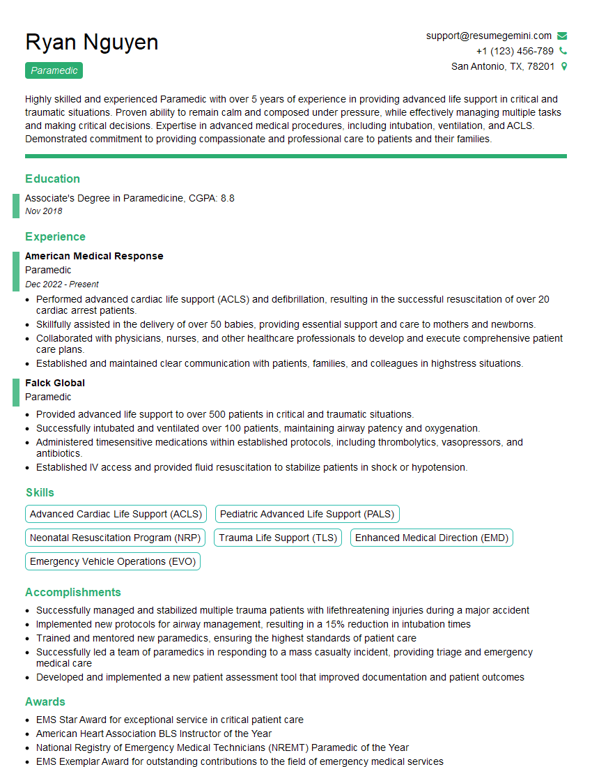
Paramedic Resume Sample
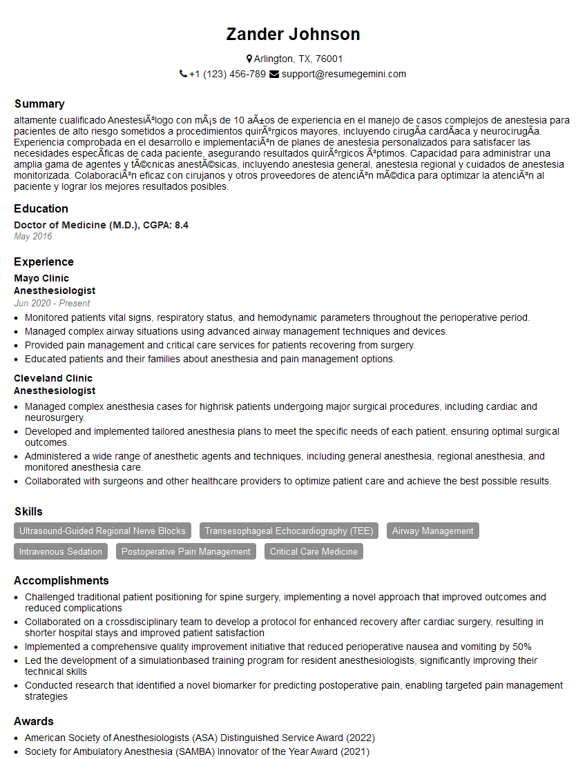
Anesthesiologist Resume Sample
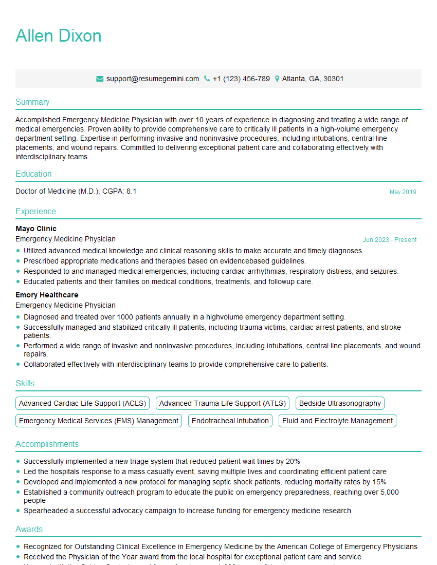
Emergency Medicine Physician Resume Sample
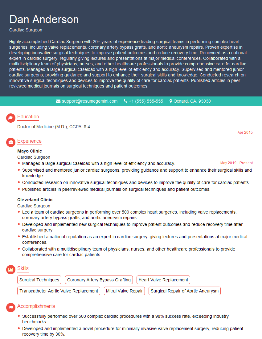
Cardiac Surgeon Resume Sample
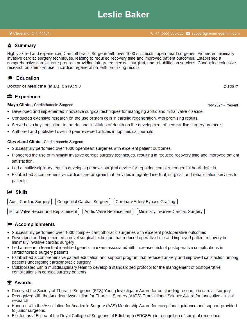
Cardiothoracic Surgeon Resume Sample
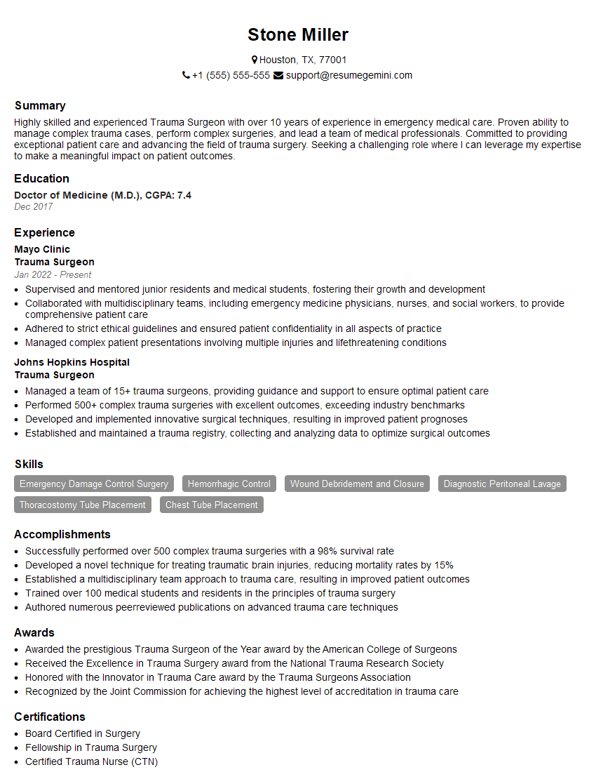
Trauma Surgeon Resume Sample
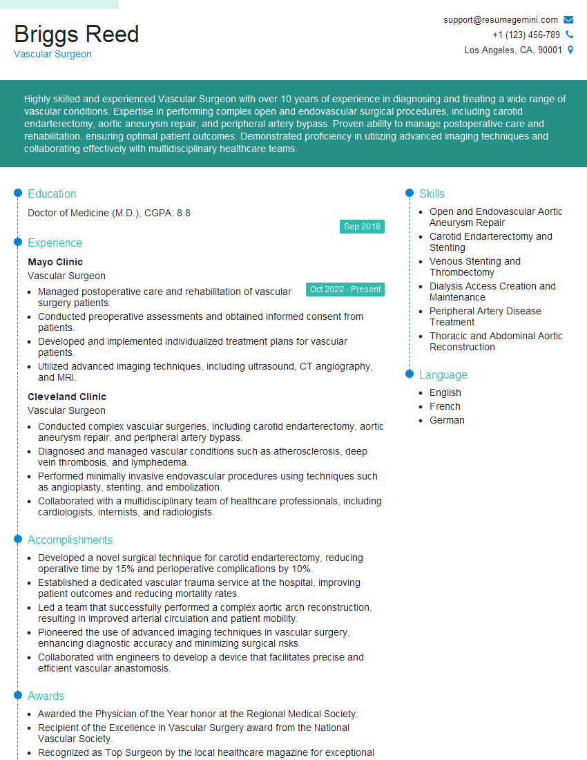
Vascular Surgeon Resume Sample
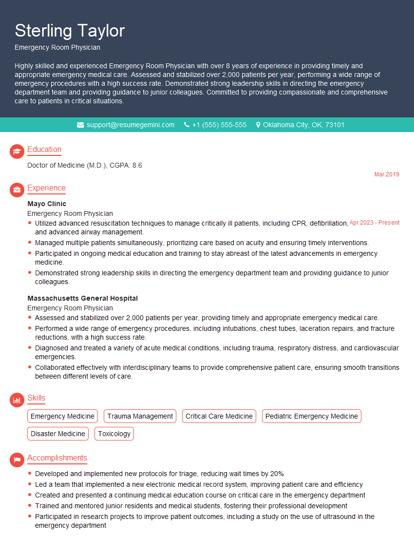
Emergency Room Physician Resume Sample
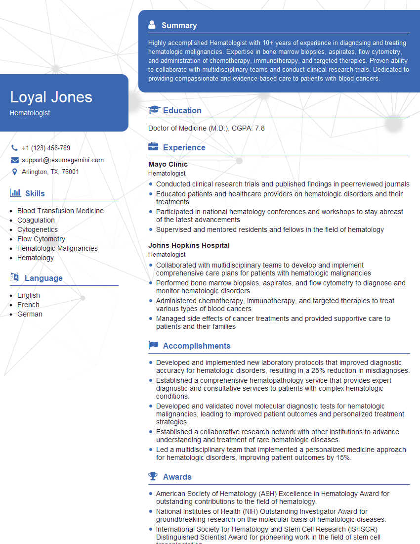
Hematologist Resume Sample
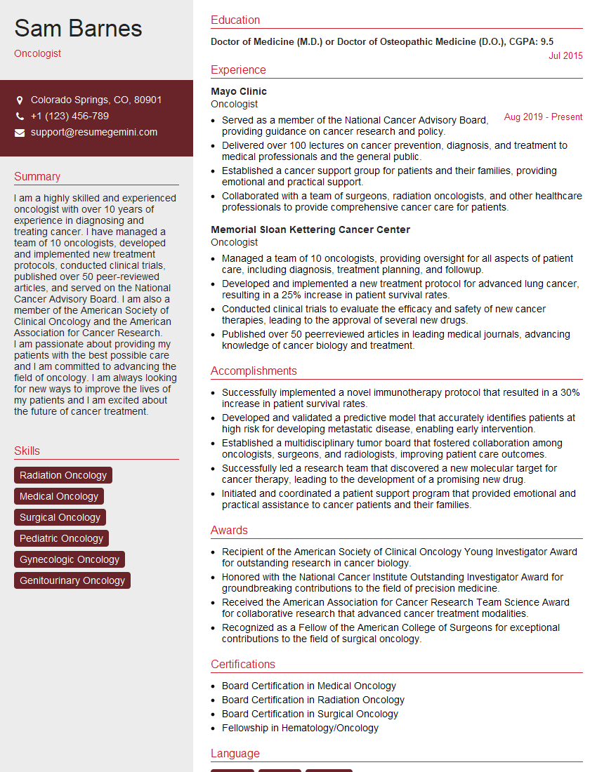
Oncologist Resume Sample
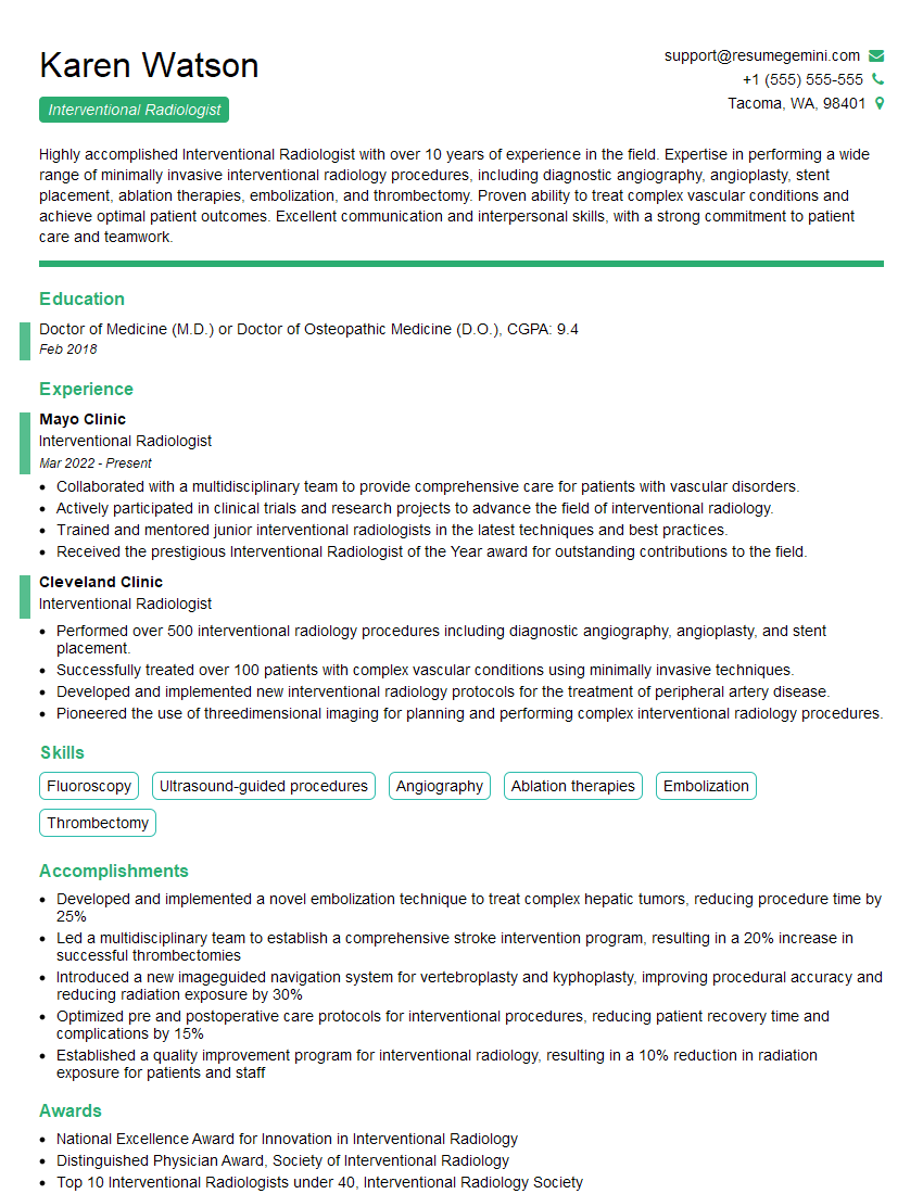
Interventional Radiologist Resume Sample
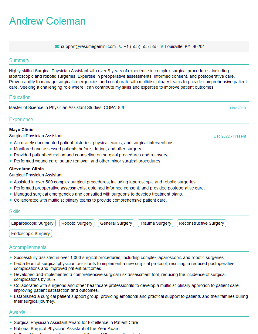
Surgical Physician Assistant Resume Sample
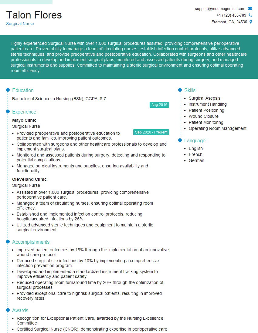
Surgical Nurse Resume Sample
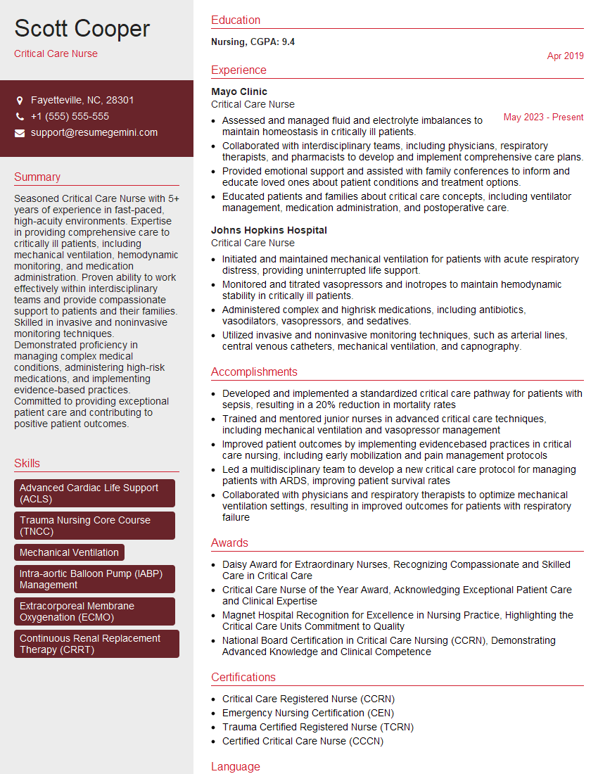
Critical Care Nurse Resume Sample
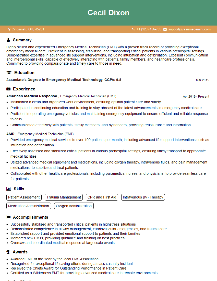
Emergency Medical Technician (EMT) Resume Sample
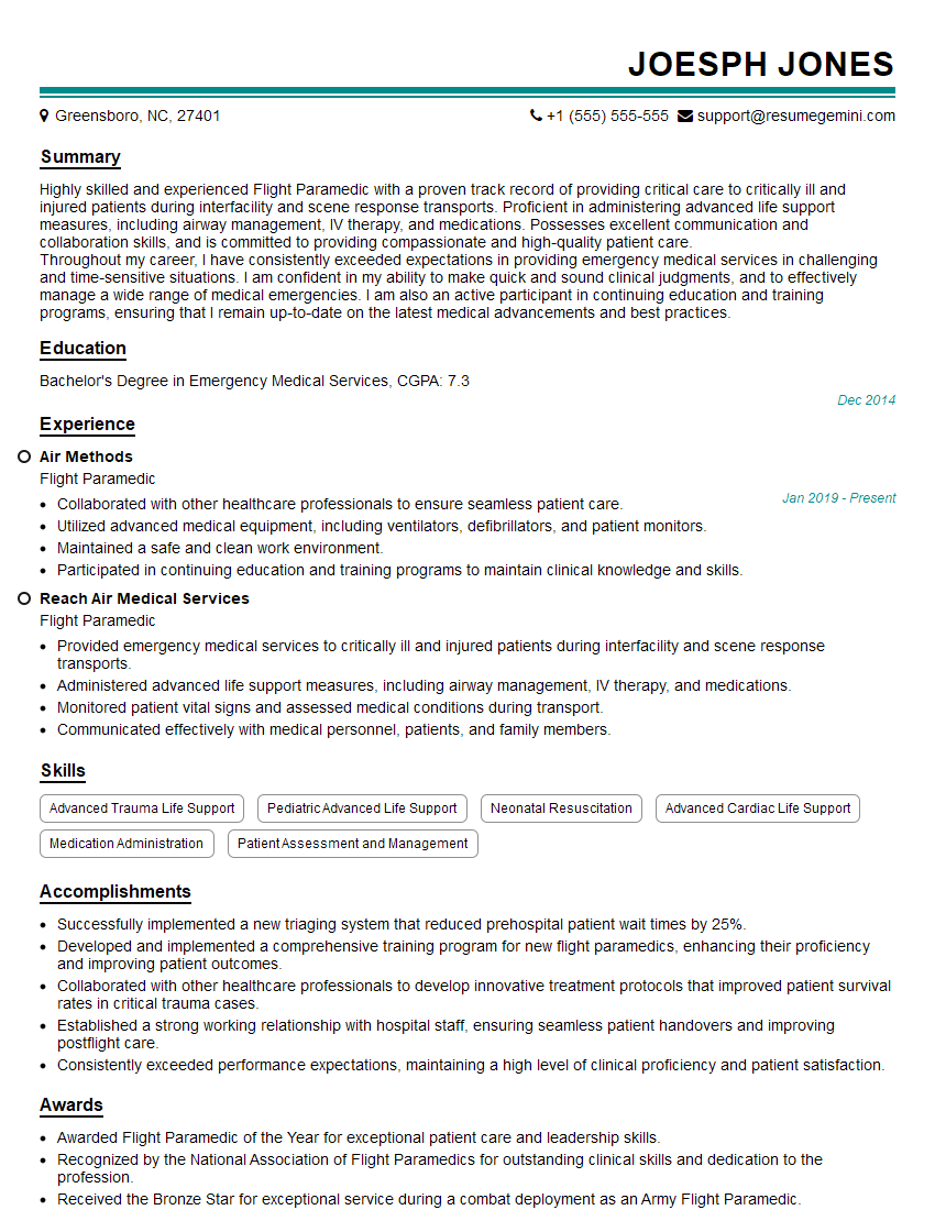
Flight Paramedic Resume Sample
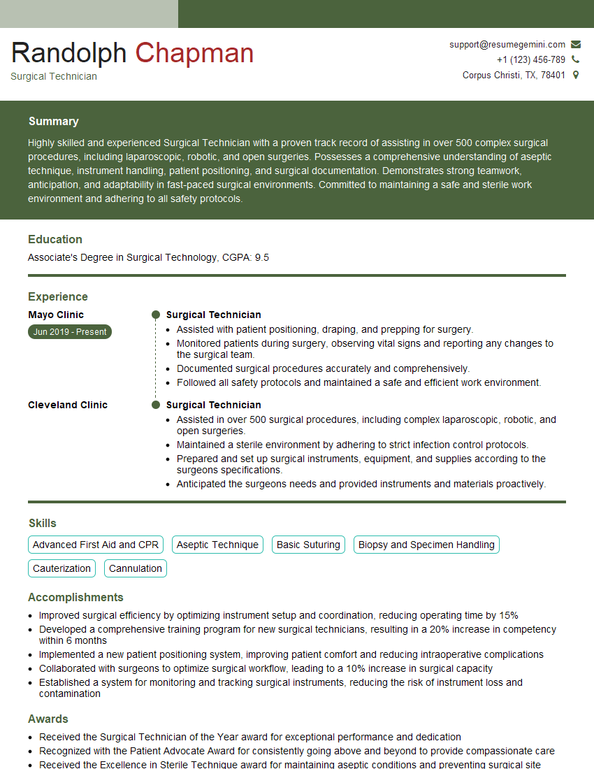
Surgical Technician Resume Sample
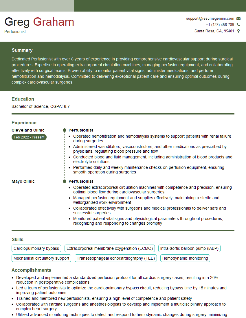
Perfusionist Resume Sample
- Paramedic
- Anesthesiologist
- Emergency Medicine Physician
- Cardiac Surgeon
- Cardiothoracic Surgeon
- Trauma Surgeon
- Vascular Surgeon
- Emergency Room Physician
- Hematologist
- Oncologist
- Interventional Radiologist
- Surgical Physician Assistant
- Surgical Nurse
- Critical Care Nurse
- Emergency Medical Technician (EMT)
- Flight Paramedic
- Surgical Technician
- Perfusionist
Explore more articles
Users Rating of Our Blogs
Share Your Experience
We value your feedback! Please rate our content and share your thoughts (optional).
What Readers Say About Our Blog
To the interviewgemini.com Webmaster.
Very helpful and content specific questions to help prepare me for my interview!
Thank you
To the interviewgemini.com Webmaster.
This was kind of a unique content I found around the specialized skills. Very helpful questions and good detailed answers.
Very Helpful blog, thank you Interviewgemini team.