Interviews are opportunities to demonstrate your expertise, and this guide is here to help you shine. Explore the essential Necropsy Performance interview questions that employers frequently ask, paired with strategies for crafting responses that set you apart from the competition.
Questions Asked in Necropsy Performance Interview
Q 1. Describe the proper procedure for performing a complete necropsy on a canine.
A canine necropsy, also known as a post-mortem examination, is a systematic dissection to determine the cause of death. It’s crucial to maintain sterility and meticulous documentation throughout the process. Think of it like a very detailed, investigative puzzle to solve the animal’s final story.
- External Examination: Begin with a thorough visual inspection, noting any external injuries, masses, or abnormalities. Record body weight, posture, and any evidence of trauma.
- Incision and Evisceration: A Y-shaped incision is typically made from the sternum to the pubis, allowing for access to the thoracic and abdominal cavities. Organs are carefully removed and examined individually, noting any changes in size, color, or texture. We use specific instruments like scalpel blades, forceps, and scissors to carefully dissect the tissues.
- Individual Organ Examination: Each organ is meticulously examined for abnormalities. For example, the heart is weighed, and the chambers examined for abnormalities. The lungs are inspected for pneumonia or other lesions. The liver is palpated and sampled for histology. The gastrointestinal tract is opened and its contents noted.
- Sampling: Samples of tissues and fluids are collected for histopathology (microscopic examination), microbiology (bacterial, fungal, and viral cultures), toxicology (detecting toxins), and other specialized testing, depending on the suspected cause of death.
- Documentation: Throughout the process, detailed observations are meticulously recorded, including photographs. A complete necropsy report summarizes the findings, and provides a possible diagnosis and cause of death.
For instance, I once performed a necropsy on a dog that presented with sudden death. The external exam was unremarkable, but internal examination revealed a massive pulmonary thromboembolism, the sudden blockage of a main artery to the lungs.
Q 2. What are the key differences between a necropsy and an autopsy?
While both involve the systematic examination of a deceased body, a necropsy is performed on animals, while an autopsy is performed on humans. The legal and regulatory frameworks surrounding these procedures differ significantly. The techniques are largely similar; however, the level of detail, specific tests performed, and the focus of the examination may vary depending on the species and circumstances.
For instance, in a canine necropsy, we might focus heavily on assessing cardiovascular and musculoskeletal systems, while a human autopsy would potentially involve more detailed neurological examination and forensic analysis of potential trauma.
Q 3. Explain the importance of proper tissue collection and preservation during a necropsy.
Proper tissue collection and preservation are paramount for accurate diagnoses. Improper handling can lead to artifacts and false negatives, compromising the diagnostic value of the necropsy.
- Sterile Technique: Maintaining a sterile environment minimizes contamination and prevents misleading results in microbiology analyses. Using sterile instruments and gloves is essential.
- Appropriate Fixatives: Tissues are typically preserved in 10% neutral buffered formalin, a fixative that hardens the tissue and prevents putrefaction. The tissue-to-fixative ratio is crucial for optimal preservation, usually about 1:10.
- Labeling and Storage: Each sample should be clearly labeled with the animal’s identification, tissue source, and date. Samples are typically stored in appropriately labeled containers and stored at a suitable temperature until analyzed.
- Specialized Preservation: Certain tissues require specialized preservation depending on the specific tests required. For example, samples for virology may require freezing at -80°C.
Imagine trying to assemble a puzzle with missing pieces or pieces that have been damaged; proper preservation ensures we have all the necessary pieces for a complete picture.
Q 4. How would you approach a necropsy on a suspected case of infectious disease?
In suspected infectious disease cases, strict biosafety protocols are paramount. Think of it as a crime scene investigation, only with microscopic culprits. Special precautions are taken to protect personnel and prevent the spread of potential pathogens.
- Personal Protective Equipment (PPE): The use of PPE, including gloves, gowns, masks, and eye protection, is mandatory. Depending on the suspected agent, additional precautions like respirators may be needed.
- Specialized Instruments: Single-use instruments are ideal to avoid cross-contamination, but reusable instruments require thorough sterilization.
- Environmental Controls: The necropsy should ideally be performed in a biosafety cabinet or a dedicated facility designed for handling infectious agents.
- Sampling for Microbiology: Multiple samples from various organs and body fluids are collected and sent to a microbiology laboratory for specific tests, including bacterial cultures, viral isolation, and PCR to detect specific pathogens.
- Waste Disposal: All waste materials, including tissues, instruments, and PPE, require careful disposal to prevent the spread of the infection.
For example, I once handled a suspected case of canine distemper. We employed maximal biosafety protocols, handling tissues with extreme care and ensuring all contaminated material was properly incinerated.
Q 5. What are the common postmortem changes and how do they affect necropsy interpretation?
Postmortem changes, the alterations occurring after death, can significantly affect necropsy interpretation. Understanding these changes is crucial for distinguishing normal postmortem findings from disease-related lesions. It’s like identifying natural decay from injuries in a crime scene.
- Rigor Mortis: Stiffening of muscles after death, typically sets in within a few hours and resolves after a day or two.
- Livor Mortis: Gravitational pooling of blood, causing discoloration of the dependent tissues. This can mimic bruising.
- Algor Mortis: Cooling of the body after death.
- Autolysis: Self-digestion of tissues by enzymes released from cells. This can mimic certain disease processes.
- Putrefaction: Bacterial decomposition of tissues, leading to gas formation, discoloration, and foul odor. This can obscure the presence of certain disease processes.
The timing of the postmortem changes impacts the findings. For example, early putrefaction can make identification of subtle organ lesions difficult.
Q 6. Describe your experience with different necropsy techniques (e.g., avian, rodent, large animal).
My experience encompasses a wide range of species, requiring adaptation of techniques depending on size, anatomy, and the specific questions driving the necropsy. Each species presents unique challenges and rewards.
- Avian Necropsies: These require delicate handling due to the fragile bones and small organs. Special attention is paid to the respiratory system, which is highly susceptible to infections. Using smaller instruments and careful dissection techniques are crucial.
- Rodent Necropsies: These are often performed for research purposes. The emphasis lies on maintaining a sterile environment and collecting samples for specific tests, including histological and microbiological analyses.
- Large Animal Necropsies: These necessitate different tools and techniques, often requiring specialized equipment for handling large organs and carcasses. Safety is of utmost importance given the sheer size and potential hazards associated with the procedure.
For instance, I’ve conducted necropsies on everything from tiny mice in a research lab to a 1,000-pound cow on a farm, each requiring a completely different approach, but always guided by the same principles of precision and accuracy.
Q 7. How do you handle unexpected findings during a necropsy?
Unexpected findings during a necropsy are not uncommon. A key skill is to remain objective, meticulously document the unexpected observations, and adapt the approach accordingly. These findings can sometimes be crucial to reaching the correct diagnosis. It’s like solving a mystery with unexpected clues.
- Thorough Documentation: Meticulous recording of all findings, including those that are unexpected, is paramount. This is critical for future analysis and to consult with colleagues.
- Additional Testing: Unexpected findings may necessitate additional tests, including specialized histology, microbiology, or toxicology studies.
- Consultation: Consultation with colleagues or experts in related fields may be essential to interpret unexpected findings and determine their significance.
- Revised Differential Diagnoses: The initial differential diagnoses may need to be revised and expanded based on the unexpected findings.
I recall a necropsy where I found a large parasitic mass in the abdominal cavity of a dog – an unexpected finding that led to an investigation into the dog’s environment and lifestyle which ultimately changed the cause of death conclusion.
Q 8. Explain your knowledge of common necropsy instruments and their proper usage.
Necropsy, or animal autopsy, requires a specialized set of instruments for precise dissection and sample collection. Proper usage ensures accurate findings and minimizes contamination. Key instruments include:
- Scalpels: Various sizes and blades (e.g., #10, #15) for making precise incisions. A #10 is generally used for initial incisions, while a #15 is better for finer work.
- Scissors: Dissecting scissors (blunt/blunt or sharp/blunt) are crucial for cutting through tissues. Metzenbaum scissors are excellent for delicate tissues, while Mayo scissors are stronger for thicker areas.
- Forceps: Adson forceps (with teeth) provide a secure grip on tissues, while Brown-Adson forceps (without teeth) are gentler on delicate structures. We also utilize tissue forceps with serrated tips for grasping and removing organs.
- Retractors: Various sizes and types allow for proper visualization during dissection, including hand-held retractors and self-retaining retractors like Weitlaner retractors.
- Bone cutters/Bone shears: Essential for accessing the skeletal system, especially in cases where bone lesions are suspected.
- Rulers and measuring tapes: Accurate measurement of lesions and organs is vital for documentation.
- Dissecting bowls and trays: For organized sample collection and to maintain sterility.
Proper usage involves careful handling of instruments to prevent damage to tissues and maintain sterility. Instruments should be sterilized before and after each necropsy. Sharp instruments should be handled with caution to prevent injury.
For example, during a necropsy of a bird, the use of fine-tipped scissors and forceps is essential for careful dissection of delicate pectoral muscles and air sacs, avoiding accidental damage to underlying tissues.
Q 9. Describe your experience with histological sample preparation.
Histological sample preparation is a critical step in necropsy, enabling microscopic examination of tissues to identify cellular changes indicative of disease. My experience encompasses the entire process, from tissue fixation to sectioning and staining.
The process typically involves:
- Tissue fixation: Immersion in 10% neutral buffered formalin is the most common method, preventing tissue autolysis and preserving cellular architecture. Fixation time depends on tissue size and type.
- Tissue processing: This involves dehydration using graded alcohols, clearing with xylene, and infiltration with paraffin wax. This prepares the tissue for sectioning.
- Embedding: The paraffin-infiltrated tissue is embedded in a paraffin block to provide support during sectioning.
- Sectioning: A microtome is used to cut thin sections (typically 4-5 μm) of the tissue block. These sections are mounted on glass slides.
- Staining: Hematoxylin and eosin (H&E) staining is a routine method used to visualize tissue morphology. Special stains like silver stains (for fungi) or PAS stains (for carbohydrates) may also be used based on the suspected disease.
I have extensive experience troubleshooting issues in each step of the process, such as ensuring proper fixation to prevent artifacts, optimizing processing times for different tissue types, and performing various staining techniques to achieve high-quality microscopic slides. For instance, I’ve had to adapt processing times for very dense tissues, like bone, to ensure complete paraffin infiltration without introducing artifacts.
Q 10. How do you ensure the safety and biosecurity protocols are followed during a necropsy?
Safety and biosecurity are paramount during necropsies. Strict adherence to protocols minimizes risk to personnel and the environment. My approach centers around these key aspects:
- Personal Protective Equipment (PPE): This includes gloves, lab coats, eye protection, and face masks. For cases involving potentially zoonotic diseases, more comprehensive PPE, such as respirators and full-body suits, may be necessary.
- Disinfection and Sterilization: Instruments and work surfaces are meticulously disinfected before and after each necropsy using appropriate disinfectants like 10% bleach solution. Instruments are sterilized using autoclaving or other suitable methods.
- Waste Management: All biological waste, including tissues, organs, and fluids, are treated as biohazardous material and disposed of following institutional protocols. This often involves autoclaving and incineration.
- Environmental Control: The necropsy area should be well-ventilated and equipped with appropriate safety equipment, such as sharps containers and spill kits.
- Sample Handling: Care is taken to prevent accidental aerosol generation, especially during procedures that might produce aerosols, like opening the thoracic or abdominal cavity. Procedures are performed with care to prevent potential splashes of tissue or fluids.
For example, when handling a case suspected of avian influenza, we would employ stringent biosecurity measures, including negative pressure environments and the use of specialized PPE, to prevent the accidental release of the virus.
Q 11. What are the ethical considerations in performing necropsies?
Ethical considerations in performing necropsies are crucial and always underpin my work. These include:
- Minimizing animal suffering: If the animal is still alive, appropriate euthanasia must be performed humanely before the necropsy begins. This is typically accomplished using methods approved by the institution and/or governing bodies.
- Respect for the animal: Necropsies are performed with respect for the animal, recognizing its inherent worth. Procedures are conducted in a professional and dignified manner.
- Informed consent: Whenever possible, informed consent should be obtained from the owner or responsible party before performing a necropsy.
- Confidentiality: Information gathered during the necropsy should be kept confidential and handled according to ethical guidelines and data privacy laws.
- Appropriate use of findings: Results should only be used for their intended purpose, such as advancing veterinary medicine or informing the owner of the cause of death, not for personal gain or unauthorized research.
I always try to balance the need for obtaining useful information with minimizing any potential harm or distress to the animal or its owner.
Q 12. Describe your experience with various fixation techniques for tissue samples.
Fixation is critical for preserving tissue architecture and preventing autolysis. Different tissues require different fixation approaches for optimal results. My experience includes various techniques, including:
- Formalin fixation: 10% neutral buffered formalin (NBF) is the most widely used fixative. It cross-links proteins, preventing enzymatic degradation. The immersion time varies based on tissue size and density (larger samples require longer fixation).
- Formalin-free fixation: Alternatives like glutaraldehyde and alcohol-based fixatives are increasingly used to minimize formaldehyde exposure. These offer advantages in reducing the risks associated with formaldehyde, but they may have limitations in preserving certain tissue components.
- Perfusion fixation: In research settings, perfusion fixation (injecting fixative directly into the circulatory system) is preferred for preserving the overall organ architecture, especially in cases where rapid fixation is crucial.
- Microwave fixation: Accelerated fixation is achieved using microwaves, reducing processing time. However, this method may introduce artifacts in some tissues.
The choice of fixation method depends on the type of tissue, the diagnostic questions, and the downstream applications (e.g., histology, immunohistochemistry, electron microscopy). For example, electron microscopy requires specific fixation protocols using glutaraldehyde and osmium tetroxide to preserve ultrastructural details.
Q 13. How do you interpret gross lesions observed during a necropsy?
Interpreting gross lesions—the visible changes in organs and tissues—is a crucial aspect of necropsy. It involves a systematic approach, combining observation with knowledge of anatomy and pathology.
I utilize a systematic approach to examine each organ system, noting:
- Location: Precise location of the lesion within the organ.
- Size: Measurement of the lesion in millimeters or centimeters.
- Shape: Description of the lesion’s shape (e.g., round, oval, irregular).
- Color: Note the lesion’s color (e.g., red, yellow, white, black).
- Texture: Description of the lesion’s texture (e.g., firm, soft, friable).
- Consistency: Whether the lesion is fluid-filled, solid, or mixed.
- Distribution: If multiple lesions are present, note their distribution (e.g., diffuse, focal, multifocal).
The combination of these observations, alongside the animal’s history and clinical signs, helps generate a preliminary diagnosis. For instance, observing a large, firm, white nodule in the lung of a dog could suggest a tumor, while observing multiple small, pale foci in the liver could suggest a disseminated infection.
However, it’s crucial to remember that gross lesions alone are often insufficient for a definitive diagnosis, and that histopathological examination and laboratory testing are often necessary to confirm the findings.
Q 14. What are the key steps in documenting a necropsy?
Accurate and detailed documentation is essential for a complete necropsy report. My documentation process typically includes:
- Case Information: Detailed information about the animal (species, breed, age, sex, signalment), including the history of the case, clinical findings, and reason for necropsy.
- Gross Findings: A comprehensive description of all gross lesions observed during the examination, using precise anatomical terminology. This typically includes photographic documentation of lesions and measurements.
- Sample Collection: A detailed list of all tissues and fluids collected for further testing, including histological examination, microbiology, toxicology, etc. The collection method for each specimen should be documented.
- Histopathology Results: Once microscopic examination is complete, the findings are carefully integrated into the final report.
- Diagnostic Interpretation: A concise summary of the findings, including a tentative or definitive diagnosis and comments on the cause of death.
- Photographs: High-quality photographs are essential and provide visual documentation of the lesions observed during the procedure.
A well-structured report allows for easy access to the collected data and supports clear communication of the results to clients and other veterinary professionals. For example, images can be added to the necropsy report using the software used to compose the report. This ensures that all information is stored in one place. Digital images are typically better than hand-drawn diagrams and can be easily shared.
Q 15. How do you differentiate between ante-mortem and post-mortem lesions?
Differentiating between ante-mortem (before death) and post-mortem (after death) lesions is crucial for accurate diagnosis in necropsy. Ante-mortem lesions show evidence of a healing process, like inflammation, repair, or scarring. For instance, a healed fracture will display callus formation, a clear sign it occurred before death. Post-mortem lesions lack these features; they’re changes occurring after death due to processes like autolysis (self-digestion) or putrefaction (decomposition). A good example is post-mortem lividity (blood pooling), which doesn’t show the inflammatory response characteristic of ante-mortem bruising.
- Ante-mortem: Shows signs of the body’s attempt to heal (inflammation, fibrosis, etc.).
- Post-mortem: Absence of healing response; changes are due to decomposition processes.
Imagine finding a lung with a consolidated area: If there’s surrounding inflammation and fibrosis, it’s likely ante-mortem pneumonia. If it’s just a discolored area with no inflammation, it’s probably a post-mortem artifact. Careful observation and understanding of the disease processes are essential for this differentiation.
Career Expert Tips:
- Ace those interviews! Prepare effectively by reviewing the Top 50 Most Common Interview Questions on ResumeGemini.
- Navigate your job search with confidence! Explore a wide range of Career Tips on ResumeGemini. Learn about common challenges and recommendations to overcome them.
- Craft the perfect resume! Master the Art of Resume Writing with ResumeGemini’s guide. Showcase your unique qualifications and achievements effectively.
- Don’t miss out on holiday savings! Build your dream resume with ResumeGemini’s ATS optimized templates.
Q 16. Explain your understanding of different types of tissue samples required for different diagnostic tests.
The type of tissue sample collected depends heavily on the suspected cause of death and the specific diagnostic test required. For example:
- Histopathology (microscopic examination): Requires formalin-fixed, paraffin-embedded tissue blocks from multiple organs (liver, kidney, lung, heart, spleen, etc.). The size and number of samples depend on the case complexity; suspected neoplasia (cancer) requires larger and more numerous samples.
- Microbiology (bacterial/fungal cultures): Sterile swabs or tissue fragments are needed for culture. Aseptic technique is critical to avoid contamination.
- Toxicology: Samples of blood, liver, kidney, and sometimes vitreous humor are collected and sent to a toxicology laboratory for analysis of drugs, pesticides, heavy metals, etc. Proper sample preservation is crucial to prevent degradation.
- Molecular Pathology (PCR): Fresh or frozen tissue samples are needed for DNA/RNA extraction to detect specific pathogens or genetic mutations.
Consider a case of suspected poisoning. We’d collect blood and liver for toxicology, ensuring samples are properly labeled and preserved to prevent degradation. Simultaneously, we would take tissue samples for histopathology to examine organ damage.
Q 17. How do you deal with difficult or challenging cases during necropsy?
Difficult cases require a systematic approach. First, I thoroughly document all observations, even seemingly insignificant details. Then, I consult relevant literature and veterinary pathologists or specialists if necessary. Sometimes, advanced imaging techniques like CT or MRI scans help clarify ambiguous findings. For example, I once encountered a case with severe autolysis. We employed advanced imaging techniques to visualize the organs better, allowing us to conclude the cause of death.
Collaboration is key. Discussions with clinicians who treated the animal before death often yield critical insights. If a definitive diagnosis remains elusive, acknowledging the limitations of the necropsy and suggesting further investigation is crucial for professional responsibility.
Q 18. Describe your experience working with different diagnostic laboratories.
Throughout my career, I’ve worked with numerous diagnostic laboratories, both large, internationally renowned facilities and smaller specialized labs. My experience spans diverse testing modalities, including histopathology, microbiology, toxicology, and molecular pathology. Effective collaboration requires understanding the strengths and limitations of each lab and their specific requirements for sample submission (e.g., specific containers, fixatives, and accompanying documentation).
Building strong relationships with laboratory personnel is crucial. It ensures efficient communication, prompt results, and helpful consultations when interpreting complex results. For example, a close relationship with a specialized toxicology lab helped us rapidly identify a rare toxin in a wildlife necropsy case.
Q 19. What is your experience with digital imaging and documentation in necropsy?
Digital imaging and documentation are integral to modern necropsy. I utilize high-resolution cameras and specialized software to capture detailed images and videos of gross lesions, organ weights, and tissue samples. This digital record provides a comprehensive and permanent archive of the necropsy findings, facilitating communication with other professionals, enhancing report quality, and supporting future research.
Digital imaging also facilitates the efficient sharing of images with collaborators and specialists for consultation. I use software that allows for annotations and measurements directly on images, improving accuracy and ease of report generation. The digital approach enhances the overall quality and clarity of the necropsy findings while reducing the reliance on potentially lost physical records.
Q 20. How do you prioritize findings during a necropsy to provide a comprehensive report?
Prioritizing findings involves a careful assessment of the lesions’ significance and their potential to explain the cause of death. Potentially life-threatening lesions, like severe organ damage or significant hemorrhage, take precedence. I systematically document all findings, but the report highlights those considered most important. A systematic approach is crucial; beginning with a detailed external examination, followed by organ system evaluation, enables a logical flow in the report.
For instance, if a dog presents with cardiac rupture, this finding overshadows other less severe lesions, even if multiple abnormalities are detected. The report focuses on the cardiac rupture as the primary cause of death and then systematically describes other significant or incidental findings.
Q 21. Describe your experience with specialized necropsy techniques such as toxicology or microbiology.
My experience with specialized necropsy techniques includes extensive work in toxicology and microbiology. In toxicology, I’m proficient in collecting and handling samples for various analyses, ensuring sample integrity and proper chain of custody. This includes selecting appropriate organs and using validated techniques to avoid contamination or degradation.
In microbiology, I’m adept at performing aseptic techniques for collecting and processing tissue samples for bacterial and fungal cultures. I have extensive experience with interpreting culture results, correlating them with gross and microscopic lesions to provide a comprehensive diagnosis. A recent case involved isolating a rare fungal infection, requiring specialized culture media and techniques for accurate identification.
Q 22. How familiar are you with various regulatory guidelines relevant to necropsy procedures?
My familiarity with regulatory guidelines for necropsy procedures is extensive. I’m well-versed in regulations such as those from the USDA (United States Department of Agriculture) for animal necropsies, the CDC (Centers for Disease Control and Prevention) guidelines for infectious disease handling, and OSHA (Occupational Safety and Health Administration) standards for workplace safety. These regulations cover aspects like biosecurity, proper handling of hazardous materials (including potentially infectious agents), waste disposal, and record-keeping. I understand the importance of adhering to these guidelines to ensure the safety of personnel, the accuracy of findings, and compliance with legal requirements. For example, working with animals suspected of having a zoonotic disease requires strict adherence to CDC guidelines to prevent the spread of infection. Similarly, proper sharps disposal as outlined by OSHA prevents accidental needle-stick injuries.
Q 23. How do you maintain accurate and detailed necropsy records?
Maintaining accurate and detailed necropsy records is crucial for the validity and usefulness of the procedure. My approach involves a systematic documentation process. I use a standardized necropsy report form that captures all relevant information. This includes the animal’s identification (species, age, sex, breed, etc.), a detailed description of the external examination (including lesions and abnormalities), the internal examination findings (organ weights, macroscopic observations of each organ system, description of any lesions), and the collection of samples for further testing (histology, microbiology, toxicology). I use clear and concise language, avoiding subjective interpretations. Photographs and sketches are included whenever possible to supplement the written descriptions. All observations are meticulously recorded, even seemingly insignificant findings, as they can become relevant later. Finally, the completed report is reviewed for completeness and accuracy before archiving.
Q 24. Describe your experience with quality control and quality assurance in a necropsy setting.
Quality control and quality assurance (QC/QA) are paramount in a necropsy setting. My experience includes implementing and following protocols to ensure the accuracy and reliability of the findings. This involves regular calibration and maintenance of equipment (scales, instruments), rigorous adherence to aseptic techniques during the procedure to prevent contamination of samples, and regular proficiency checks of my own skills and knowledge. We conduct internal audits to review our processes and identify areas for improvement. For instance, blinded proficiency testing is used periodically where a known sample is sent to different labs and results are compared to assess the consistency and accuracy of our findings. We also maintain a detailed log of equipment maintenance, reagent batches, and any deviations from established protocols, providing traceability and accountability. Participation in professional development programs and external quality assurance schemes further enhances our quality control measures.
Q 25. How do you handle conflicting information or findings during a necropsy?
Handling conflicting information or findings during a necropsy requires a systematic and objective approach. The first step is to carefully review all the evidence, including the macroscopic findings, microscopic findings, and any laboratory results. If the conflict is between macroscopic and microscopic findings, I correlate the observations. Sometimes, one finding might be better explained by a second more subtle finding. It is also possible there are two co-existing diseases or injuries. If the conflict remains, I consult relevant literature, seek a second opinion from a colleague, or utilize additional diagnostic techniques to resolve the discrepancy. Open communication and collaboration with other specialists are often vital. In summary, thoroughness and critical thinking are essential in resolving such conflicts. Thorough documentation of the process allows for peer review and transparency.
Q 26. What are your strengths and weaknesses in performing necropsies?
My strengths include a meticulous approach, a strong understanding of anatomy and pathology, and the ability to interpret complex findings. I have considerable experience in performing necropsies on a wide variety of species and handling different types of cases. My systematic documentation skills ensure that my reports are comprehensive and easy to understand. One of my weaknesses is that I sometimes spend too long on details, which can occasionally affect turnaround times. I’m actively working on improving my time management skills to balance thoroughness with efficiency. I’m also always looking to improve my experience with less commonly encountered species or specialized diagnostic techniques.
Q 27. How do you stay updated on advancements in necropsy techniques and technologies?
Staying updated on advancements in necropsy techniques and technologies is crucial. I achieve this through several methods. I actively participate in professional organizations such as the American Association of Veterinary Laboratory Diagnosticians (AAVLD), attending conferences and workshops to learn about the latest techniques and technologies. I regularly read scientific literature, including peer-reviewed journals and online resources, focusing on advancements in necropsy methodology and diagnostic tools. I also participate in continuing education courses to maintain my certification and professional competence. This combination keeps me at the forefront of my field.
Q 28. Describe a challenging necropsy case and how you approached it.
A particularly challenging case involved a young dog presenting with acute respiratory distress. The initial macroscopic examination revealed minimal findings. However, microscopic examination of lung tissue revealed the presence of unusual fungal hyphae. Identifying the specific fungal species proved difficult. I consulted with a mycologist, and we utilized advanced molecular techniques (PCR testing) to confirm the diagnosis as a rare invasive fungal infection. The challenge lay in the subtle nature of the initial findings and the need to employ specialized diagnostic techniques for definitive diagnosis. This case highlighted the importance of collaboration and the utilization of advanced technologies to tackle complex diagnostic challenges in necropsy.
Key Topics to Learn for Necropsy Performance Interview
- Specimen Handling and Preservation: Understanding proper techniques for collecting, preserving, and transporting samples for accurate necropsy results.
- Necropsy Techniques and Procedures: Mastering the systematic approach to performing necropsies, including external examination, organ dissection, and sample collection. This includes familiarity with various species and their unique anatomical features.
- Histopathology and Microscopic Examination: Knowledge of tissue processing, staining techniques, and interpretation of microscopic findings to support necropsy diagnoses.
- Diagnostic Interpretation and Reporting: Developing skills in analyzing necropsy findings, correlating them with clinical history, and preparing comprehensive and accurate reports.
- Safety and Biosecurity Protocols: Understanding and adhering to strict safety protocols to prevent exposure to biohazards and maintain a sterile work environment.
- Data Analysis and Record Keeping: Utilizing accurate record-keeping practices and data analysis methods to draw meaningful conclusions from necropsy results and contribute to research and disease surveillance.
- Troubleshooting and Problem-Solving: Developing the ability to identify and resolve technical challenges that may arise during the necropsy process.
- Ethical Considerations in Necropsy: Understanding the ethical implications of necropsy procedures and maintaining the highest professional standards.
Next Steps
Mastering necropsy performance opens doors to exciting career opportunities in veterinary pathology, research, and forensic science, offering significant contributions to animal health and human understanding. To maximize your job prospects, crafting an ATS-friendly resume is crucial. ResumeGemini is a trusted resource that can help you build a professional and impactful resume. We provide examples of resumes tailored to Necropsy Performance to guide you. Take this opportunity to showcase your skills and experience effectively and confidently pursue your career goals.
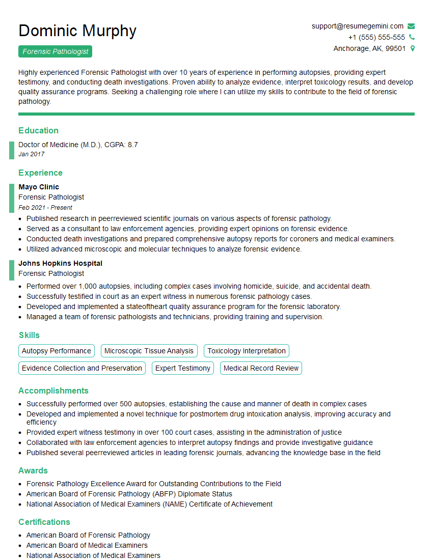
Forensic Pathologist Resume Sample
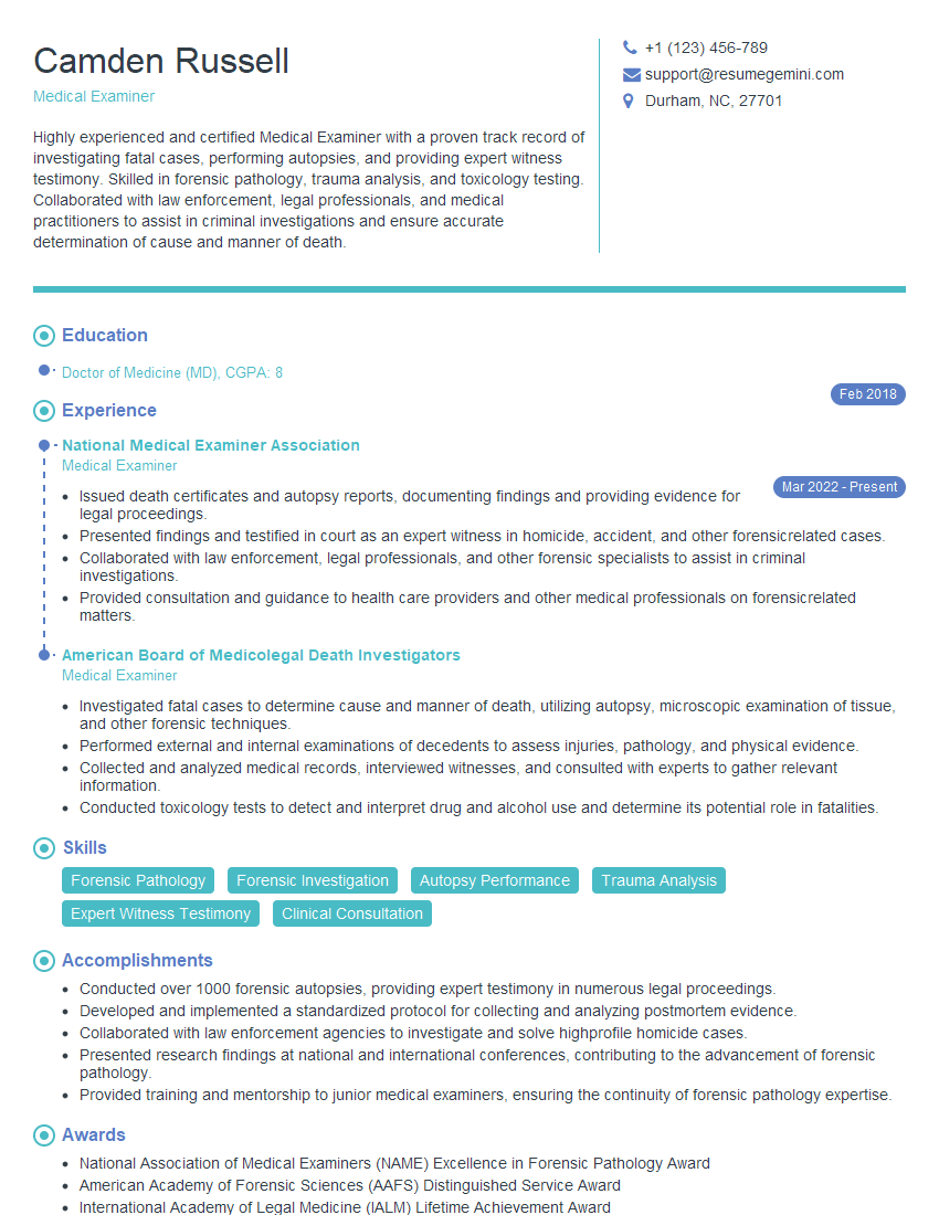
Medical Examiner Resume Sample
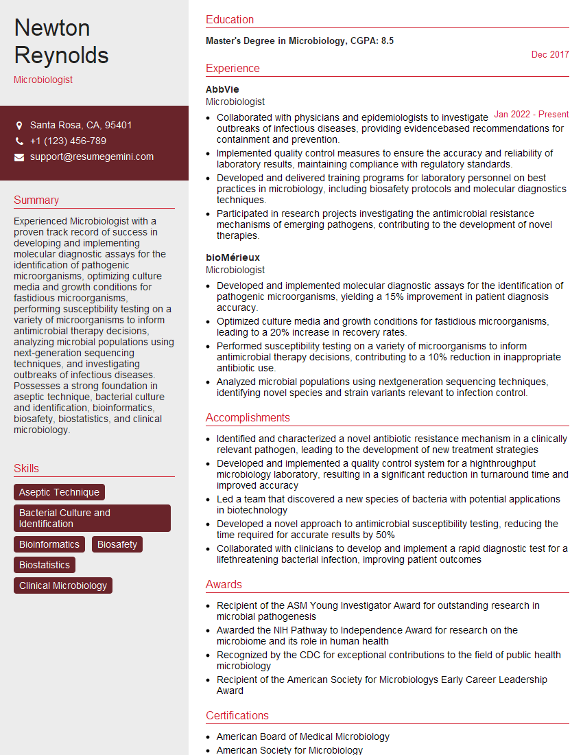
Microbiologist Resume Sample
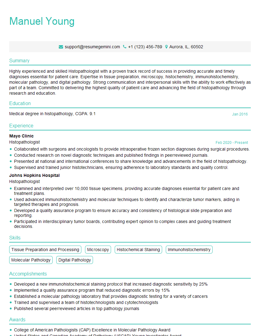
Histopathologist Resume Sample
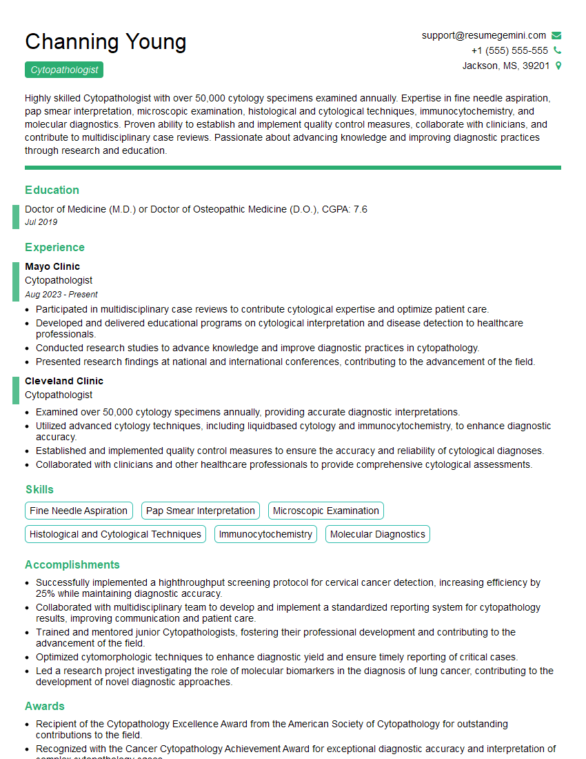
Cytopathologist Resume Sample
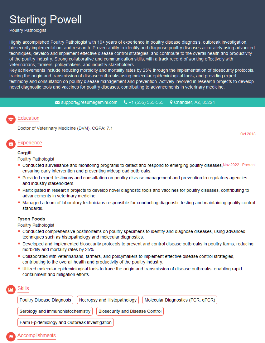
Poultry Pathologist Resume Sample
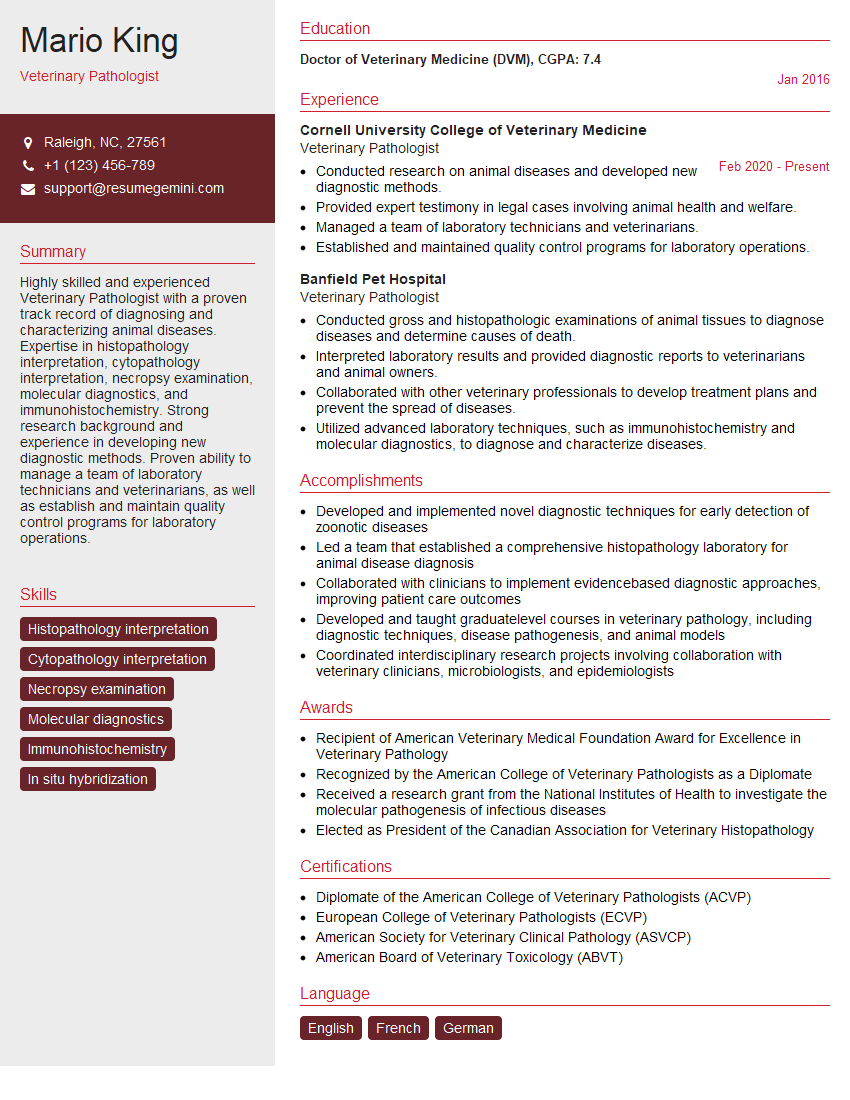
Veterinary Pathologist Resume Sample
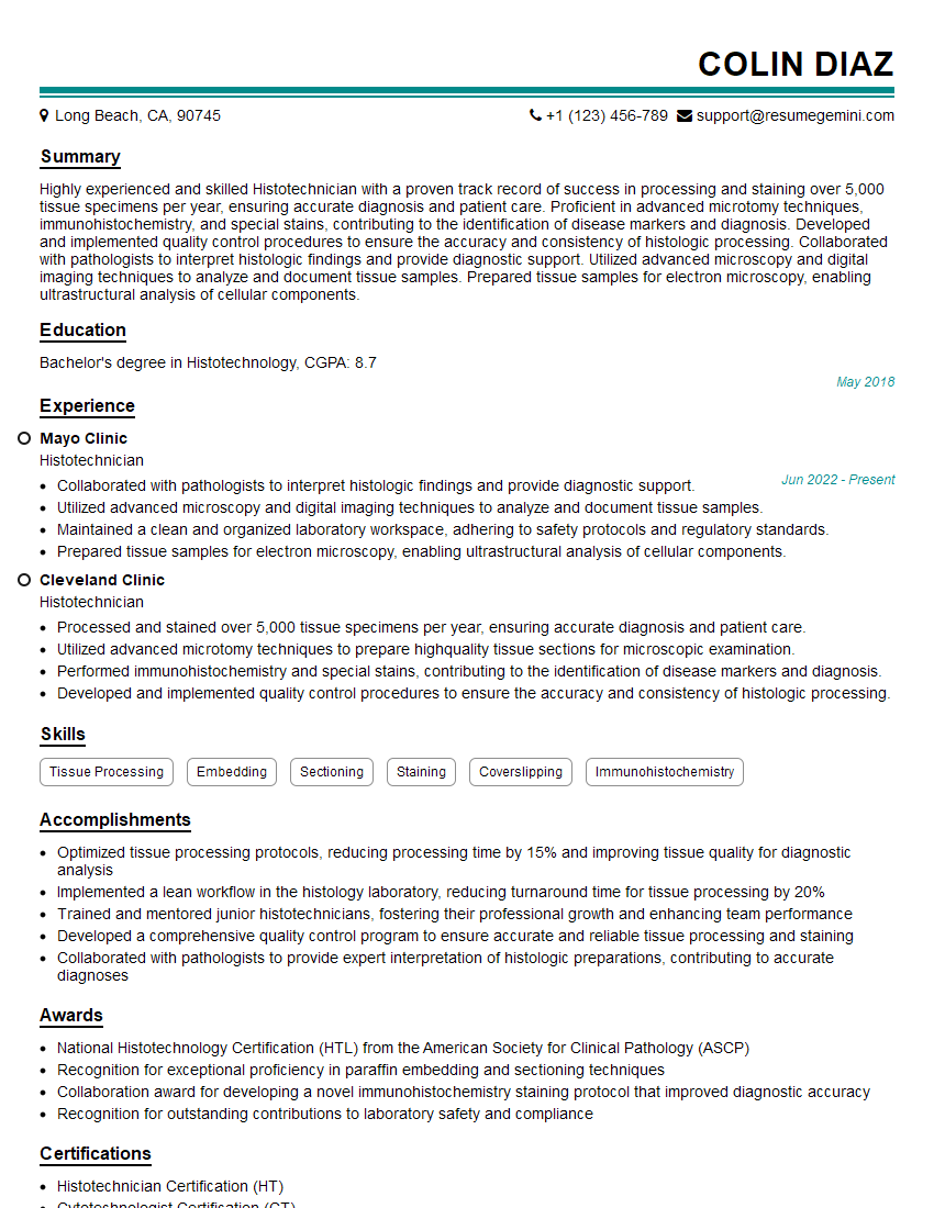
Histotechnician Resume Sample
Animal Health Technician Resume Sample
Explore more articles
Users Rating of Our Blogs
Share Your Experience
We value your feedback! Please rate our content and share your thoughts (optional).
What Readers Say About Our Blog
To the interviewgemini.com Webmaster.
Very helpful and content specific questions to help prepare me for my interview!
Thank you
To the interviewgemini.com Webmaster.
This was kind of a unique content I found around the specialized skills. Very helpful questions and good detailed answers.
Very Helpful blog, thank you Interviewgemini team.