Preparation is the key to success in any interview. In this post, we’ll explore crucial Orthopedic Trauma Management interview questions and equip you with strategies to craft impactful answers. Whether you’re a beginner or a pro, these tips will elevate your preparation.
Questions Asked in Orthopedic Trauma Management Interview
Q 1. Describe your experience managing open fractures.
Managing open fractures, also known as compound fractures, requires a multi-faceted approach prioritizing infection control and fracture stabilization. My experience encompasses the entire spectrum of care, from initial assessment and resuscitation in the trauma bay to definitive surgical fixation and long-term follow-up. This includes:
- Immediate Wound Care: Debridement of devitalized tissue is crucial to minimize infection risk. I meticulously remove all foreign material and thoroughly irrigate the wound with copious amounts of saline solution.
- Antibiotic Prophylaxis: Broad-spectrum antibiotics are administered promptly to prevent infection. The choice of antibiotic depends on local antibiotic sensitivity patterns and the severity of contamination.
- Fracture Stabilization: The method of stabilization depends on the fracture pattern, location, and patient’s overall condition. Options range from external fixation for unstable fractures in polytraumatized patients to internal fixation with plates, screws, or intramedullary nails once the patient is hemodynamically stable.
- Wound Closure: Delayed primary closure is often preferred, allowing for adequate wound healing and reducing the risk of infection. Sometimes, negative pressure wound therapy (NPWT) is utilized to promote granulation tissue formation before closure.
- Postoperative Management: Postoperative care involves pain management, meticulous wound care, regular monitoring for signs of infection, and appropriate physical therapy to promote recovery.
For instance, I recently managed a Grade III open tibia fracture in a young man involved in a motorcycle accident. After initial resuscitation and wound debridement, we utilized an external fixator for initial stabilization due to associated soft tissue injuries. Once the soft tissues had improved, we proceeded with intramedullary nailing for definitive fixation.
Q 2. Explain the principles of damage control orthopedics.
Damage control orthopedics (DCO) is a surgical strategy used in severely injured patients where addressing all injuries simultaneously would put the patient at unacceptable risk. The core principle is to perform life-saving interventions first, stabilizing the patient’s hemodynamic status and addressing life-threatening injuries before definitive skeletal repair. It prioritizes temporary stabilization over perfect anatomical reduction.
- Hemodynamic Stabilization: Addressing hemorrhage and shock is paramount. This often involves blood transfusions, fluid resuscitation, and potentially damage control surgery.
- Temporary Stabilization: Fractures are stabilized using techniques like external fixation, allowing for easy access to other injuries and minimizing operative time. This avoids prolonged anesthesia and blood loss.
- Wound Debridement: Minimally invasive debridement and wound closure are performed, focusing on preventing infection without delaying the process excessively.
- Delayed Definitive Fixation: Once the patient is stabilized, a second-stage operation is planned for definitive fracture fixation. This allows for better soft tissue conditions and improved healing.
Imagine a patient with multiple injuries including a pelvic fracture, a femur fracture, and a pneumothorax after a high-speed motor vehicle collision. DCO would involve immediate stabilization of the pneumothorax, addressing hemorrhage, and then applying external fixators to the femur and pelvis. Definitive fixation would be planned after the patient is stabilized and their overall condition improves.
Q 3. How do you assess the stability of a fracture?
Assessing fracture stability involves a combination of clinical examination and imaging studies. We consider the fracture pattern, the integrity of the surrounding soft tissues, and the forces acting on the fracture.
- Clinical Examination: This involves assessing the patient’s pain, range of motion, and any signs of deformity or malalignment. Gentle palpation can help assess for crepitus (a grating sensation indicating bone fragments rubbing together).
- Imaging: X-rays are essential for visualizing the fracture and assessing its location, pattern, and displacement. Computed tomography (CT) scans provide more detailed three-dimensional images, particularly helpful for complex fractures. Sometimes, MRI scans are used to assess soft tissue injuries.
- Fracture Classification: Fracture classification systems (e.g., AO/OTA classification) provide a standardized way to describe fractures and guide treatment decisions. Understanding the fracture classification helps predict the stability and likelihood of healing.
For example, a simple, undisplaced transverse fracture of the tibia is generally considered stable and may heal well with non-operative management, whereas a comminuted, displaced fracture of the femur is unstable and typically requires surgical intervention for fixation.
Q 4. What are the indications for external fixation?
External fixation is indicated in various scenarios where internal fixation may not be ideal. It provides temporary or definitive stabilization depending on the situation. Common indications include:
- Severe Soft Tissue Injury: When substantial soft tissue damage is present, external fixation allows for initial stabilization while allowing for wound management and improving the soft tissue environment before internal fixation.
- Polytrauma Patients: In patients with multiple injuries, external fixation minimizes operating time and blood loss, allowing for simultaneous attention to life-threatening issues.
- Infection: External fixation can be used in cases of open fractures or post-operative infections, allowing for access to the wound for debridement and treatment.
- Fractures in Difficult Locations: External fixation can be beneficial for fractures in areas difficult to reach with internal fixation, such as distal tibia fractures with significant soft tissue damage.
- Inadequate Bone Stock: When there is insufficient bone stock for internal fixation, external fixation can provide temporary stability before bone grafting or other reconstructive procedures.
A good example is a severely mangled lower extremity with an open fracture. Initially, external fixation would allow management of the open wound, preserving circulation and limb viability. Once the wounds are cleaned and healthy, the external fixator could be removed and the fracture stabilised with a plate and screws or intramedullary nail.
Q 5. What are the complications associated with intramedullary nailing?
Intramedullary nailing, while a powerful technique for fracture fixation, is associated with several potential complications:
- Infection: Infection remains a significant concern, particularly in open fractures or cases where surgical technique is compromised.
- Malunion/Nonunion: Improper reduction or inadequate fixation can lead to malunion (healing in a malaligned position) or nonunion (failure of the fracture to heal).
- Implant Failure: Fracture can occur at the nail or bending of the nail can occur.
- Nerve or Vessel Injury: Damage to surrounding nerves or blood vessels during surgery is possible.
- Fat Embolism: Although rare, fat embolism can occur, particularly with femoral fractures.
- Stress Shielding: The implant may bear too much of the weight bearing, leading to bone loss around the implant site.
For example, inadequate reaming of the medullary canal during intramedullary nailing can lead to stress shielding, reducing bone density around the implant site, increasing the risk of fracture and implant loosening. Careful surgical technique and postoperative weight-bearing restrictions help mitigate these risks.
Q 6. Describe your experience with plating techniques.
My experience with plating techniques is extensive, encompassing various plate designs and applications depending on the specific fracture pattern, bone quality, and patient factors. I’m proficient in both open reduction and internal fixation (ORIF) using plates and screws for a wide range of fractures, including:
- Choosing the Right Plate: Selection of the appropriate plate involves considering factors such as the fracture location, bone morphology, and the forces acting on the fracture. Different plate designs (e.g., dynamic compression plates, reconstruction plates, locking plates) are tailored for specific situations.
- Surgical Technique: Accurate reduction and stable fixation are crucial. This involves meticulous exposure of the fracture site, precise reduction of the fracture fragments, and secure placement of screws to achieve stable fixation.
- Biomechanics: A strong understanding of fracture biomechanics is essential for optimal plate placement to prevent complications. This includes knowing how forces are transmitted and how the plate should be placed to counteract them.
- Postoperative Management: Postoperative care includes pain management, wound care, and early mobilization with appropriate weight-bearing restrictions, guided by fracture stability and patient’s healing progress.
For instance, I recently used a locking plate for a comminuted fracture of the distal radius. Locking plates are particularly beneficial in osteoporotic bones, where screw pullout is a major concern. The locking mechanism enhances the stability of fixation, allowing for early mobilization.
Q 7. How do you manage compartment syndrome?
Compartment syndrome is a serious condition characterized by increased pressure within a confined anatomical space (compartment) compromising blood supply to the muscles and nerves within that compartment. Early recognition and prompt treatment are vital to prevent permanent damage.
- Clinical Assessment: The hallmark signs of compartment syndrome are the six Ps: Pain (disproportionate to the injury), Pallor (pale skin), Paresthesia (numbness or tingling), Pulselessness (decreased or absent pulse), Paralysis (muscle weakness or loss of function), and Poikilothermia (coldness of the extremity). However, early detection is critical, as these signs may not always be present in the early stages.
- Measuring Compartment Pressure: Compartment pressure measurement using a Stryker pressure monitor is crucial for objective assessment. Elevated compartment pressure (>30 mmHg difference between diastolic blood pressure and compartment pressure) is a strong indication for fasciotomy.
- Fasciotomy: Surgical decompression (fasciotomy) is the definitive treatment for compartment syndrome. This involves making incisions to relieve pressure within the affected compartment. The incisions are left open to ensure adequate decompression and allow the swelling to subside before secondary closure.
- Postoperative Care: Postoperative care involves close monitoring, wound care, and appropriate pain management. Physical therapy is essential for rehabilitation once the swelling has subsided and the wound has healed.
I recall a case where a patient presented with severe pain and swelling in the leg after a tibial plateau fracture. Compartment pressure measurements confirmed compartment syndrome, requiring urgent fasciotomy to prevent irreversible damage. This demonstrates that clinical examination and pressure measurements must be combined for timely and appropriate management.
Q 8. Discuss your approach to treating pelvic fractures.
Treating pelvic fractures requires a systematic approach prioritizing hemodynamic stability and then addressing the fracture pattern. Initial management focuses on resuscitation – ABCs (airway, breathing, circulation) are paramount. Significant blood loss is common, so rapid fluid resuscitation and blood transfusion may be necessary. Pelvic stabilization, often with a pelvic binder, is crucial to minimize further bleeding.
Once stabilized, imaging (CT scan) is essential to classify the fracture. The Young-Burgess classification is frequently used, categorizing fractures based on their mechanism and location. This guides treatment decisions. Management options range from conservative (non-operative) with bed rest and pain management for stable fractures to complex surgical interventions for unstable injuries involving significant displacement or disruption of the pelvic ring. These surgical options can include external fixation, internal fixation with plates and screws, or a combination of both.
For example, a patient with a stable, minimally displaced acetabular fracture might be managed conservatively, while a patient with a severely unstable, vertically unstable fracture pattern might require immediate surgery to prevent life-threatening hemorrhage. Post-operative care involves meticulous pain management, early mobilization (as tolerated), and close monitoring for complications such as infection, non-union, and malunion. The long-term rehabilitation will depend on the fracture pattern and surgical treatment
Q 9. Explain your understanding of the Gustilo-Anderson classification of open fractures.
The Gustilo-Anderson classification system categorizes open fractures (fractures where the bone communicates with the external environment) based on the severity of soft tissue injury. This classification is crucial in guiding treatment decisions and predicting outcomes.
- Type I: Clean wound, less than 1 cm in length, minimal soft tissue damage. These fractures generally have a good prognosis.
- Type II: Wound less than 10cm long with moderate soft tissue injury; contusion or laceration, minimal periosteal stripping.
- Type III: Extensive soft tissue damage. This is further subdivided:
- Type IIIA: Extensive soft tissue damage but adequate soft tissue coverage for primary closure.
- Type IIIB: Extensive soft tissue loss requiring muscle flaps or skin grafts for coverage.
- Type IIIC: Associated with arterial injury requiring vascular repair.
Understanding this classification allows surgeons to tailor treatment plans, considering factors like the need for debridement (surgical removal of damaged tissue), antibiotic prophylaxis, and the timing of definitive fracture fixation. For instance, a Type IIIB fracture demands a more aggressive approach involving extensive soft tissue reconstruction along with fracture stabilization, while a Type I might only need wound cleansing and simple fracture fixation.
Q 10. How do you manage a patient with multiple trauma injuries?
Managing a patient with multiple trauma injuries necessitates a systematic and prioritized approach, often following the Advanced Trauma Life Support (ATLS) protocol. This involves a rapid assessment and stabilization of life-threatening injuries first. The initial focus is on the ABCDEs: Airway, Breathing, Circulation, Disability (neurological assessment), and Exposure (complete physical examination).
Once immediate threats are addressed, a more thorough secondary survey is conducted, including imaging (CT scan, X-rays) to identify all injuries. Damage control surgery, a staged surgical approach that prioritizes life-saving interventions, might be employed. For example, controlling hemorrhage and stabilizing the spine are often prioritized over definitive fracture repair in initially unstable patients.
A multidisciplinary approach is crucial, involving orthopedic surgeons, neurosurgeons, intensivists, and other specialists. The goal is to optimize patient outcomes through coordinated care and meticulous monitoring. Effective communication within the medical team is paramount, ensuring everyone is informed about the patient’s condition and treatment plan.
Q 11. What are the different types of bone grafts and their applications?
Bone grafts are used to promote bone healing in situations where the body’s natural healing process is insufficient. They provide a scaffold for new bone formation and contain osteoinductive (stimulates bone formation) and/or osteoconductive (provides a pathway for bone growth) properties.
- Autografts: Bone harvested from the patient’s own body (e.g., iliac crest). These are considered the gold standard due to their excellent osteoinductive and osteoconductive properties, but they have limitations such as donor site morbidity and limited quantity.
- Allografts: Bone obtained from a deceased donor. These are readily available but carry a risk of disease transmission and may have lower osteoinductive potential compared to autografts.
- Xenografts: Bone from another species (e.g., bovine). These are less commonly used due to potential immunogenicity and inferior osteointegration properties.
- Synthetic bone grafts: These are manufactured materials designed to mimic the properties of natural bone. Examples include calcium phosphate ceramics and bioactive glasses. They have good osteoconductivity but lack osteoinductivity in many cases.
The choice of bone graft depends on factors such as the size of the defect, the patient’s overall health, the surgeon’s preference, and the availability of resources. For example, a large segmental bone defect might require a combination of autograft and allograft, while a smaller defect might be adequately addressed with a synthetic bone graft.
Q 12. Describe your experience with arthroscopy in trauma cases.
Arthroscopy plays a significant role in the management of certain trauma cases, particularly in the evaluation and treatment of intra-articular fractures (fractures involving a joint). It allows for minimally invasive diagnostic and therapeutic interventions.
In the setting of trauma, arthroscopy can be used to assess the extent of articular injury, remove loose fragments of bone or cartilage, and reduce and fixate articular fractures. For example, in a displaced intra-articular fracture of the distal humerus, arthroscopy allows for precise reduction and internal fixation, minimizing the risk of post-traumatic arthritis. In addition, it’s used in the treatment of ligament tears or meniscus injuries associated with fracture.
The advantages of arthroscopy include smaller incisions, reduced pain, faster recovery time, and reduced risk of infection compared to open surgery. However, it is not suitable for all trauma cases. For example, severely comminuted (shattered) fractures with extensive soft tissue damage might necessitate an open surgical approach.
Q 13. How do you assess the neurovascular status of an extremity?
Assessing the neurovascular status of an extremity after trauma is crucial to prevent long-term complications such as nerve palsy or limb ischemia. This assessment involves a systematic examination, including:
- Sensory function: Testing light touch, pinprick, and temperature sensation in the dermatomes supplied by the nerves that innervate the extremity. Any deficits indicate potential nerve injury.
- Motor function: Assessing the strength of muscle groups innervated by the relevant nerves. Weakness or paralysis suggests nerve damage.
- Pulses: Palpating peripheral pulses (e.g., radial, ulnar, dorsalis pedis, posterior tibial) to assess blood flow. Absent or diminished pulses suggest compromised arterial circulation.
- Capillary refill: Assessing capillary refill time (CRT) by pressing on the nail bed and observing how quickly the color returns. Prolonged CRT indicates reduced blood flow.
- Skin color and temperature: Observing the skin for pallor (pale skin), cyanosis (bluish discoloration), or coolness, all indicative of reduced perfusion.
- Edema: Checking for swelling, which can compress blood vessels and nerves.
Detailed documentation of the neurovascular examination is essential, with repeat assessments performed regularly to detect any changes. Any signs of neurovascular compromise warrant immediate intervention to prevent irreversible damage, which might involve surgical exploration, fasciotomy (incision of the fascia to relieve pressure), or vascular repair.
Q 14. Explain your approach to managing a patient with a humeral shaft fracture.
Management of a humeral shaft fracture depends on several factors, including fracture pattern, patient age, bone quality, and overall health. The treatment goals are to restore anatomical alignment, achieve stable fixation, and allow for early functional recovery.
Conservative management with a well-applied cast or splint may be considered for certain minimally displaced fractures in low-demand patients. However, most humeral shaft fractures require surgical intervention for optimal healing and functional recovery. Surgical options include intramedullary nailing (inserting a rod inside the bone marrow canal) or plate fixation (applying plates and screws to the outside of the bone). Intramedullary nailing is generally preferred for its minimally invasive nature, reduced soft tissue disruption, and improved early range of motion.
Post-operative care involves pain management, early mobilization of the elbow and shoulder (within limits dictated by the surgical approach and fixation), and physical therapy to regain strength and function. Potential complications include malunion (healing in a non-anatomical position), non-union (failure to heal), infection, and radial nerve palsy. Regular follow-up appointments are crucial to monitor healing progress and address any complications that may arise.
Q 15. How do you manage a patient with a femoral neck fracture?
Management of a femoral neck fracture depends heavily on the patient’s age, overall health, and the fracture pattern. It’s a challenging fracture because of its precarious blood supply. Essentially, we aim for fracture reduction (realigning the broken bone) and stable fixation to promote healing.
In younger, healthier patients with displaced fractures, we typically perform open reduction and internal fixation (ORIF). This involves surgically exposing the fracture, realigning the bone fragments, and securing them with screws and/or plates. This provides excellent stability and promotes union. We carefully select the implant to minimize stress shielding and optimize healing.
For older, less active patients with undisplaced or minimally displaced fractures, non-surgical management with close monitoring, weight-bearing restrictions, and pain management might be considered. This approach carries a higher risk of avascular necrosis (bone death due to lack of blood supply), but it’s a viable option for certain individuals. Regular radiographic monitoring is crucial in this scenario to ensure the fracture heals properly.
In cases of severe comminution (fragmentation of the bone) or significant displacement, total hip arthroplasty (THA) might be the best option. This is a more extensive procedure, but it provides immediate pain relief and restores function, especially in older patients with poor bone quality.
Career Expert Tips:
- Ace those interviews! Prepare effectively by reviewing the Top 50 Most Common Interview Questions on ResumeGemini.
- Navigate your job search with confidence! Explore a wide range of Career Tips on ResumeGemini. Learn about common challenges and recommendations to overcome them.
- Craft the perfect resume! Master the Art of Resume Writing with ResumeGemini’s guide. Showcase your unique qualifications and achievements effectively.
- Don’t miss out on holiday savings! Build your dream resume with ResumeGemini’s ATS optimized templates.
Q 16. Describe your experience with total hip arthroplasty in trauma patients.
My experience with total hip arthroplasty (THA) in trauma patients centers around managing complex fractures where ORIF is not feasible or carries a high risk of complications. For instance, I’ve performed THAs in elderly patients with severe comminuted femoral neck fractures and significant osteoporosis. In these cases, the extensive bone loss and poor bone quality make traditional fixation strategies unreliable, resulting in a high risk of non-union or avascular necrosis.
THA offers a definitive solution, providing immediate pain relief and restoring function. However, it’s crucial to meticulously plan the surgery, considering factors such as bone stock, soft tissue condition, and the patient’s overall health. Post-operative rehabilitation is critical for optimal outcomes. We tailor the rehabilitation program to the patient’s individual needs, starting with gentle range of motion exercises and gradually progressing to weight-bearing activities.
One case stands out: an 85-year-old female with a displaced femoral neck fracture and severe osteoporosis. ORIF was deemed high-risk due to the fragility of her bones. THA provided excellent pain relief, restored her mobility, and significantly improved her quality of life. She was able to return home within weeks and regained independent ambulation.
Q 17. What are the principles of fracture healing?
Fracture healing is a complex biological process involving several stages: inflammation, soft callus formation, hard callus formation, and bone remodeling. Think of it like building a house.
- Inflammation: This is the initial stage, where the body’s immune response is activated to clean up the fracture site. It’s characterized by swelling, pain, and redness.
- Soft Callus Formation: Fibrous tissue and cartilage begin to form, bridging the gap between the fractured bone fragments. This provides initial stability.
- Hard Callus Formation: The soft callus is gradually replaced by bone, creating a larger, more substantial callus that provides significant stability.
- Bone Remodeling: The excess bone is gradually resorbed, and the fracture site is remodeled to its original shape and density. This stage can take months or even years.
Several factors influence fracture healing, including the patient’s age, overall health, fracture type, and the adequacy of reduction and fixation. Adequate blood supply is crucial throughout the process. Nutritional deficiencies, such as vitamin D deficiency, can also impair healing.
Q 18. How do you manage nonunion and malunion of fractures?
Nonunion and malunion are two common complications of fractures. Nonunion is the failure of the fracture to heal completely, while malunion is healing in a deformed position.
Management strategies vary depending on the specific situation. For nonunion, we might try conservative measures like bone stimulators or medication to stimulate bone growth. If this fails, surgical intervention is often necessary, which may involve bone grafting (adding bone from another site), internal fixation to stabilize the fracture, or a combination of both. Vascularized bone grafts are particularly useful for challenging cases.
Malunion is managed by considering the functional limitations imposed by the deformity. If the deformity significantly affects function, corrective osteotomy (surgical cutting and realignment of the bone) may be necessary. Sometimes, if the deformity is minor and functional limitations are negligible, observation alone might be the appropriate approach.
It’s important to emphasize that patient selection for surgical vs. non-surgical management of both nonunion and malunion is crucial and requires careful assessment of risks, benefits, and patient preferences.
Q 19. Describe your experience with the use of biological augmentation in fracture healing.
My experience with biological augmentation in fracture healing is quite extensive. Biological agents are used to enhance and accelerate the natural healing process. Bone morphogenetic proteins (BMPs), for instance, are growth factors that stimulate bone formation. They can be delivered locally to the fracture site, often incorporated into a collagen scaffold.
I have used BMPs in cases of challenging nonunions and segmental bone defects, particularly in situations where bone grafting alone might not be sufficient. While BMPs offer significant promise, their use is carefully considered due to potential complications like heterotopic ossification (formation of bone in inappropriate locations) and the associated high cost.
Other biological augmentation techniques include platelet-rich plasma (PRP) and autologous bone marrow aspirate concentrate (BMAC), which contain various growth factors that promote bone healing. I have found these particularly useful in promoting healing in osteoporotic fractures and in patients with limited bone graft options.
The key is a careful selection of the appropriate biological augmentation method tailored to the specific clinical scenario and patient characteristics. Furthermore, thorough follow-up and imaging are essential to monitor the healing progress and identify any potential complications.
Q 20. Explain your understanding of the different types of bone substitutes.
Bone substitutes serve as scaffolds to facilitate bone regeneration. They come in various forms, each with its own properties and applications.
- Autografts: These are bone grafts harvested from the patient’s own body. They represent the gold standard due to their osteoinductive and osteoconductive properties (stimulating bone formation and providing a scaffold for bone growth, respectively). However, they have limitations relating to donor site morbidity and limited quantity.
- Allografts: Bone grafts harvested from deceased donors. They are readily available but carry a risk of disease transmission and may not always be osteoinductive.
- Xenografts: Bone grafts derived from other species, often bovine. They are osteoconductive and offer a readily available alternative, but they are slower to resorb (break down) than autografts.
- Synthetic Bone Substitutes: These are manufactured materials designed to mimic the properties of natural bone. Examples include calcium phosphate ceramics (hydroxyapatite, tricalcium phosphate), and bioactive glasses. They are readily available, biocompatible, and osteoconductive but are not osteoinductive.
The choice of bone substitute depends on factors such as the size and location of the bone defect, the patient’s overall health, and the surgeon’s experience. Often, a combination of different substitutes is employed to achieve optimal results.
Q 21. What are the principles of infection control in orthopedic trauma?
Infection control in orthopedic trauma is paramount to prevent devastating complications such as osteomyelitis (bone infection), which can lead to limb loss or even death. Our approach involves a multi-faceted strategy incorporating several key principles.
- Surgical Technique: Maintaining a sterile surgical field during fracture fixation is crucial. This includes meticulous surgical preparation, appropriate use of sterile drapes and instruments, and minimizing operative time.
- Prophylactic Antibiotics: Administering prophylactic antibiotics before surgery is standard practice to reduce the risk of postoperative infection. The choice of antibiotics is tailored to the patient’s risk factors and the local bacterial flora.
- Wound Care: Post-operative wound care is crucial. This involves meticulous cleaning and dressing changes, using sterile techniques to prevent contamination. Wound cultures should be obtained if there is any suspicion of infection.
- Postoperative Monitoring: Close monitoring of the patient for signs and symptoms of infection, such as fever, localized pain, erythema (redness), and purulent drainage, is critical. Prompt intervention is essential to treat infections effectively.
- Implants: The selection of implants also plays a role in infection control. Certain implant materials are more susceptible to infection than others.
In summary, a comprehensive approach integrating surgical asepsis, appropriate antibiotic prophylaxis, meticulous wound care, and vigilant postoperative monitoring is vital in minimizing the risk of infection in orthopedic trauma patients.
Q 22. How do you manage post-operative pain in orthopedic trauma patients?
Post-operative pain management in orthopedic trauma is crucial for patient comfort, recovery, and preventing complications. Our approach is multimodal and individualized, focusing on preemptive analgesia, and a balanced approach to different pain medication classes.
- Preemptive Analgesia: We begin pain management before surgery, using techniques like regional nerve blocks (e.g., femoral nerve block for hip fractures) to reduce the need for opioids post-operatively. This minimizes the risk of opioid-related side effects.
- Multimodal Analgesia: We combine different pain relief methods to achieve optimal pain control with fewer side effects. This often includes non-opioid analgesics (like NSAIDs or acetaminophen), adjuvant medications (such as gabapentin or pregabalin for neuropathic pain), and local anesthetic infiltration at the surgical site. Opioids are used judiciously and only when necessary, with a focus on minimizing their duration and dosage.
- Patient-Controlled Analgesia (PCA): PCA pumps allow patients to self-administer pain medication, providing greater control and potentially reducing overall opioid consumption.
- Regular Pain Assessments: We frequently assess pain levels using validated scales (like the Numeric Rating Scale or Visual Analog Scale) to adjust the treatment plan accordingly. We also carefully monitor for signs of side effects from pain medication.
- Physical Therapy: Early mobilization and physical therapy play a vital role in pain management by improving range of motion, reducing stiffness, and restoring function.
For example, a patient with a tibial plateau fracture might receive a femoral nerve block before surgery, followed by a combination of acetaminophen, ibuprofen, and a PCA pump with morphine. Regular physiotherapy would commence soon after surgery to aid mobility and pain reduction.
Q 23. Explain your experience with the use of advanced imaging techniques in orthopedic trauma.
Advanced imaging is indispensable in orthopedic trauma. My experience encompasses the use of various techniques to accurately assess injuries and guide treatment.
- Computed Tomography (CT) scans: These are crucial for visualizing fractures, especially complex ones involving multiple fragments or articular surfaces. CT scans provide excellent bone detail and are particularly useful in planning surgical approaches.
- Magnetic Resonance Imaging (MRI): MRI is superior for evaluating soft tissues, including ligaments, tendons, muscles, and cartilage. It helps identify injuries that may not be apparent on X-rays or CT scans, such as ligament sprains or meniscus tears associated with fractures.
- Fluoroscopy: This real-time X-ray imaging is essential during surgery to guide fracture reduction (putting the bone fragments back into place) and internal fixation (using plates, screws, or rods to stabilize the fracture).
- 3D Reconstruction: Software can generate three-dimensional models of fractures from CT scans, allowing surgeons to pre-plan complex surgeries and create custom implants if necessary.
For instance, a patient presenting with a suspected acetabular fracture (a fracture of the hip socket) would undergo CT scanning to precisely assess the fracture pattern and plan the surgical approach. Intra-operatively, fluoroscopy would be used to confirm accurate reduction and secure fixation.
Q 24. What are your strategies for improving patient outcomes in orthopedic trauma?
Improving patient outcomes in orthopedic trauma requires a holistic approach encompassing several key strategies:
- Early fracture stabilization: Timely surgical intervention, where indicated, reduces complications such as nonunion (failure of the fracture to heal), malunion (healing in a poor position), and infection.
- Minimally invasive techniques: When appropriate, minimally invasive surgical approaches result in smaller incisions, less tissue trauma, reduced pain, faster recovery, and improved cosmetic outcomes.
- Evidence-based treatment protocols: Adhering to established guidelines ensures that patients receive the most effective treatments, maximizing the chance of a successful outcome.
- Multidisciplinary care: A team approach involving orthopedic surgeons, anesthesiologists, nurses, physical therapists, and other specialists ensures comprehensive patient care.
- Infection prevention: Strict adherence to sterile techniques during surgery and post-operative care is critical in minimizing the risk of infection.
- Patient education and rehabilitation: Empowering patients with knowledge about their condition, treatment, and recovery process is crucial for adherence to treatment plans and optimal outcomes.
For example, adopting a protocol for early surgical fixation of displaced femoral neck fractures has been shown to significantly reduce the incidence of avascular necrosis (bone death) and improve functional outcomes.
Q 25. Describe your approach to patient education and rehabilitation after orthopedic trauma.
Patient education and rehabilitation are cornerstones of successful orthopedic trauma recovery. Our approach involves a multi-faceted strategy:
- Pre-operative education: Before surgery, we explain the nature of the injury, the surgical procedure (if applicable), potential risks and complications, and the expected recovery timeline. We address patient anxieties and answer questions thoroughly.
- Post-operative instructions: Clear instructions are provided regarding wound care, pain management, medication, weight-bearing restrictions, and physical therapy exercises. We emphasize the importance of compliance with these instructions.
- Rehabilitation program: A tailored rehabilitation program is developed in collaboration with physical therapists, focusing on regaining range of motion, strength, and functional mobility. We set achievable goals and provide regular progress updates.
- Follow-up appointments: Regular follow-up appointments allow us to monitor healing progress, address any concerns, and make adjustments to the rehabilitation program as needed.
- Patient resources: We provide patients with educational materials, such as brochures, videos, and websites, to supplement the information provided during consultations.
For a patient with a fractured humerus, for example, pre-operative education would cover the fracture type, surgical technique, post-operative pain management, and the importance of physiotherapy. Post-operative instructions would include specific exercises for regaining shoulder motion and the timing of weight-bearing restrictions.
Q 26. How do you manage a patient with a complex regional pain syndrome (CRPS)?
Complex Regional Pain Syndrome (CRPS) is a debilitating condition characterized by persistent pain, swelling, changes in skin temperature and color, and decreased function. Management is challenging and requires a multidisciplinary approach:
- Early diagnosis and intervention: Prompt recognition and treatment are critical, as early intervention is associated with better outcomes.
- Pain management: A combination of medications, including analgesics, antidepressants, anticonvulsants, and possibly opioids (used cautiously), may be employed. Regional nerve blocks or sympathetic nerve blocks can provide temporary or longer-term relief.
- Physical and occupational therapy: Graded motor imagery, mirror therapy, and other specialized techniques aim to improve function and reduce pain. Regular exercise, avoiding prolonged immobilization, is important.
- Psychological support: CRPS can have a significant psychological impact, leading to depression and anxiety. Cognitive behavioral therapy (CBT) and other psychological interventions can help patients cope with the condition.
- Other interventions: In some cases, other therapies such as spinal cord stimulation or intravenous regional anesthesia may be considered.
A patient with CRPS might receive a combination of gabapentin for neuropathic pain, a selective serotonin reuptake inhibitor (SSRI) for depression, and regular physical therapy sessions, including mirror therapy, tailored to the specific limitations and symptoms. We would monitor response closely and adjust the treatment plan as needed.
Q 27. How do you stay current with the latest advancements in orthopedic trauma management?
Staying current in orthopedic trauma management requires continuous learning and engagement with the latest advancements. My strategies include:
- Professional organizations: Active membership in organizations such as the American Academy of Orthopaedic Surgeons (AAOS) provides access to journals, conferences, and educational resources.
- Peer-reviewed journals: Regularly reviewing leading journals like the Journal of Bone and Joint Surgery and Clinical Orthopaedics and Related Research ensures I am informed about new research and treatment techniques.
- Conferences and workshops: Attending national and international conferences and workshops allows for direct engagement with leading experts and the latest developments in the field.
- Continuing medical education (CME): Participating in CME activities ensures maintenance of my license and continuous updating of my knowledge base.
- Collaboration and mentorship: Collaboration with colleagues and participation in mentorship programs provide opportunities for knowledge sharing and continuous improvement.
For example, I regularly attend the AAOS annual meeting, read articles in the Journal of Bone and Joint Surgery, and actively participate in local and national CME activities to ensure I’m aware of innovative surgical techniques, new implant technologies, and the latest evidence-based practices in fracture care.
Key Topics to Learn for Orthopedic Trauma Management Interview
- Fracture Management: Understanding different fracture classifications (e.g., AO classification), principles of reduction and fixation (open vs. closed), and the selection of appropriate implants.
- Trauma Principles: Mastering the ATLS (Advanced Trauma Life Support) principles, including initial assessment, resuscitation, and damage control orthopedics.
- Imaging Interpretation: Developing proficiency in interpreting X-rays, CT scans, and other imaging modalities to accurately diagnose fractures and associated injuries.
- Surgical Techniques: Familiarizing yourself with common surgical procedures for fracture fixation, including intramedullary nailing, plating, and external fixation. Understanding the indications and contraindications for each.
- Post-Operative Care: Knowing the principles of post-operative management, including pain control, infection prevention, and rehabilitation protocols.
- Complication Management: Preparing to discuss common complications such as nonunion, malunion, infection, and compartment syndrome, including their diagnosis and treatment.
- Biomechanics of Trauma: Understanding the biomechanical principles underlying fracture patterns and the rationale behind different treatment strategies.
- Patient Communication & Decision Making: Developing skills in effectively communicating with patients and their families, explaining treatment options, and making informed clinical decisions.
Next Steps
Mastering Orthopedic Trauma Management opens doors to exciting career opportunities in a field that demands both technical expertise and compassionate care. To maximize your job prospects, crafting a compelling and ATS-friendly resume is crucial. ResumeGemini can significantly enhance your resume-building experience, helping you present your skills and experience effectively to potential employers. Take advantage of the opportunity to create a standout resume, and explore the examples of resumes tailored to Orthopedic Trauma Management provided to give you a head start. Your success in this competitive field begins with a powerful first impression.
Check Out Resume Samples at ResumeGemini

Check Out Resume Samples at ResumeGemini
Check Out Resume Samples at ResumeGemini

Check Out Resume Samples at ResumeGemini
Check Out Resume Samples at ResumeGemini
Check Out Resume Samples at ResumeGemini
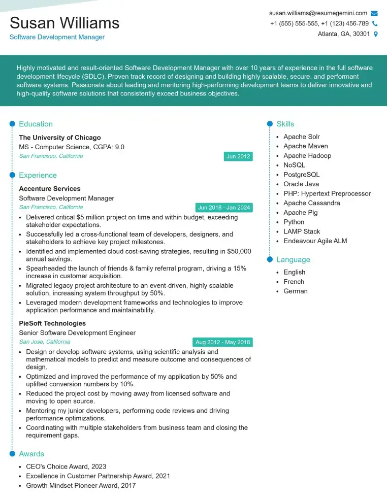
Check Out Resume Samples at ResumeGemini
Explore more articles
Users Rating of Our Blogs
Share Your Experience
We value your feedback! Please rate our content and share your thoughts (optional).
What Readers Say About Our Blog
To the interviewgemini.com Webmaster.
Very helpful and content specific questions to help prepare me for my interview!
Thank you
To the interviewgemini.com Webmaster.
This was kind of a unique content I found around the specialized skills. Very helpful questions and good detailed answers.
Very Helpful blog, thank you Interviewgemini team.