The thought of an interview can be nerve-wracking, but the right preparation can make all the difference. Explore this comprehensive guide to Proficient in musculoskeletal ultrasound for diagnosis and guided procedures interview questions and gain the confidence you need to showcase your abilities and secure the role.
Questions Asked in Proficient in musculoskeletal ultrasound for diagnosis and guided procedures Interview
Q 1. Describe your experience performing musculoskeletal ultrasound examinations.
My experience with musculoskeletal ultrasound encompasses over [Number] years, during which I’ve performed thousands of examinations across a wide range of MSK conditions. This includes both routine screenings and complex cases requiring detailed assessment. I’m proficient in evaluating various anatomical structures, from superficial tendons and ligaments to deep-seated muscles and joints. My experience extends to various patient populations, including athletes, elderly individuals with age-related degeneration, and patients with acute and chronic injuries. I routinely utilize high-resolution ultrasound systems and maintain a meticulous approach to image acquisition and interpretation. This allows for precise diagnosis and guides treatment strategies.
For example, I recently diagnosed a subtle partial tear of the supraspinatus tendon in a professional tennis player, which was initially missed on MRI. The high-resolution ultrasound clearly visualized the disruption of the tendon fibers, leading to appropriate management and a faster return to play. Another example includes the detection of early-stage osteoarthritis in a 65-year-old patient, allowing us to implement preventative measures and delay the need for more invasive intervention.
Q 2. What imaging protocols do you utilize for various MSK conditions (e.g., rotator cuff tears, carpal tunnel syndrome)?
Imaging protocols are tailored to the specific MSK condition. A systematic approach ensures comprehensive evaluation.
- Rotator Cuff Tears: I utilize longitudinal and transverse scans of the supraspinatus, infraspinatus, teres minor, and subscapularis tendons, assessing their echogenicity, thickness, and integrity. I carefully examine the subacromial-subdeltoid bursa for fluid.
- Carpal Tunnel Syndrome: I perform longitudinal and transverse scans of the median nerve at the carpal tunnel, measuring its cross-sectional area and evaluating for compression signs such as increased echogenicity or flattening. I also assess for tenosynovitis.
- Other conditions: Similar standardized protocols exist for other conditions, including plantar fasciitis (assessing thickness and echogenicity of the plantar fascia), tennis elbow (evaluating the common extensor tendon origin), and various joint effusions (assessing joint space and synovial fluid characteristics).
The protocol always includes comparing the affected side to the contralateral (unaffected) side to establish a baseline and identify any asymmetry.
Q 3. Explain the difference between grayscale and Doppler ultrasound in MSK imaging.
Grayscale ultrasound displays the anatomical structures based on their acoustic impedance—the resistance to the sound waves. Different tissues reflect sound waves differently, creating variations in grayscale. Tendons appear as hyperechoic (bright) structures due to their fibrous composition, while fluid-filled structures like bursae or hematomas appear anechoic (black) because they don’t reflect sound.
Doppler ultrasound, on the other hand, assesses blood flow within vessels. Color Doppler assigns colors to blood flow (red for flow toward the transducer, blue for flow away) while Power Doppler increases sensitivity to detect even slow blood flow. In MSK imaging, Doppler is crucial for detecting vascularity in lesions, assessing for inflammation, and identifying potential bleeding.
Think of grayscale as a static image of the anatomy, while Doppler adds a dynamic layer showing the movement of blood within that anatomy. Together they provide a complete picture.
Q 4. How do you assess tendon integrity using ultrasound?
Assessing tendon integrity using ultrasound involves evaluating several key features:
- Echogenicity: Normal tendons appear hyperechoic (bright white) and homogenous. Increased echogenicity or heterogeneous appearance suggests degeneration or inflammation.
- Thickness: Tendons thicken with inflammation or tendinosis. Measuring tendon thickness and comparing it to the contralateral side helps assess the extent of involvement.
- Fiber disruption: Full-thickness tears will show complete disruption of the tendon fibers, creating a gap or defect. Partial tears show focal areas of disruption or thinning.
- Fluid: Peritendinous fluid collections (around the tendon) suggest inflammation or recent trauma.
These findings are assessed in both longitudinal and transverse planes to fully characterize the tendon’s condition. Combining these features with clinical information provides a complete picture of the tendon’s integrity.
Q 5. Describe your experience with ultrasound-guided injections (e.g., corticosteroids, hyaluronic acid).
I have extensive experience performing ultrasound-guided injections, including corticosteroids and hyaluronic acid. My experience includes injections into various locations, such as joints, bursae, and tendon sheaths. The procedure involves real-time visualization of the needle as it approaches the target area, minimizing the risk of accidental injection into non-target tissues. I meticulously follow aseptic techniques to prevent infection.
For instance, I recently performed an ultrasound-guided corticosteroid injection into the subacromial-subdeltoid bursa of a patient with shoulder impingement syndrome. Using real-time ultrasound guidance, I precisely targeted the bursa and confirmed the injection’s placement. The patient reported significant pain relief shortly after the procedure.
Q 6. What are the potential complications of ultrasound-guided injections?
While generally safe, ultrasound-guided injections carry potential complications:
- Infection: Strict aseptic techniques are crucial to minimize this risk.
- Bleeding: This is more likely in patients on anticoagulants or with vascular abnormalities. Careful assessment of the area prior to injection is essential.
- Nerve injury: Needle placement close to nerves can cause pain or paresthesia. Ultrasound guidance significantly reduces this risk, but it’s still possible.
- Joint damage: Incorrect placement of the needle can damage cartilage or ligaments.
- Allergic reaction: This is rare but possible with the injected substance.
Proper patient selection, meticulous technique, and post-procedure monitoring are crucial for minimizing complications.
Q 7. How do you manage a patient with difficult vascular access during an ultrasound-guided procedure?
Managing patients with difficult vascular access during ultrasound-guided procedures requires a methodical approach. I start by carefully identifying the target area and surrounding vasculature with ultrasound. If the superficial veins are unsuitable, I explore deeper structures or alternative injection sites. If necessary, I use smaller gauge needles or specialized catheters to facilitate access. I also adjust my needle insertion angle and approach to optimize visualization and needle passage.
In certain cases, I may consult with a vascular access specialist for assistance. Patient hydration and pre-injection assessment of vascular anatomy play a critical role in improving the chances of successful cannulation. Documenting all attempts, including the reasons for failure and alternative strategies employed, is crucial for maintaining a comprehensive patient record.
Q 8. How do you interpret ultrasound findings in the presence of artifacts?
Interpreting ultrasound findings in the presence of artifacts requires a systematic approach. Artifacts are echoes or visual distortions on the ultrasound image that don’t represent actual anatomical structures. They can be confusing, but understanding their characteristics helps differentiate them from real pathology.
My approach involves first identifying the type of artifact. Common artifacts in MSK ultrasound include shadowing (a dark area behind a highly reflective structure like bone), reverberation (multiple repeating echoes), acoustic enhancement (increased brightness behind a fluid-filled structure), and anisotropy (changes in the appearance of a structure depending on the angle of the sound beam).
Once identified, I consider the clinical context. For example, shadowing behind a calcified tendon may be expected, but shadowing in an unexpected location might indicate a fracture or mass. I carefully correlate the ultrasound findings with the patient’s history, physical examination, and other imaging modalities (if available). I may also adjust the transducer angle, frequency, or depth to try and minimize the artifact or obtain a different view to better understand the underlying anatomy.
Essentially, it’s a process of elimination and careful correlation. I wouldn’t make a diagnosis based solely on the presence of an artifact. Instead, I focus on the actual anatomical structures visible despite the artifact and how they relate to the clinical picture.
Q 9. Explain your approach to image optimization and artifact reduction.
Image optimization and artifact reduction are crucial for accurate MSK ultrasound. My approach is multi-pronged and involves optimizing both the machine settings and the scanning technique.
- Transducer Selection: Choosing the appropriate transducer is the first step. High-frequency transducers provide better resolution for superficial structures, while lower-frequency transducers are better for deeper structures.
- Gain and Time Gain Compensation (TGC): Adjusting gain controls the overall brightness of the image, while TGC adjusts the amplification at different depths. Proper adjustment is critical to ensure good visualization of both superficial and deep structures and reduce artifacts.
- Focal Zones: Focusing the ultrasound beam at the depth of interest improves resolution and reduces artifacts, particularly reverberation.
- Transducer Angle and Pressure: Applying optimal pressure and adjusting the angle of the transducer is essential. Too much pressure can distort the image and create artifacts, while too little might result in poor acoustic coupling.
- Gel Coupling: Ensuring proper gel coupling between the transducer and the skin eliminates air gaps that cause significant artifacts.
- Harmonics and Compound Imaging: Using harmonic imaging or compound imaging techniques can improve image quality and reduce certain types of artifacts.
Furthermore, I utilize standardized scanning protocols to ensure consistency and reduce the likelihood of missing important information. This includes systematic sweeps through each area of interest, using specific anatomical landmarks as guides.
Q 10. Describe your knowledge of different ultrasound transducer types and their applications in MSK imaging.
Different transducer types offer unique advantages in MSK imaging. The selection depends on the specific anatomical location and the clinical question.
- Linear Transducers: These provide high-resolution images and are ideal for superficial structures like tendons, muscles, and nerves in the extremities. Their rectangular footprint gives a wider field of view and better visualization of these structures in their entirety.
- Curvilinear (Convex) Transducers: These are used for deeper structures, such as the hip joint, shoulder joint, or muscles of the back. Their curved surface allows for a wider field of view, essential for visualizing larger areas.
- Phased-array Transducers: These are often used for cardiac imaging, but can also be used for MSK imaging, especially for intercostal views of the chest wall or in limited acoustic windows.
- High-frequency Linear Transducers: These are used for superficial structures requiring excellent resolution, like in the assessment of small nerves or in hand and foot imaging.
For instance, a linear transducer is ideal for assessing a suspected lateral epicondylitis (tennis elbow), while a curvilinear transducer would be more appropriate for examining the rotator cuff tendons of the shoulder. Selecting the correct transducer is paramount to acquiring high-quality images and reducing ambiguity in diagnosis.
Q 11. How do you ensure patient safety during ultrasound-guided procedures?
Patient safety is my utmost priority during ultrasound-guided procedures. My approach encompasses several key aspects:
- Sterile Technique: I meticulously follow sterile techniques for all procedures, minimizing the risk of infection. This includes appropriate hand hygiene, sterile gloves, drapes, and the use of sterile needles and equipment.
- Patient Consent: Informed consent is obtained before any procedure, explaining the risks and benefits in clear and understandable terms.
- Real-time Monitoring: I constantly monitor the patient’s vital signs and observe for any signs of discomfort or adverse reactions during the procedure.
- Needle Visualization: I carefully visualize the needle’s trajectory in real-time using ultrasound guidance to avoid accidental puncture of vessels, nerves, or other critical structures.
- Proper Anesthesia: When indicated, local anesthetic is administered to minimize patient discomfort and anxiety during the procedure. This reduces the likelihood of unexpected patient movement during the procedure.
- Post-Procedure Monitoring: After the procedure, I monitor the patient for bleeding, infection, or other complications.
I regularly review and update my knowledge of safety protocols and best practices to ensure I provide the safest possible care to my patients. Incident reporting is a critical aspect of continuous improvement in safety measures.
Q 12. What is your experience with different types of needle guidance techniques?
My experience encompasses various needle guidance techniques, including:
- In-plane technique: The needle is advanced in the same plane as the ultrasound beam. This provides excellent visualization of needle tip location relative to the target and surrounding structures. It’s commonly used for injections into tendons or joints.
- Out-of-plane technique: The needle is advanced perpendicular to the ultrasound beam. This technique is helpful when the target is deep or when access is limited. It requires a more skilled understanding of spatial relationships.
- Hydro-dissection: This involves injecting fluid to create a plane between tissue layers, facilitating needle passage and better visualization. It’s particularly useful when working in complex anatomical areas.
The choice of technique depends on factors such as the target location, its depth, surrounding anatomy, and the type of procedure. I always select the technique that offers the best safety profile and optimizes the accuracy of the procedure. Each technique presents specific challenges and the operator must be fully competent in their application.
Q 13. How do you document your ultrasound findings and procedures?
Documentation of ultrasound findings and procedures is critical for patient care and legal reasons. My documentation includes:
- Patient demographics and clinical history: This ensures that the findings are appropriately contextualized.
- Images: High-quality ultrasound images are stored digitally and often printed for the patient’s chart. These images are labeled and accurately documented.
- Detailed descriptions of the findings: I provide precise descriptions of the structures visualized, including their size, shape, echogenicity, and location. Any abnormalities or artifacts are also carefully noted.
- Procedure details: For guided procedures, I document the technique used, needle location, amount and type of injected substance, and any complications encountered.
- Impression and recommendations: I summarize my findings and provide a concise interpretation, relating these to the initial clinical questions and offering recommendations for further management.
All documentation adheres to institutional standards and regulations. Accurate and complete documentation ensures that my findings are easily understood and can be used by other healthcare providers for continuity of care.
Q 14. Describe your understanding of the anatomical structures relevant to MSK ultrasound.
A thorough understanding of MSK anatomy is foundational to successful MSK ultrasound. This includes detailed knowledge of:
- Bones: Their cortical and cancellous components, variations in shape and density, and relationship to adjacent soft tissues.
- Tendons: Their fibrous structure, echogenicity (hypoechoic with hyperechoic fibrils), and relationship to surrounding muscles and bone.
- Muscles: Their fiber orientation, echogenicity, and relationship to tendons and nerves.
- Ligaments: Their fibrous structure and echogenicity, often visualized as hypoechoic bands.
- Bursae: Their fluid-filled nature, appearing anechoic with posterior acoustic enhancement.
- Nerves: Their hypoechoic appearance and relationship to muscles and vessels.
- Blood vessels: Their echogenicity (hypoechoic), pulsatile nature, and anatomical location.
- Joint structures: Including cartilage, synovium, and joint fluid.
A strong grasp of the normal anatomical appearance of each structure is essential for identifying pathology. This requires not only textbook knowledge but also extensive hands-on experience in visualizing these structures in various planes and orientations, using anatomical landmarks and contextual knowledge to ensure proper identification.
Q 15. Explain the role of ultrasound in the diagnosis of various musculoskeletal disorders.
Musculoskeletal (MSK) ultrasound plays a crucial role in diagnosing a wide range of disorders by providing real-time images of muscles, tendons, ligaments, joints, and bones. It’s a non-invasive technique that allows for a detailed assessment of soft tissue structures, identifying abnormalities that might not be visible on X-rays or other imaging modalities.
- Tendinopathies: Ultrasound can clearly visualize tendon tears, inflammation (tendinitis), and degenerative changes. For example, we can differentiate a partial rotator cuff tear from a complete tear based on the extent of the disruption of the tendon fibers.
- Bursitis and Tenosynovitis: The ultrasound’s ability to assess fluid collections helps diagnose bursitis (inflammation of the bursae) and tenosynovitis (inflammation of the tendon sheath). We can see the fluid accumulation and assess the extent of inflammation within these structures.
- Muscle Injuries: Ultrasound can detect muscle strains, tears, and hematomas (blood clots). The different echogenicity patterns – reflecting the sound waves differently – allow us to differentiate between different types of muscle damage.
- Joint Effusions and Arthritis: Ultrasound can identify fluid within joints (effusions) and assess the synovial lining for signs of inflammation, crucial in evaluating conditions like arthritis. We can also visualize joint cartilage and evaluate its integrity.
- Nerve Entrapments: Ultrasound helps evaluate nerves, identifying compression or abnormalities leading to conditions such as carpal tunnel syndrome. We can assess the nerve diameter and look for signs of compression or changes in its surrounding tissues.
In summary, MSK ultrasound offers a comprehensive, dynamic assessment, guiding treatment decisions and monitoring response to therapy.
Career Expert Tips:
- Ace those interviews! Prepare effectively by reviewing the Top 50 Most Common Interview Questions on ResumeGemini.
- Navigate your job search with confidence! Explore a wide range of Career Tips on ResumeGemini. Learn about common challenges and recommendations to overcome them.
- Craft the perfect resume! Master the Art of Resume Writing with ResumeGemini’s guide. Showcase your unique qualifications and achievements effectively.
- Don’t miss out on holiday savings! Build your dream resume with ResumeGemini’s ATS optimized templates.
Q 16. How do you differentiate between various soft tissue lesions using ultrasound?
Differentiating soft tissue lesions using ultrasound relies on a combination of factors: echogenicity (how the tissue reflects sound waves), texture, shape, size, and location. Specific features help distinguish between different pathologies.
- Echogenicity: A lesion appearing hypoechoic (darker) might represent a fluid collection (e.g., ganglion cyst), while a hyperechoic (brighter) lesion may be a calcification or a fibroma. Comparing echogenicity to the surrounding tissue provides crucial clues.
- Texture: Homogenous lesions have a uniform appearance, while heterogeneous lesions show varied echogenicity patterns, hinting at complex internal structure, as seen in some tumors.
- Vascularity: Doppler ultrasound, a component of many MSK ultrasound systems, assesses blood flow within the lesion. Highly vascular lesions, for example, might suggest an inflammatory process or a malignant neoplasm.
- Margins: Well-defined margins suggest a benign lesion, whereas poorly defined or irregular margins may indicate a malignant process. For instance, a clearly defined ganglion cyst has different margins than a poorly defined sarcoma.
- Shape: The shape of the lesion—round, oval, irregular—can be suggestive of particular conditions. A lipoma, for example, frequently appears as an oval hypoechoic lesion.
Accurate differentiation often requires correlating ultrasound findings with the patient’s clinical presentation, and sometimes, further investigations like biopsy are needed to confirm the diagnosis.
Q 17. What are the limitations of musculoskeletal ultrasound?
While MSK ultrasound is a powerful diagnostic tool, it does have limitations:
- Operator Dependence: Image quality and interpretation heavily depend on the sonographer’s skill and experience. It requires extensive training to become proficient at reading the images and interpreting the findings.
- Limited Penetration Depth: Ultrasound struggles to visualize deep structures clearly, making it less effective for evaluating conditions in areas like the pelvis or deep within the spine.
- Obesity and Body Habitus: Excess adipose tissue can interfere with sound wave penetration, obscuring underlying structures and reducing image quality.
- Gas Interference: Air within tissues (e.g., bowel gas) strongly attenuates ultrasound waves, often making accurate assessment impossible in those areas.
- Cannot Visualize Bone Detail: While ultrasound can evaluate the surrounding soft tissues, it’s not suitable for detailed bone assessment. X-rays remain the gold standard for assessing bone integrity.
- Inability to Determine Specific Tissue Composition: While ultrasound can suggest certain tissue types based on echogenicity, other advanced imaging techniques such as MRI are required for definitive tissue characterization and diagnosis.
These limitations necessitate using ultrasound in conjunction with other imaging modalities and clinical information for a comprehensive diagnosis.
Q 18. Describe your experience with ultrasound-guided biopsies.
I have extensive experience performing ultrasound-guided biopsies, including both needle aspirations and core needle biopsies. The process begins with meticulous patient preparation, obtaining informed consent, and performing a thorough ultrasound examination to identify the target lesion precisely. I then use real-time imaging to guide the needle to the lesion under strict sterile conditions. The entire procedure is monitored on the ultrasound screen, ensuring accurate needle placement and minimizing risk to the patient.
For example, I regularly perform ultrasound-guided biopsies of musculoskeletal masses to obtain tissue samples for histopathological analysis to confirm the diagnosis, ranging from benign lesions like lipomas to more concerning lesions that require prompt assessment. Real-time guidance ensures that we obtain a representative sample, reducing the need for repeat procedures. We also use ultrasound to assist in biopsies of fluid collections, providing a fast and effective way to analyze the fluid content and potentially provide rapid diagnosis.
Patient safety is paramount. I always prioritize proper sterile technique, meticulous needle placement, and post-procedure monitoring to detect and manage any complications effectively. The procedure’s success rate is quite high due to the real-time visualization provided by ultrasound, however, even with high success, I always discuss the associated risks and benefits with the patient prior to the procedure.
Q 19. How do you handle challenging patient interactions during procedures?
Handling challenging patient interactions during procedures requires empathy, clear communication, and a patient-centered approach. I always take time to explain the procedure clearly, answering all questions patiently and honestly. I address patient anxieties and concerns directly, providing reassurance and addressing any misunderstandings. Pain management is crucial; I use appropriate techniques to minimize discomfort during the procedure.
For instance, if a patient expresses significant anxiety, I might take extra time to discuss their fears, and in certain cases, we may even use mild sedation to ensure their comfort and cooperation. If a patient is claustrophobic, the procedure room should be prepared appropriately with extra space and considerations for ventilation, perhaps even providing reassurance breaks during the procedure to limit the claustrophobic experience. If language barriers exist, I use interpreters or ensure that my communication is clear and simple. Building rapport and trust is key to a successful and comfortable experience for both the patient and myself.
Q 20. How do you maintain and troubleshoot ultrasound equipment?
Maintaining and troubleshooting ultrasound equipment is a critical aspect of my practice. This involves regular checks of the machine’s functionality, including transducer performance, image quality, and Doppler capabilities. We conduct regular preventative maintenance according to manufacturer’s guidelines, cleaning the transducers and system, checking for loose wires, and ensuring the system is correctly operating within the parameters. I also perform regular quality control checks using test phantoms to validate image quality and measurement accuracy.
Troubleshooting typically involves systematic problem-solving. For example, if I encounter poor image quality, I’ll first check the transducer connection, then the gel application, and then consider whether there’s an issue with the machine’s settings. If the issue persists, I document the problem and contact the biomedical engineering department for repair or maintenance. We also keep detailed logs of any maintenance performed, errors observed, and troubleshooting actions taken to maintain comprehensive records.
Q 21. What quality assurance measures do you implement in your MSK ultrasound practice?
Quality assurance in my MSK ultrasound practice is paramount and focuses on several key aspects:
- Regular Equipment Maintenance and Calibration: This includes preventative maintenance, quality control checks with test phantoms to ensure consistent image quality, and adherence to manufacturer’s guidelines.
- Continuing Medical Education (CME): I regularly attend conferences, workshops, and online courses to keep abreast of the latest advancements in MSK ultrasound techniques and interpretation.
- Peer Review: I participate in regular peer review sessions with colleagues to review challenging cases and ensure consistent diagnostic accuracy. The peer review sessions allow for a critical discussion, and the exchange of different perspectives helps improve overall diagnostic accuracy and consistency across the team.
- Image Archiving and Storage: Images and reports are archived electronically, adhering to HIPAA guidelines, ensuring easy access to previous studies for comparison and review.
- Quality Control Monitoring: The usage of the machines is monitored using a systematic approach, ensuring proper utilization and calibration of the machines for effective and efficient procedures.
- Documentation and Record-Keeping: Meticulous documentation of examinations, findings, and any interventions performed is crucial for patient care and quality assurance.
By implementing these measures, I strive to maintain a high standard of care and ensure accurate and reliable MSK ultrasound services.
Q 22. Describe your experience with reporting and interpreting ultrasound findings for clinicians.
Reporting and interpreting musculoskeletal (MSK) ultrasound findings requires a meticulous approach. It’s not just about identifying structures; it’s about correlating the images with the patient’s clinical presentation to arrive at a meaningful diagnosis and guide management. My reports are structured to provide clinicians with clear, concise, and actionable information.
I begin by describing the technical aspects of the exam, including the transducer used and the imaging planes acquired. Then, I systematically describe the findings, focusing on the anatomy assessed and any abnormalities detected (e.g., tendon tears, bursitis, joint effusions). I use standardized terminology to ensure consistency and clarity, referencing specific anatomical locations and quantifying findings whenever possible (e.g., ‘partial-thickness tear of the supraspinatus tendon, measuring 1.5 cm in length’). The report concludes with a differential diagnosis and recommendations for further investigation or management, always emphasizing the limitations of the ultrasound examination.
For example, in a case of suspected rotator cuff tear, my report would detail the integrity of each tendon (supraspinatus, infraspinatus, subscapularis, teres minor), noting any tears (location, size, and morphology), presence of bursal fluid, and any other relevant findings, like calcific tendinitis or subacromial impingement signs. I would then offer a concise differential diagnosis considering other potential causes of shoulder pain and provide suggestions for the referring physician.
Q 23. How do you stay updated with the latest advancements in musculoskeletal ultrasound?
Staying current in MSK ultrasound is crucial given the rapid advancements in technology and techniques. My approach is multifaceted:
- Continuing Medical Education (CME): I regularly attend conferences, workshops, and webinars focused on musculoskeletal ultrasound. These events offer opportunities to learn from leading experts and network with colleagues.
- Peer-reviewed Publications: I subscribe to relevant journals (e.g., Skeletal Radiology, Ultrasound in Medicine & Biology) and actively read articles on new techniques, research findings, and emerging applications of MSK ultrasound.
- Professional Societies: Membership in professional organizations like the American Institute of Ultrasound in Medicine (AIUM) and the American College of Rheumatology (ACR) provides access to resources, guidelines, and continuing education opportunities.
- Online Resources: I utilize reputable online resources, including educational videos and interactive modules, to enhance my knowledge and skills.
- Case Review: Regularly reviewing challenging or unusual cases with colleagues helps refine diagnostic skills and expands my understanding of different clinical presentations.
Q 24. Describe a time you had to troubleshoot a technical issue during a procedure.
During a guided injection procedure for a patient with trochanteric bursitis, the ultrasound transducer’s connection unexpectedly failed mid-procedure. The image froze, and I immediately lost real-time guidance. This was concerning, as precise needle placement is crucial to avoid complications.
My first step was to remain calm and explain the situation to the patient. I then systematically troubleshooted the problem. I checked the transducer cable for any damage, ensuring it was securely connected to both the machine and the transducer. I also confirmed the machine’s power supply and tried a different transducer, but the problem persisted. I recognized that the issue might lie within the ultrasound machine itself.
Fortunately, I had a backup ultrasound machine in the room. I swiftly switched to the backup machine, carefully reassessed the anatomy, and successfully completed the procedure without further delay or compromise to patient safety. This experience highlighted the importance of having contingency plans and backup equipment, especially during guided procedures.
Q 25. How do you adapt your techniques based on patient size, body habitus, or clinical presentation?
Adapting techniques based on patient factors is essential for optimal image quality and patient comfort. Patient size and body habitus significantly influence transducer selection and scanning technique. For example, a larger patient may require a lower-frequency transducer to achieve adequate penetration, while a smaller patient might benefit from a higher-frequency transducer for improved resolution.
For patients with significant obesity, I often use a curved array transducer to navigate through subcutaneous fat. I might also employ compression techniques to improve image quality, but always prioritizing patient comfort. In patients with significant muscle atrophy, such as those with advanced neuromuscular diseases, I might adjust my scanning technique to compensate for the reduced tissue echogenicity.
Clinical presentation also guides my approach. If a patient presents with focal tenderness over a specific anatomical region, I will focus my attention on that area and use higher resolution settings to visualize fine details. If the clinical suspicion is a diffuse process, I will perform a more comprehensive examination, covering a broader anatomical area.
Q 26. What is your experience with image archiving and communication systems (PACS)?
I have extensive experience with Picture Archiving and Communication Systems (PACS). PACS is an integral part of my workflow. I routinely upload MSK ultrasound images into the PACS system for easy access, storage, and sharing among the healthcare team. This ensures efficient communication and collaboration, enabling seamless integration of ultrasound findings with other diagnostic tests and clinical notes.
I am proficient in using PACS to review prior studies, compare images from different modalities, and generate reports that are integrated directly into the patient’s electronic medical record (EMR). Familiarity with PACS features such as image annotation, measurement tools, and report generation is essential for efficient and accurate documentation and communication.
Q 27. Describe your proficiency in using different ultrasound software packages.
My proficiency extends to several ultrasound software packages, including [List specific software packages used, e.g., GE Logiq, Philips Epiq, etc.]. This experience allows me to adapt to various systems and optimize image acquisition and processing based on the specific capabilities of each software package. I am adept at adjusting machine settings, optimizing image quality (brightness, contrast, gain, depth), and utilizing advanced imaging modes like elastography, when appropriate, for specific diagnostic challenges.
Understanding the nuances of different software packages allows me to effectively utilize their features such as color Doppler, power Doppler, M-mode, and other specialized functionalities to obtain the best images and information possible. This ensures that the images I capture accurately reflect the patient’s condition and facilitate a precise diagnosis.
Q 28. Explain your understanding of radiation safety principles and their relevance to MSK ultrasound.
MSK ultrasound, unlike X-ray, CT, or MRI, does not involve ionizing radiation. This is a significant advantage, allowing for repeated scans without concerns about radiation exposure. However, radiation safety principles remain relevant in the context of MSK ultrasound, albeit in a different way.
My understanding of radiation safety focuses on the principles of ALARA (As Low As Reasonably Achievable). While there’s no direct radiation from the ultrasound machine, other aspects of patient safety must be considered. These include:
- Infection Control: Strict adherence to sterile technique during guided procedures is crucial to minimize the risk of infection. Proper hand hygiene and the use of sterile gloves, drapes, and needles are essential.
- Patient Positioning and Comfort: Proper patient positioning is necessary to reduce the risk of injury, especially during prolonged procedures. Maintaining patient comfort minimizes the chance of discomfort, stress, or unexpected movement, which could negatively impact the procedure.
- Bioeffects: While generally considered safe, prolonged exposure to high-intensity ultrasound could have potential bioeffects. The ALARA principle suggests keeping ultrasound exposure time to a minimum by efficiently performing the scan and obtaining necessary information as quickly and effectively as possible.
Key Topics to Learn for Proficient in Musculoskeletal Ultrasound for Diagnosis and Guided Procedures Interview
- Image Acquisition Techniques: Mastering optimal probe placement, transducer selection, and image optimization for various musculoskeletal structures (e.g., tendons, ligaments, muscles, joints).
- Anatomy and Sonoanatomy: Demonstrate a thorough understanding of normal musculoskeletal anatomy and its corresponding ultrasound appearance. Practice identifying key anatomical landmarks and variations.
- Pathology Identification: Develop the ability to identify common musculoskeletal pathologies (tears, strains, inflammation, cysts, tumors) through ultrasound imaging, including differentiating normal from abnormal findings.
- Guided Procedures: Explain your experience and understanding of performing ultrasound-guided injections (e.g., corticosteroids, anesthetics), biopsies, and aspirations. Discuss safety protocols and potential complications.
- Measurement and Documentation: Practice accurately measuring structures and documenting findings, including creating concise and informative reports.
- Equipment Operation and Maintenance: Demonstrate familiarity with various ultrasound machines and their functionalities, including troubleshooting basic issues.
- Patient Interaction and Communication: Highlight your skills in effectively communicating with patients, explaining procedures, and addressing their concerns.
- Ethical Considerations and Safety Protocols: Understand and articulate the ethical implications and safety precautions associated with musculoskeletal ultrasound procedures.
- Problem-Solving and Critical Thinking: Prepare examples demonstrating your ability to analyze complex ultrasound images, interpret findings, and formulate appropriate diagnoses and treatment plans.
- Emerging Technologies and Advancements: Stay updated on the latest advancements in musculoskeletal ultrasound technology and techniques.
Next Steps
Mastering musculoskeletal ultrasound for diagnosis and guided procedures opens doors to exciting career opportunities and establishes you as a highly skilled professional in a growing field. To maximize your job prospects, creating a strong, ATS-friendly resume is crucial. ResumeGemini is a trusted resource that can help you build a professional resume that showcases your skills and experience effectively. Examples of resumes tailored to highlight proficiency in musculoskeletal ultrasound for diagnosis and guided procedures are available, providing you with a valuable template and inspiration for creating your own compelling application.

Check Out Resume Samples at ResumeGemini

Check Out Resume Samples at ResumeGemini

Check Out Resume Samples at ResumeGemini

Check Out Resume Samples at ResumeGemini

Check Out Resume Samples at ResumeGemini
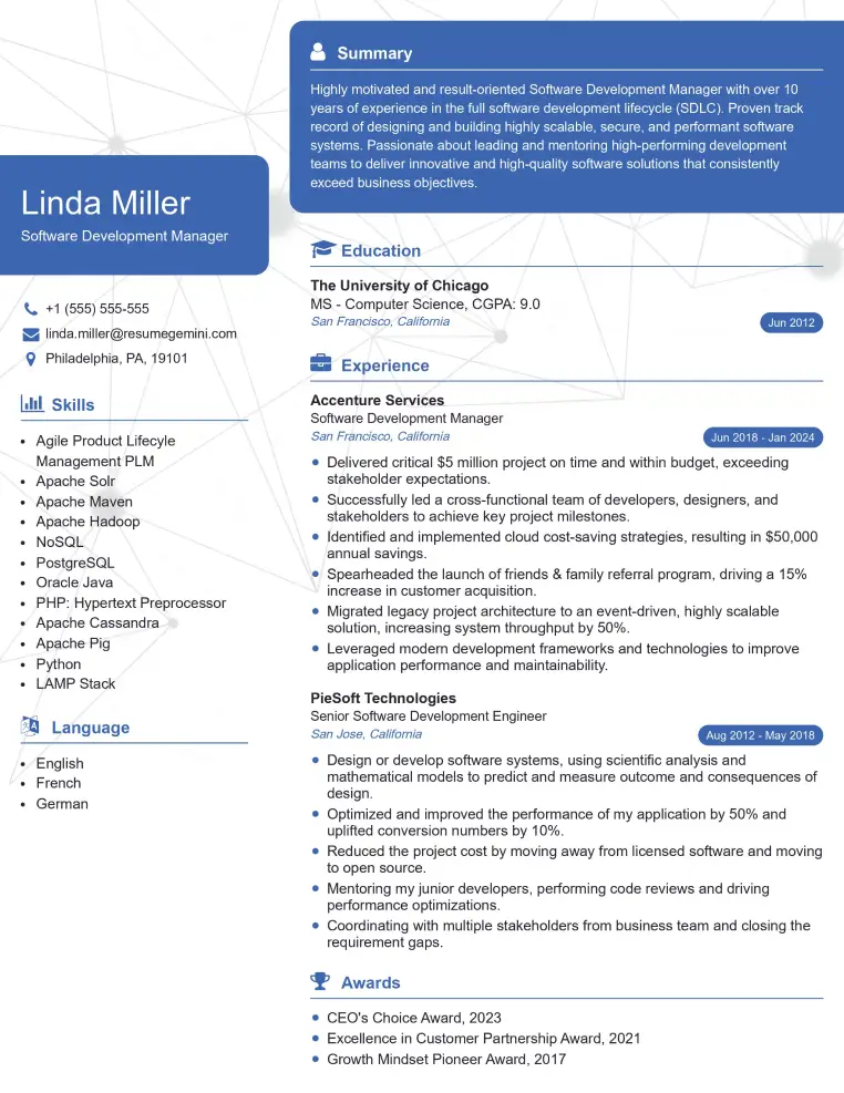
Check Out Resume Samples at ResumeGemini

Check Out Resume Samples at ResumeGemini
Explore more articles
Users Rating of Our Blogs
Share Your Experience
We value your feedback! Please rate our content and share your thoughts (optional).
What Readers Say About Our Blog
To the interviewgemini.com Webmaster.
Very helpful and content specific questions to help prepare me for my interview!
Thank you
To the interviewgemini.com Webmaster.
This was kind of a unique content I found around the specialized skills. Very helpful questions and good detailed answers.
Very Helpful blog, thank you Interviewgemini team.