Interviews are opportunities to demonstrate your expertise, and this guide is here to help you shine. Explore the essential Splenectomy interview questions that employers frequently ask, paired with strategies for crafting responses that set you apart from the competition.
Questions Asked in Splenectomy Interview
Q 1. Describe the different surgical approaches for splenectomy (e.g., laparoscopic, open).
Splenectomy, the surgical removal of the spleen, can be performed via several approaches, each with its own advantages and disadvantages. The two primary methods are open splenectomy and laparoscopic splenectomy.
- Open Splenectomy: This is the traditional approach, involving a larger incision in the abdomen to directly access and remove the spleen. It’s typically used in cases where the spleen is significantly enlarged, damaged, or difficult to access laparoscopically. Think of it like a mechanic needing to replace a large, hard-to-reach engine part; direct access is crucial.
- Laparoscopic Splenectomy: This minimally invasive technique utilizes small incisions and specialized instruments, including a laparoscope (a thin, lighted tube with a camera), to visualize and remove the spleen. It’s associated with less pain, shorter hospital stays, and quicker recovery times compared to open surgery. Imagine a technician using miniature tools to fix a delicate piece of electronics – precise, smaller-scale intervention.
Other less common approaches may involve robotic-assisted laparoscopic splenectomy (combining laparoscopy with robotic precision) or splenic artery embolization, which is a non-surgical procedure that blocks blood flow to the spleen, causing it to atrophy. The choice of surgical approach depends on various factors, including the patient’s overall health, the size and location of the spleen, and the reason for the splenectomy.
Q 2. What are the indications for splenectomy?
Splenectomy is indicated in several clinical situations where the spleen is damaged, diseased, or causing harm. These include:
- Trauma: A ruptured spleen, often resulting from a car accident or blunt force trauma, is a life-threatening emergency requiring immediate splenectomy.
- Hematologic Disorders: Conditions like hypersplenism (overactive spleen causing destruction of blood cells), thalassemia, and certain types of leukemia may necessitate splenectomy to improve blood cell counts and reduce symptoms.
- Infections: Severe infections like overwhelming post-splenectomy sepsis (OPSI) can occur, especially in children, leading to the need for prophylactic splenectomy.
- Tumors: Benign or malignant tumors in the spleen may require surgical removal.
- Congenital Abnormalities: Certain developmental anomalies involving the spleen may require splenectomy.
The decision to perform a splenectomy is made on a case-by-case basis, considering the risks and benefits for the individual patient. It’s a significant decision, as the spleen plays an important role in the immune system.
Q 3. Explain the potential complications associated with splenectomy.
While splenectomy is a life-saving procedure in many cases, it carries potential complications. These include:
- Infection: The spleen plays a crucial role in fighting infections, and its absence increases the risk of serious infections, particularly encapsulated bacteria (like Streptococcus pneumoniae, Haemophilus influenzae, and Neisseria meningitidis). This is why post-splenectomy vaccination is crucial.
- Bleeding: Intra-abdominal bleeding is possible, especially during and immediately after the surgery. Meticulous surgical technique minimizes this risk.
- Thrombocytopenia (low platelet count): The spleen’s role in regulating platelet numbers can result in transient or persistent low platelet counts post-splenectomy.
- Overwhelming Post-Splenectomy Infection (OPSI): This is a severe, potentially fatal infection that can occur following splenectomy. Prophylactic antibiotics and vaccinations are vital in mitigating this risk.
- Pancreatitis: Injury to the pancreas during surgery is a rare but serious complication.
- Abscess: Formation of a collection of pus in the surgical site.
The frequency and severity of complications vary depending on factors such as the surgical technique used, the patient’s overall health, and the underlying reason for the splenectomy. Post-operative monitoring and proactive management are key to minimizing these risks.
Q 4. How do you manage post-operative pain after splenectomy?
Post-operative pain management after splenectomy is crucial for patient comfort and recovery. A multimodal approach is usually employed, combining several pain relief strategies:
- Analgesics: Opioid and non-opioid analgesics are frequently used, tailored to the individual patient’s needs and pain level. The goal is to provide adequate pain relief while minimizing side effects.
- Regional Anesthesia: Techniques such as epidural or paravertebral blocks can effectively reduce pain in the immediate postoperative period.
- Non-Pharmacological Methods: Strategies like deep breathing exercises, ice packs, and patient-controlled analgesia (PCA) pumps can significantly improve pain management.
The choice of pain management strategy depends on several factors, including the type of surgery performed (open vs. laparoscopic), the patient’s medical history, and their response to treatment. Regular assessment of pain levels and adjustment of analgesics are crucial for optimizing patient comfort and promoting a speedy recovery.
Q 5. What are the key steps in the perioperative management of a splenectomy patient?
Perioperative management of a splenectomy patient encompasses the entire process, from pre-operative assessment and preparation through post-operative monitoring and recovery. Key steps include:
- Pre-operative Assessment: A thorough medical history and physical examination, including blood tests, coagulation studies, and imaging, are essential to evaluate the patient’s fitness for surgery and identify any potential risks.
- Pre-operative Counseling: Educating the patient about the procedure, potential complications, and post-operative care is crucial for informed consent and managing expectations.
- Intra-operative Care: Careful surgical technique, meticulous hemostasis (control of bleeding), and gentle handling of tissues minimize the risk of complications during the procedure.
- Post-operative Monitoring: Close monitoring of vital signs, pain levels, and potential complications (e.g., bleeding, infection) is vital in the immediate post-operative period.
- Post-operative Education: Instructing the patient on wound care, pain management, diet, and activity restrictions is essential for successful recovery.
- Immunization: Patients undergoing splenectomy require vaccination against encapsulated bacteria (pneumococcal, meningococcal, and Haemophilus influenzae type b) to reduce the risk of OPSI.
Effective perioperative management is crucial for a successful outcome, minimizing complications and ensuring a smooth recovery for the patient.
Q 6. Discuss the role of imaging (e.g., CT, ultrasound) in the diagnosis and planning of splenectomy.
Imaging plays a vital role in the diagnosis and planning of splenectomy. CT scans and ultrasounds are the most commonly used imaging modalities.
- CT Scan: Provides detailed anatomical information about the spleen, including its size, shape, and any lesions or abnormalities. This helps assess the extent of splenic injury or disease and aids in surgical planning.
- Ultrasound: Useful in evaluating the spleen’s size and texture, and in detecting abnormalities. It can be particularly helpful in trauma cases, allowing for rapid assessment of splenic injury.
Imaging helps determine the optimal surgical approach (open vs. laparoscopic), identifies potential complications, and assists in assessing the post-operative outcome. For example, a CT scan post-splenectomy may be used to detect any residual splenic tissue or potential collections of fluid or blood.
Q 7. How do you assess a patient’s suitability for a laparoscopic splenectomy?
Assessing a patient’s suitability for a laparoscopic splenectomy involves considering several factors:
- Size and Location of the Spleen: A significantly enlarged spleen or one with unusual anatomy may make laparoscopic splenectomy technically challenging or impossible. Open surgery might be a better choice in such cases.
- Patient’s Overall Health: Patients with severe co-morbidities or significant cardiovascular or respiratory compromise may not tolerate laparoscopic surgery.
- Previous Abdominal Surgery: Extensive prior abdominal surgery, particularly adhesions (scar tissue), can make laparoscopic surgery difficult and increase the risk of complications.
- Presence of Infection: An active infection involving the spleen or surrounding structures contraindicates laparoscopic splenectomy.
- Surgeon’s Experience: The surgeon’s expertise in laparoscopic techniques is crucial for a successful outcome. This is a skill developed over time and practice.
A comprehensive pre-operative assessment by the surgeon, along with imaging studies, helps determine whether a laparoscopic approach is feasible and safe for a given patient.
Q 8. What are the specific challenges of performing a splenectomy in a patient with severe adhesions?
Severe adhesions, bands of scar tissue that bind organs together, present a significant challenge during splenectomy. These adhesions can obscure the surgical field, making identification of the splenic vessels and surrounding structures difficult and increasing the risk of bleeding and injury to adjacent organs like the pancreas, stomach, or left kidney. Imagine trying to untangle a tightly knotted ball of yarn – that’s what it can be like for the surgeon. The dense adhesions require meticulous dissection, often requiring the use of specialized instruments like blunt dissection scissors and ultrasonic shears to carefully separate the adhesions without damaging underlying structures. This increased complexity leads to longer operative times, increased blood loss, and a higher risk of complications like injury to adjacent organs or postoperative ileus (bowel obstruction).
For example, if a surgeon encounters dense adhesions around the splenic hilum (where the splenic vessels enter and exit the spleen), it may take considerably longer to control bleeding from these vessels, increasing the risk of significant blood loss. In such cases, the surgeon might utilize advanced techniques such as laparoscopic ultrasound or indocyanine green fluorescence imaging to better visualize the vascular anatomy.
Q 9. Describe the procedure for managing a bleeding complication during a splenectomy.
Managing bleeding during a splenectomy is paramount. The initial step involves identifying the source of the bleeding. This often requires careful exploration of the splenic hilum and surrounding tissues. Once the bleeding vessel is identified, various methods are employed to control it. These include direct pressure, the use of surgical clips or ligatures (surgical ties), and cauterization (using heat to seal the vessel). For more significant bleeding, a surgical sponge may be packed temporarily to provide pressure while further steps are taken. In rare cases, where bleeding is uncontrollable, the surgeon may need to use more advanced techniques, like applying a vascular clamp to the main splenic artery or vein. Ultimately, the goal is to achieve hemostasis (stop the bleeding) quickly and safely to prevent complications like hypovolemic shock (due to significant blood loss).
For instance, if bleeding occurs from a small, easily accessible vessel, a simple surgical clip may suffice. However, if the bleeding stems from a larger vessel within the hilum, requiring more complex maneuvering, laparoscopic or open surgical techniques might be needed to expose the area, enabling the application of a vascular clamp or careful ligation of the bleeding vessel.
Q 10. What are the potential risks of leaving a splenic remnant?
Leaving a splenic remnant (a portion of the spleen) after splenectomy, while sometimes unavoidable or intentionally performed in partial splenectomies, carries potential risks. The most significant risk is the potential for the remnant to become dysfunctional or infected, potentially leading to recurrent pain, bleeding or abscess formation. The retained tissue can continue to have its own blood supply and is thus susceptible to the same pathological processes that may have necessitated the splenectomy in the first place. Furthermore, the remnant may not provide sufficient immune function, leaving the patient susceptible to infections, particularly encapsulated bacteria like Streptococcus pneumoniae, Haemophilus influenzae, and Neisseria meningitidis.
For example, a patient with a large splenic remnant might still experience episodes of left upper quadrant pain, possibly due to congestion or infection in the remaining tissue. This could require additional interventions including repeat surgery or image-guided drainage. It’s crucial to carefully assess the extent of the remnant and provide appropriate postoperative monitoring to identify any issues early on.
Q 11. How do you counsel a patient about the risks and benefits of splenectomy?
Counseling patients about splenectomy requires a thoughtful and thorough approach. I begin by explaining the underlying medical condition necessitating the procedure in clear, non-technical terms. I then outline the benefits of the surgery, such as alleviating symptoms or resolving a life-threatening condition, emphasizing the positive impact it could have on their quality of life. Following this, I discuss the risks associated with the procedure, which include bleeding, infection, injury to surrounding organs, and the long-term risk of overwhelming post-splenectomy infection (OPSI). I use visuals and analogies to enhance understanding, providing examples of similar surgical procedures or scenarios that are easier to grasp. The discussion also covers the possibility of requiring blood transfusions and the need for lifelong prophylactic antibiotics.
For example, when explaining OPSI, I’d explain that removing the spleen affects the body’s ability to fight certain types of bacteria, making it vital for patients to receive vaccinations and prophylactic antibiotics to minimize this risk. The conversation should conclude with opportunities for the patient to ask questions, ensure their concerns are addressed, and empower them to make an informed decision.
Q 12. What are the post-operative monitoring parameters after splenectomy?
Post-operative monitoring after splenectomy is critical. It typically includes frequent assessments of vital signs (heart rate, blood pressure, respiratory rate, temperature), monitoring for signs of bleeding (hematocrit and hemoglobin levels), and assessing for signs of infection (white blood cell count). Pain management is also a crucial aspect, ensuring the patient’s comfort and minimizing the risk of complications. Regular monitoring of abdominal examination will also help check for any distension, tenderness or rigidity indicating potential complications like ileus or peritonitis. The patient’s fluid balance, including intake and output, is closely monitored to detect signs of dehydration or fluid overload. The imaging studies may be used, such as an ultrasound or CT scan if signs of complications are suspected.
For instance, a sudden drop in blood pressure or a rise in temperature could indicate bleeding or infection, requiring prompt intervention. Regular blood tests help track the recovery of the patient’s hemoglobin levels, ensuring sufficient blood oxygen-carrying capacity. Finally, assessing the patient’s bowel function, paying attention to when the bowel sounds return, is critical to assess recovery from any surgical-induced ileus.
Q 13. Discuss the management of post-splenectomy infection.
Post-splenectomy infection management focuses on prompt diagnosis and treatment. The primary concern is the development of serious infections caused by encapsulated bacteria. The initial approach involves assessing the patient’s clinical status, conducting blood cultures to identify the causative organism, and initiating broad-spectrum antibiotic therapy based on the suspected pathogen. The antibiotics are selected based on antibiotic sensitivity testing. If an abscess or other localized infection is present, it may require drainage, either percutaneously (through the skin using imaging guidance) or surgically. Close monitoring of the patient’s response to treatment is vital, adjusting the antibiotics or surgical approach as needed.
For example, a patient presenting with fever, chills, and abdominal pain post-splenectomy might require immediate hospitalization, blood cultures, and intravenous antibiotics. Imaging studies would help identify the location of the infection and guide any necessary drainage procedures.
Q 14. Explain the concept of overwhelming post-splenectomy infection (OPSI).
Overwhelming post-splenectomy infection (OPSI) is a life-threatening complication characterized by a rapidly progressing, severe infection, often caused by encapsulated bacteria. These bacteria normally have their virulence neutralized by the spleen. The absence of the spleen significantly compromises the body’s ability to effectively combat these organisms. OPSI typically presents with septic shock (a life-threatening condition caused by the body’s overwhelming response to an infection), disseminated intravascular coagulation (DIC, abnormal blood clotting), and multi-organ failure. It’s a medical emergency requiring aggressive and immediate treatment. Early recognition and prompt initiation of broad-spectrum antibiotics, along with supportive measures like fluid resuscitation and vasopressor support, are crucial for improving survival rates.
Imagine the body’s immune system as a well-coordinated army. The spleen acts as a critical base, providing a crucial line of defense. Without it, the army is less effective, leaving the patient highly vulnerable to an overwhelming attack by certain types of bacteria. Early recognition of symptoms like rapid onset of fever, chills, hypotension, and altered mental status is vital for prompt intervention and improved survival rates.
Q 15. What preventative measures are taken to reduce the risk of OPSI?
Overwhelming Post-Splenectomy Infection (OPSI) is a serious complication following splenectomy, characterized by severe sepsis caused by encapsulated bacteria like Streptococcus pneumoniae, Haemophilus influenzae, and Neisseria meningitidis. The spleen’s crucial role is in clearing these bacteria from the bloodstream. Therefore, preventing OPSI focuses on preemptively protecting against these infections.
Vaccination: Before splenectomy, patients receive vaccinations against S. pneumoniae, H. influenzae type b, and N. meningitidis. This is paramount and significantly reduces the risk of OPSI. The timing of these vaccinations is crucial and should be discussed with the patient well in advance of the surgery.
Prophylactic Antibiotics: Post-splenectomy, many surgeons prescribe prophylactic antibiotics for a period of time, typically several years, to further decrease the risk of infection. The choice of antibiotic and duration depends on factors like the patient’s overall health and the reason for the splenectomy. Regular follow-up is crucial to adjust the antibiotic regimen as needed.
Patient Education: Thorough patient education is key. Patients must be aware of the increased risk of infection and learn to recognize the early signs of sepsis (fever, chills, rapid heart rate, etc.) and seek immediate medical attention if they develop these symptoms. This includes understanding the importance of hand hygiene and avoiding close contact with individuals who are ill.
Think of it like this: the spleen acts as a filter, removing dangerous bacteria. After splenectomy, this filter is gone, so we need to bolster the body’s defenses with vaccinations and antibiotics to compensate for this loss.
Career Expert Tips:
- Ace those interviews! Prepare effectively by reviewing the Top 50 Most Common Interview Questions on ResumeGemini.
- Navigate your job search with confidence! Explore a wide range of Career Tips on ResumeGemini. Learn about common challenges and recommendations to overcome them.
- Craft the perfect resume! Master the Art of Resume Writing with ResumeGemini’s guide. Showcase your unique qualifications and achievements effectively.
- Don’t miss out on holiday savings! Build your dream resume with ResumeGemini’s ATS optimized templates.
Q 16. How do you manage a splenic rupture?
Management of splenic rupture depends heavily on the severity of the injury and the patient’s overall condition. It ranges from conservative management to immediate surgery.
Non-operative Management: For minor splenic lacerations without significant hemorrhage, conservative management, involving close monitoring of vital signs, serial abdominal examinations, and imaging (CT scan), might be chosen. This approach focuses on close observation and supportive care, with the goal of allowing the spleen to heal naturally. This approach is often reserved for stable patients with minimal bleeding.
Operative Management: If there is significant hemorrhage or hemodynamic instability (low blood pressure, rapid heart rate), immediate surgical intervention is necessary. This could involve splenorrhaphy (repairing the spleen) if possible, or splenectomy (removal of the spleen) if the injury is too severe to repair. In some cases, partial splenectomy – removing only the damaged part – may be feasible.
Imagine the spleen as a delicate sponge. A minor tear might heal on its own with rest (conservative management), but a major tear causing significant bleeding necessitates immediate repair or removal (operative management).
Q 17. What is the role of splenic artery embolization in the management of splenic trauma?
Splenic artery embolization (SAE) is a minimally invasive interventional radiology procedure used to control bleeding from splenic trauma. It’s a valuable alternative or adjunct to surgery in selected patients.
The procedure involves a radiologist inserting a catheter into the femoral artery and guiding it to the splenic artery. Then, using imaging guidance (fluoroscopy), embolic agents (tiny particles or coils) are deployed into the branches of the splenic artery, blocking blood flow to the injured area of the spleen. This stops bleeding and allows the injured area to heal.
SAE is particularly useful in:
Hemodynamically unstable patients who are at high risk for surgery.
Patients with minor to moderate splenic injuries where surgical intervention may not be necessary.
Patients unsuitable for surgery due to other medical conditions.
It is crucial to remember that SAE is not appropriate for all splenic injuries. The decision to use SAE should be made on a case-by-case basis by a multidisciplinary team, taking into account the patient’s clinical status, the extent of the injury, and the availability of interventional radiology services. Think of it as using a tiny plug to stop a leak instead of replacing the entire pipe.
Q 18. Describe the anatomy of the spleen and its surrounding vasculature.
The spleen is a relatively soft, purplish organ located in the left upper quadrant of the abdomen, nestled behind the stomach and below the diaphragm. Its size and shape vary but it is generally described as being about the size of a fist.
Anatomy: The spleen has a unique structure, with a hilum where blood vessels and lymphatics enter and exit. Internally, it is composed of red pulp (involved in filtering blood and removing old red blood cells) and white pulp (involved in immune function). It’s encapsulated in a relatively thin capsule, making it susceptible to rupture in trauma.
Vasculature: The splenic artery, a branch of the celiac artery, is the main blood supply to the spleen. It branches extensively within the splenic parenchyma. The splenic vein drains the spleen and joins with the superior mesenteric vein to form the portal vein, which carries blood to the liver.
Understanding the splenic artery’s branching pattern is critical during procedures like splenic artery embolization, as precise targeting is required to stop bleeding without causing unintended damage to other structures.
Q 19. What are the different types of splenic cysts and their management?
Splenic cysts can be broadly classified as parasitic, non-parasitic (true or pseudocysts), and potentially malignant.
Parasitic cysts: These are usually caused by Echinococcus infection (hydatid cysts) and require specific anti-parasitic treatment along with surgical intervention or percutaneous drainage, depending on the cyst’s size and location.
Non-parasitic (true) cysts: These are lined by epithelium, are often asymptomatic, and are mostly observed incidentally. Management depends on size and symptoms. Small, asymptomatic cysts often require no intervention, while larger cysts causing discomfort or complications might warrant surgical resection or cyst fenestration (creating an opening to drain the fluid).
Non-parasitic (pseudocysts): These are collections of fluid without an epithelial lining. They often occur after trauma or inflammation. Management depends on the symptoms, with small, asymptomatic pseudocysts needing no intervention while larger, symptomatic pseudocysts can be managed by surgical excision, percutaneous drainage, or sclerotherapy (injecting a substance to collapse the cyst).
The management approach must always consider the specific type of cyst, its size, symptoms, and the patient’s overall health. Each case must be considered individually, often necessitating a multidisciplinary discussion involving surgeons, radiologists, and infectious disease specialists.
Q 20. How do you differentiate between benign and malignant splenic lesions?
Differentiating between benign and malignant splenic lesions requires a multi-faceted approach using a combination of imaging and sometimes biopsy.
Imaging: Ultrasound, CT scans, and MRI provide crucial information about the lesion’s size, location, and characteristics (e.g., solid vs. cystic, presence of calcifications). These imaging techniques can often suggest whether a lesion is benign or malignant, but they aren’t definitive.
Biopsy: In cases where imaging findings are inconclusive or suggestive of malignancy, a biopsy is usually performed to obtain tissue for microscopic examination (histology). This is the gold standard for definitive diagnosis.
Markers and Cytology: In some cases, blood tests may be used to look for tumor markers associated with certain splenic cancers. Additionally, fine-needle aspiration cytology can provide preliminary information about the cellular composition of the lesion.
Benign lesions tend to show smooth borders, a homogeneous appearance on imaging, and lack of features suggestive of malignancy. Malignant lesions often show irregular borders, heterogeneous internal structure, and potential infiltration into surrounding tissues. The clinical context (patient history, presence of symptoms) also plays a significant role in the evaluation.
Q 21. Discuss the use of minimally invasive techniques in splenectomy.
Minimally invasive techniques are increasingly used in splenectomy, offering several advantages over open surgery, including less pain, shorter hospital stays, and faster recovery times. These techniques primarily include laparoscopic splenectomy and robotic-assisted laparoscopic splenectomy.
Laparoscopic Splenectomy: This procedure involves making several small incisions in the abdomen to insert laparoscopic instruments and a camera. The spleen is dissected and removed through these small incisions. It is suitable for many patients, but its feasibility depends on the size and location of the spleen, the presence of adhesions, and the surgeon’s experience.
Robotic-Assisted Laparoscopic Splenectomy: This technique utilizes a robotic surgical system, providing enhanced visualization, dexterity, and precision. The robotic arms are controlled by the surgeon from a console, allowing for more intricate maneuvers in confined spaces within the abdomen. This approach may be especially beneficial in complex cases where laparoscopic splenectomy may be challenging.
The choice between open surgery and a minimally invasive approach is individualized based on the patient’s specific circumstances and the surgeon’s expertise. Minimally invasive techniques offer significant benefits, but are not always appropriate or feasible for every patient. Think of it as the difference between open-heart surgery and a minimally invasive valve repair— both aim to achieve the same outcome, but through different methods with varying levels of invasiveness.
Q 22. Explain the role of robotic surgery in splenectomy.
Robotic surgery has revolutionized splenectomy, offering significant advantages over traditional open surgery and even laparoscopic approaches in certain cases. The da Vinci Surgical System, for example, allows for minimally invasive procedures with enhanced dexterity and precision. The surgeon controls robotic arms through a console, providing a magnified, three-dimensional view of the surgical field. This translates to smaller incisions, reduced trauma to surrounding tissues, less blood loss, and faster recovery times. In complex cases, where the spleen is adhered to surrounding organs or there’s significant inflammation, the robotic system’s advanced articulation and visualization can be particularly beneficial, allowing for safer and more precise dissection.
Think of it like this: a traditional open splenectomy is like using a large wrench to remove a stubborn bolt. Laparoscopic surgery uses smaller tools, like a set of smaller wrenches. Robotic surgery provides the equivalent of incredibly precise, miniature robotic arms that can maneuver in tight spaces with superior control and vision, making complex procedures safer and less invasive.
Q 23. What are the advantages and disadvantages of laparoscopic versus open splenectomy?
The choice between laparoscopic and open splenectomy depends heavily on the patient’s condition and the nature of the splenic pathology. Laparoscopic splenectomy generally offers several advantages: smaller incisions leading to less pain, reduced scarring, shorter hospital stays, and faster recovery. It’s also associated with a lower risk of postoperative infections and complications. However, laparoscopic splenectomy may not always be feasible, particularly in cases of large splenomegaly, severe adhesions, or previous abdominal surgery. In such situations, an open splenectomy may be necessary to ensure complete removal of the spleen and to safely address any complications that may arise during surgery.
- Laparoscopic Advantages: Smaller incisions, less pain, reduced scarring, shorter hospital stay, faster recovery, lower infection risk.
- Laparoscopic Disadvantages: Not suitable for all cases (e.g., large spleen, adhesions), increased risk of conversion to open surgery.
- Open Advantages: Suitable for complex cases, better visualization and control during surgery.
- Open Disadvantages: Larger incisions, more pain, longer recovery time, increased risk of infection and other complications.
Q 24. Describe your experience with managing patients who have undergone splenectomy.
My experience managing post-splenectomy patients involves a multi-faceted approach focused on preventing and managing potential complications. Pre-operative counseling is critical, explaining the procedure, potential risks, and the importance of post-operative care, including vaccination against encapsulated bacteria (pneumococcal, Haemophilus influenzae type B, and meningococcal). Post-operatively, I closely monitor patients for signs of infection, bleeding, and other complications. Regular blood counts are essential to assess for any evidence of infection. Pain management is crucial, and I utilize a multi-modal approach including analgesics and, when indicated, nerve blocks. I also provide clear instructions on wound care and activity restrictions, emphasizing the importance of adhering to these guidelines to promote healing and prevent complications.
For example, I recently managed a patient who developed a mild postoperative infection. Prompt recognition and initiation of appropriate antibiotics resulted in a rapid resolution, preventing more serious complications. Another patient presented with post-operative abdominal pain, which was determined to be a small hematoma requiring conservative management.
Q 25. What are your preferred methods for controlling bleeding during a splenectomy?
Controlling bleeding during a splenectomy is paramount. My preferred methods depend on the surgical approach and the nature of the bleeding. In laparoscopic splenectomy, meticulous dissection and the use of electrocautery, ultrasonic shears, and advanced energy devices like LigaSure are crucial. Hemostatic clips are often used to secure bleeding vessels. If significant bleeding occurs, laparoscopic suturing or even conversion to open surgery may be necessary. In open splenectomy, traditional surgical techniques such as direct pressure, ligation, and suturing are employed, but again, advanced energy devices have proven effective for quicker and safer hemostasis. Prophylactic measures such as using hypotensive anesthesia and keeping the surgical field dry also help to minimize bleeding.
Q 26. How do you identify and address potential complications during and after a splenectomy?
Identifying and addressing potential complications during and after splenectomy requires vigilance and a proactive approach. Intraoperative complications such as bleeding, injury to adjacent organs (e.g., stomach, pancreas, colon, kidney), and splenic rupture need immediate attention, often involving surgical repair or conversion to open surgery if necessary. Postoperative complications include infection, bleeding (hematoma formation), pancreatitis, and thromboembolic events. Regular monitoring of vital signs, blood counts, and imaging studies (ultrasound, CT scan) helps identify these issues early. I also discuss with my patients the increased risk of overwhelming post-splenectomy infection (OPSI) and the importance of prophylactic vaccinations and prompt treatment of any infection.
Q 27. How do you manage a patient who develops a delayed complication after splenectomy?
Management of delayed complications post-splenectomy requires a thorough evaluation to determine the cause and institute appropriate treatment. This could range from managing a wound infection with antibiotics and debridement to investigating the cause of abdominal pain (e.g., abscess, hematoma) using imaging and potentially surgical intervention. Delayed complications such as OPSI require aggressive antibiotic therapy and close monitoring. The overall strategy is to determine the source of the problem, stabilize the patient, and implement targeted treatment.
For example, a patient presenting with fever and abdominal pain weeks after splenectomy would prompt imaging (CT scan) to rule out an abscess. If an abscess is found, percutaneous drainage or surgical intervention might be necessary.
Q 28. Describe a challenging splenectomy case and how you overcame the difficulties.
One particularly challenging case involved a patient with massive splenomegaly and extensive adhesions from prior abdominal surgery. Initial attempts at laparoscopic splenectomy were hampered by the difficulty in safely dissecting the spleen from its surrounding attachments. Converting to an open procedure was considered, but the potential risks associated with the patient’s size and adhesions were significant. Instead, we employed meticulous dissection with the aid of advanced energy devices and specialized laparoscopic instruments, achieving a successful laparoscopic splenectomy with minimal blood loss and without requiring conversion to an open approach. The key to overcoming the difficulties was careful planning, precise surgical technique, a multi-disciplinary approach, and a commitment to minimally invasive surgery whenever safely feasible.
Key Topics to Learn for Splenectomy Interview
- Anatomy and Physiology of the Spleen: Understand the spleen’s location, structure, blood supply, and its role in the immune system and hematologic processes. Prepare to discuss variations in anatomy and potential complications related to splenic location and size.
- Indications for Splenectomy: Master the various medical conditions requiring splenectomy, including trauma, hematologic disorders, and neoplastic diseases. Be ready to discuss the decision-making process and weighing the risks and benefits.
- Surgical Techniques: Familiarize yourself with different splenectomy approaches (open, laparoscopic, robotic). Understand the nuances of each technique, including patient selection, advantages, disadvantages, and potential complications.
- Pre-operative Assessment and Planning: Know the importance of thorough patient evaluation, including complete blood count, coagulation studies, and imaging. Be prepared to discuss risk stratification and optimization strategies.
- Post-operative Management: Understand post-operative care, including pain management, infection prevention, and monitoring for complications such as bleeding, infection, and thromboembolic events. Be able to discuss the management of overwhelming post-splenectomy infection (OPSI).
- Complications and their Management: Thoroughly review potential complications of splenectomy and their management strategies. This includes hemorrhage, infection, pancreatitis, and other related issues.
- Overwhelming Post-Splenectomy Infection (OPSI): This is a critical area. Understand the pathogenesis, risk factors, clinical presentation, prevention, and management of OPSI.
- Alternative Procedures and Non-Surgical Management: Be aware of situations where splenectomy may not be the primary treatment choice and be prepared to discuss alternative approaches.
Next Steps
Mastering the intricacies of splenectomy significantly enhances your career prospects in surgical specialties and related fields. A strong understanding of the surgical procedure, pre- and post-operative management, and potential complications is highly valued by employers. To increase your chances of securing your dream role, focus on creating an ATS-friendly resume that showcases your skills and experience effectively. ResumeGemini is a trusted resource to help you build a professional and impactful resume. Examples of resumes tailored to the Splenectomy field are available to help guide you. Use these resources to highlight your expertise and advance your career in this specialized area.
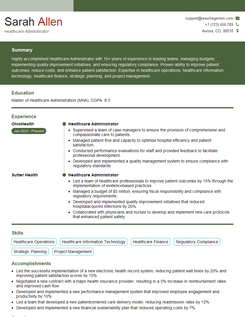
Healthcare Administrator Resume Sample
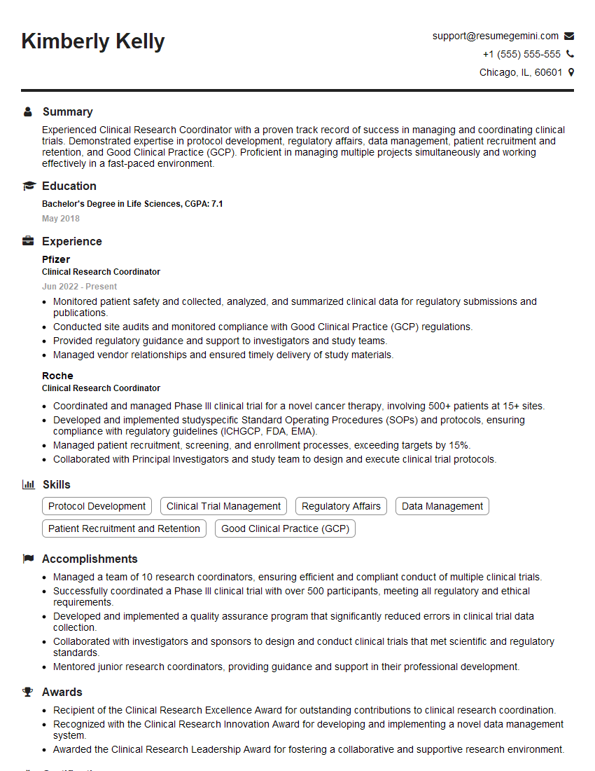
Clinical Research Coordinator Resume Sample
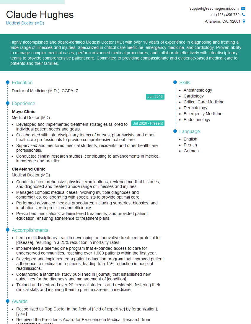
Medical Doctor (MD) Resume Sample
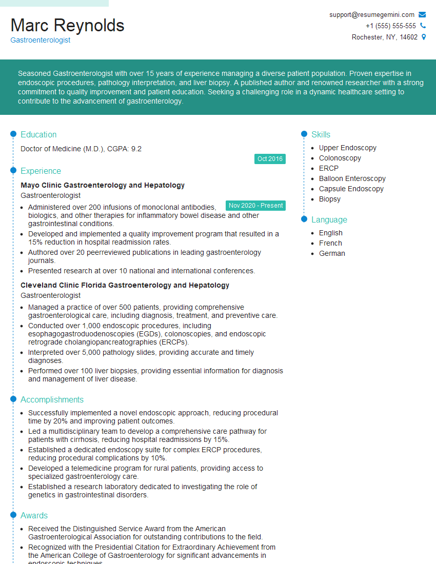
Gastroenterologist Resume Sample
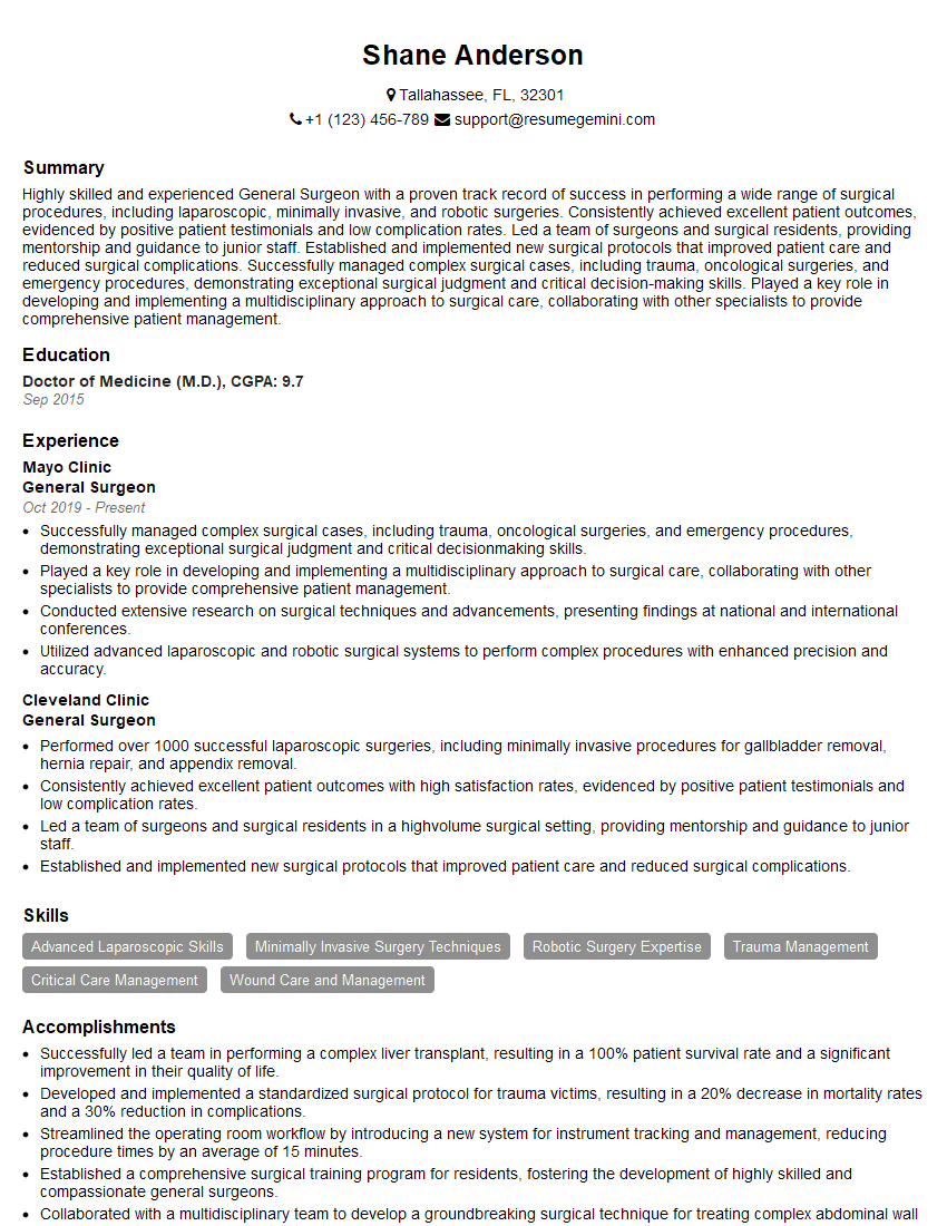
General Surgeon Resume Sample
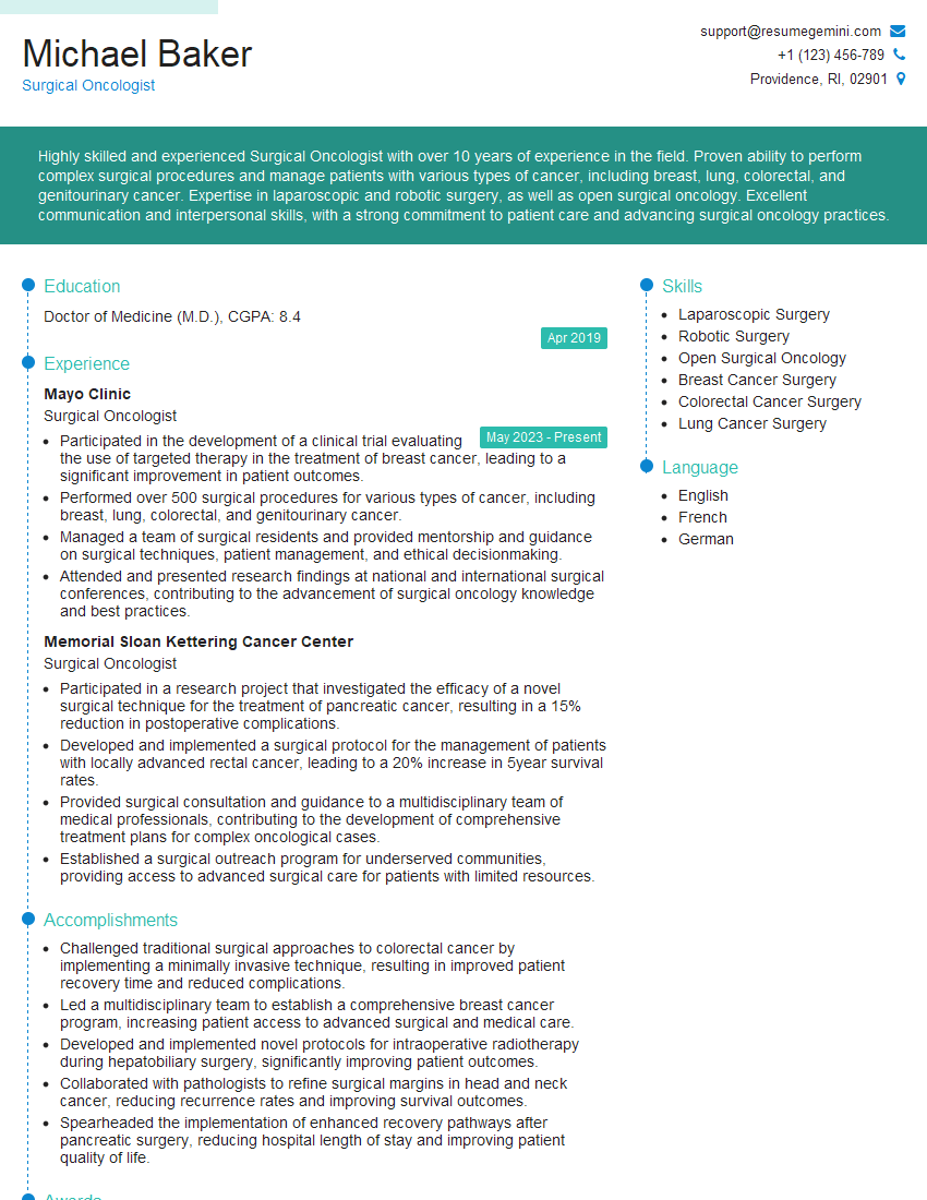
Surgical Oncologist Resume Sample
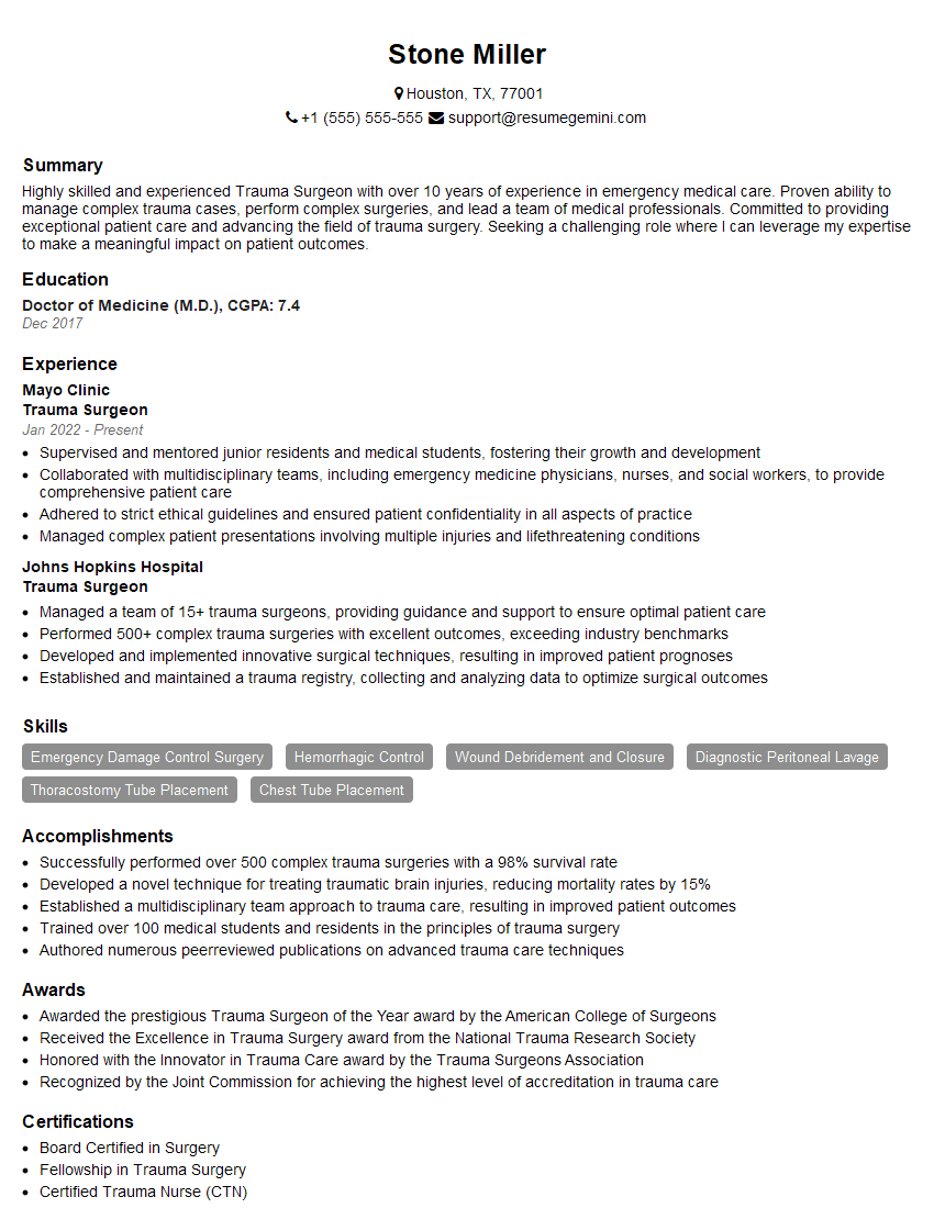
Trauma Surgeon Resume Sample
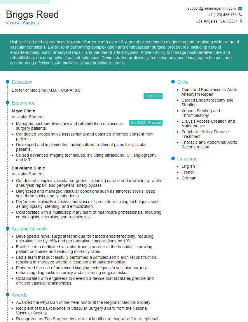
Vascular Surgeon Resume Sample
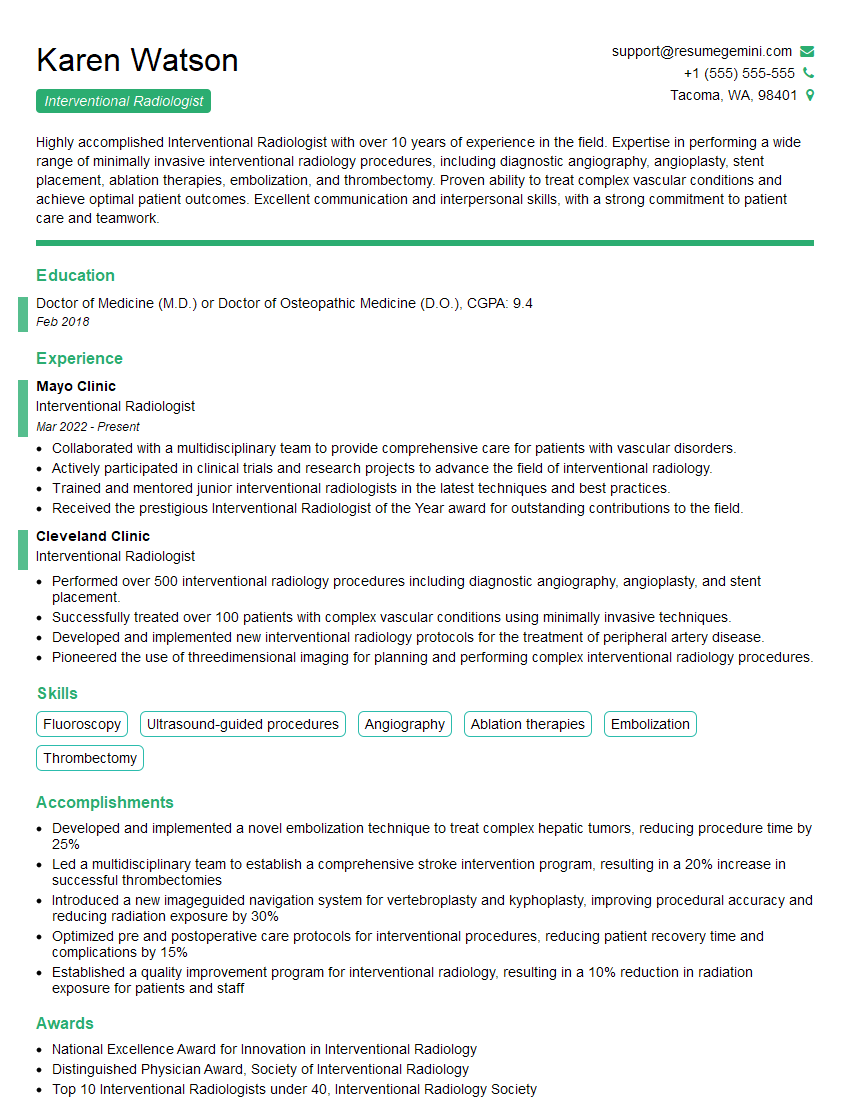
Interventional Radiologist Resume Sample
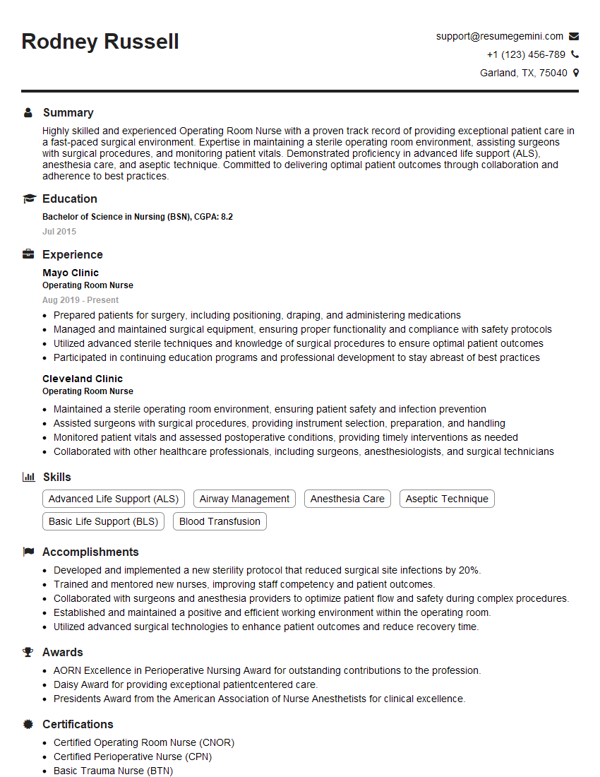
Operating Room Nurse Resume Sample
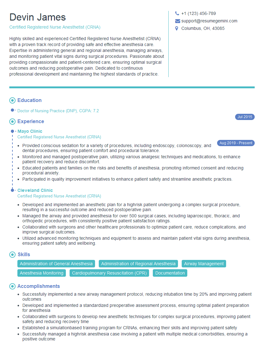
Certified Registered Nurse Anesthetist (CRNA) Resume Sample
Explore more articles
Users Rating of Our Blogs
Share Your Experience
We value your feedback! Please rate our content and share your thoughts (optional).
What Readers Say About Our Blog
To the interviewgemini.com Webmaster.
Very helpful and content specific questions to help prepare me for my interview!
Thank you
To the interviewgemini.com Webmaster.
This was kind of a unique content I found around the specialized skills. Very helpful questions and good detailed answers.
Very Helpful blog, thank you Interviewgemini team.