Preparation is the key to success in any interview. In this post, we’ll explore crucial Endoscopic Navigation interview questions and equip you with strategies to craft impactful answers. Whether you’re a beginner or a pro, these tips will elevate your preparation.
Questions Asked in Endoscopic Navigation Interview
Q 1. Describe the principles of endoscopic navigation.
Endoscopic navigation utilizes image guidance to enhance the accuracy and safety of minimally invasive surgical procedures. Essentially, it’s like having a GPS for surgery within the body. Instead of relying solely on the surgeon’s visual experience, endoscopic navigation systems integrate real-time imaging with 3D models of the anatomy, allowing the surgeon to see exactly where their instruments are located within the patient, even when out of direct view. This improves precision, reduces procedure time, and minimizes the risk of complications.
Think of it like this: imagine navigating a maze blindfolded. Endoscopic navigation is like having a detailed map and a GPS that constantly shows your location and the path to the target. This removes the uncertainty and allows for a more controlled approach.
Q 2. Explain different image-guidance modalities used in endoscopic navigation.
Several image-guidance modalities are used in endoscopic navigation, each offering unique advantages and limitations:
- Fluoroscopy: Uses X-rays to provide real-time images of the anatomy. It’s particularly useful for visualizing bony structures and identifying the location of instruments. However, it involves ionizing radiation, limiting the exposure duration.
- Ultrasound (US): Utilizes high-frequency sound waves to generate real-time images of soft tissues. It’s excellent for visualizing organs and blood vessels, but the image quality can be affected by gas or bone.
- Computed Tomography (CT): Creates detailed cross-sectional images of the anatomy using X-rays. It provides high-resolution images but is not real-time. CT images are usually used to create a pre-operative 3D model.
- Magnetic Resonance Imaging (MRI): Uses magnetic fields and radio waves to generate detailed images of soft tissues. MRI provides excellent soft tissue contrast, but it’s not real-time and can be challenging to use intra-operatively.
- Optical Coherence Tomography (OCT): This technique offers high-resolution images of tissue microstructure, providing important information at the tissue level during endoscopic procedures. It is particularly useful in gastroenterology.
The choice of modality depends on the specific procedure, the anatomical region involved, and the surgeon’s preference. Often, a combination of modalities is used for optimal visualization.
Q 3. What are the advantages and limitations of robotic-assisted endoscopic surgery?
Robotic-assisted endoscopic surgery combines the advantages of minimally invasive surgery with the enhanced dexterity and precision of robotic systems. Think of it as giving the surgeon a more refined set of tools within the confined space of the body.
- Advantages: Enhanced precision and dexterity, improved visualization (often with 3D HD vision), reduced tremor, ergonomic benefits for the surgeon, potential for smaller incisions, and increased range of motion.
- Limitations: High initial cost of the robotic system, increased procedural time due to setup and learning curve, technical expertise required for operation and maintenance, potential for system malfunction, and the need for specialized training.
For example, in complex laparoscopic procedures, robotic assistance can significantly improve the surgeon’s ability to perform intricate maneuvers, particularly in narrow or confined spaces. However, the high cost and specialized training requirements can limit its widespread adoption.
Q 4. How does 3D visualization enhance the accuracy of endoscopic procedures?
3D visualization is a game-changer in endoscopic procedures, offering significant advantages over traditional 2D views. The additional depth perception provides a more intuitive understanding of the surgical field, allowing for better spatial awareness and more precise instrument manipulation.
In a 2D view, depth perception relies on cues like shadows and relative sizes, which can be misleading. 3D visualization removes this ambiguity, offering a much clearer representation of the anatomy and the relationship between instruments and tissues. This translates to improved accuracy in target identification, dissection, and suturing, leading to better surgical outcomes and potentially fewer complications. Imagine trying to assemble a complex model from a 2D instruction sheet versus a 3D model – the 3D model is significantly easier and more accurate.
Q 5. Describe the role of fluoroscopy in endoscopic navigation.
Fluoroscopy plays a crucial role in endoscopic navigation, particularly in procedures involving bony structures or the placement of devices. It provides real-time X-ray images that allow the surgeon to visualize the location of instruments and devices relative to the anatomy. This is especially useful in procedures like biliary stent placement or vertebroplasty, where precise placement is crucial for a successful outcome.
For example, during a procedure involving stent placement in the biliary system, fluoroscopy allows the surgeon to monitor the real-time advancement of the stent, ensuring its proper positioning and preventing complications such as perforation or misplacement. However, it is important to remember that it involves ionizing radiation, and exposure should be minimized.
Q 6. Explain the concept of registration in image-guided surgery.
Registration in image-guided surgery is the process of aligning pre-operative images (like CT or MRI scans) with the real-time intraoperative images. Think of it as aligning a map with the actual terrain. This is crucial because the patient’s position and anatomy might shift slightly during surgery.
Precise registration ensures that the information from the pre-operative images is accurately superimposed on the real-time view. This allows the surgeon to plan and execute the procedure with precision, knowing the exact location of anatomical structures relative to their instruments. Different registration methods exist, ranging from manual landmark-based techniques to more sophisticated automated algorithms. Accurate registration is essential for the success of image-guided procedures.
Q 7. What are the different types of endoscopic instruments used in navigation systems?
A variety of endoscopic instruments are integrated into navigation systems. These instruments are often specifically designed to interact with the navigation system. The types of instruments used depend heavily on the surgical procedure.
- Endoscopes: These provide the visual pathway into the body. They can be rigid or flexible, depending on the procedure and anatomical site. Specialized endoscopes might have ports for the introduction of other instruments.
- Forceps: Used for grasping, manipulating, and dissecting tissue.
- Scissors: Used for cutting and dissecting tissue.
- Needles: Used for injections or suturing.
- Specialized instruments: Specific instruments are designed for particular procedures (e.g., probes, catheters, stents). Many of these instruments may incorporate sensors to provide additional real-time information to the navigation system.
Many of these instruments might have integrated sensors that feed information back to the navigation system, aiding in precise tracking and manipulation.
Q 8. How do you handle technical malfunctions during an endoscopic procedure?
Technical malfunctions during an endoscopic procedure are unfortunately a possibility, but having a systematic approach is crucial. My first step is always to assess the severity of the malfunction. Is it a minor issue like a loose connection, or something more serious like a camera failure?
For minor issues, troubleshooting is usually straightforward. I’ll check cables, connections, and power sources. I always have a backup set of instruments readily available, allowing for a quick swap if necessary. For example, if the insufflator malfunctions (the device that inflates the abdomen for better visualization), I have a secondary one immediately available to prevent any delay in the procedure.
Major malfunctions, such as camera failure, require a more decisive response. Patient safety is paramount. I would immediately stop the procedure, inform the surgical team and the patient (as appropriate), and determine the best course of action. This might involve switching to a different modality, such as laparoscopy, or postponing the procedure until the equipment is repaired. Open communication with the team is vital during such events. We conduct post-incident reviews to analyze what occurred and implement preventative measures for the future.
Q 9. Describe your experience with different endoscopic navigation software platforms.
I’ve had extensive experience with several endoscopic navigation software platforms, including Intuitive Surgical’s da Vinci system (though primarily used for robotic surgery, it has navigation capabilities) and various image-guided navigation systems specifically designed for endoscopy. Each platform has its own strengths and weaknesses. For instance, some excel at 3D reconstruction of the anatomy from pre-operative imaging, while others offer real-time tracking of the endoscope’s position.
My experience with these systems extends beyond simply using the interface; it also includes understanding their limitations. A crucial element is knowing how the software interprets anatomical data and the potential for discrepancies between the virtual model and the real-time anatomy. I am adept at compensating for these discrepancies, often using a combination of the software’s guidance and my own clinical judgment, which is critical for preventing errors.
For instance, I remember a case where the software had difficulty precisely tracking the endoscope within a highly convoluted anatomical structure. Using my own anatomical knowledge and combining the software’s general guidance, I was still able to navigate successfully and complete the procedure without compromising patient safety. This underscores the need for both technical expertise and strong anatomical knowledge in navigating these systems.
Q 10. Explain the importance of pre-operative planning in endoscopic navigation.
Pre-operative planning is absolutely fundamental for successful and safe endoscopic navigation. It lays the groundwork for the entire procedure. Imagine trying to assemble furniture without instructions – it’s chaotic! Similarly, without proper planning in endoscopic navigation, the procedure becomes significantly more difficult and risky.
The process typically involves obtaining detailed pre-operative imaging such as CT scans or MRI scans. This allows for 3D reconstruction of the anatomy, providing a virtual roadmap for the procedure. The software then allows me to plan the optimal trajectory for the endoscope, identifying potential obstacles or critical structures beforehand. This can significantly reduce procedure time and complications. We meticulously plan access points, identify target areas and potential risks (e.g., proximity to major blood vessels). Detailed surgical planning also helps to minimize the risk of accidental injury to adjacent structures.
For instance, in a complex procedure involving a deep-seated lesion, pre-operative planning allows us to anticipate the challenging angles and potential blind spots. We use the software to simulate the procedure, predicting the optimal path and identifying potential pitfalls. This reduces unexpected surprises during the actual procedure which ultimately enhances the safety and efficacy of the intervention.
Q 11. How do you ensure patient safety during procedures involving endoscopic navigation?
Patient safety is the absolute top priority in any procedure involving endoscopic navigation, and we employ multiple layers of safeguards. Firstly, thorough pre-operative assessment, including reviewing the patient’s medical history and conducting a detailed physical examination, is critical. This identifies any potential risks or contraindications to the procedure.
Secondly, rigorous adherence to sterile techniques and proper use of equipment is non-negotiable. We use checklist protocols to confirm each step is completed correctly. During the procedure itself, continuous monitoring of vital signs, including heart rate, blood pressure, and oxygen saturation, is crucial. We closely observe the patient for any signs of complications. The use of navigation software helps reduce the risk of accidental perforation or injury to adjacent structures.
Thirdly, open communication with the patient and their family helps manage expectations, addresses concerns and builds trust, vital to a positive outcome. In the event of unexpected complications, we have established protocols for prompt and effective response, including the ability to quickly transition to alternative surgical approaches if necessary.
Finally, ongoing training, professional development, and quality control measures are essential to maintain the highest standards of patient safety. We conduct regular reviews to identify areas for improvement in our protocols and technique.
Q 12. What are the common challenges faced during endoscopic navigation procedures?
Endoscopic navigation, while offering numerous advantages, presents unique challenges. One major hurdle is the often-limited field of view provided by the endoscope itself, making precise navigation challenging, especially in complex anatomical areas. This is like navigating a maze with a small peephole.
Another challenge is the inherent distortion and limitations in image quality. Image artifacts, motion artifacts (patient movement), and variations in tissue properties can all hinder the accuracy of the navigation system. Furthermore, there can be discrepancies between the pre-operative imaging and the real-time anatomy due to patient positioning changes, breathing, or tissue deformation during the procedure.
Dealing with bleeding during the procedure can also significantly complicate navigation, obscuring the field of view and interfering with accurate tracking. Finally, there’s the constant need to balance the benefits of navigation technology with the clinician’s own surgical experience and judgment; it’s not a simple case of blindly following the software. The combination of technical skill, anatomical knowledge, and good judgment is key to successful navigation and a safe surgical procedure.
Q 13. Describe your experience with different types of endoscopic cameras and their functionalities.
My experience encompasses a wide range of endoscopic cameras, from standard white-light endoscopes to those equipped with narrow-band imaging (NBI) or chromoendoscopy. Each camera type offers distinct functionalities depending on the clinical application. Standard white-light endoscopes provide basic visualization, suitable for many routine procedures. However, the improved image quality and higher resolution of newer cameras provide enhanced visualization and improved clarity.
NBI enhances the visualization of superficial vascular patterns, helping to identify subtle abnormalities in the tissue that might be missed with white light. This is particularly useful in detecting early cancerous lesions in the gastrointestinal tract. Chromoendoscopy, using dyes to highlight specific tissue features, offers yet another tool for improved visualization. For instance, methylene blue can highlight specific areas, further enhancing the identification of potentially cancerous or pre-cancerous tissue.
The choice of camera largely depends on the specific clinical indication. For example, in cases of suspected early colorectal cancer, NBI is frequently employed to enhance the detection and characterization of lesions, whereas white light endoscopy may be sufficient for other indications. The functionality of these cameras is constantly improving, with enhancements in resolution, image processing, and the availability of advanced imaging modalities offering surgeons greater precision and clearer visualization during procedures.
Q 14. How do you interpret and utilize intraoperative imaging data during a procedure?
Intraoperative imaging data is crucial for real-time guidance and decision-making during endoscopic navigation. I interpret this data in several ways. Firstly, I use it to confirm the endoscope’s position relative to the target anatomy. This constant feedback is essential for precise navigation, ensuring that I’m accurately approaching the target area without damaging surrounding structures.
Secondly, I use intraoperative imaging to assess the status of the tissues during the procedure. For example, I monitor for any signs of bleeding, perforation, or inflammation. The ability to view these changes in real-time allows me to take immediate corrective measures if needed. This provides real-time feedback that the standard endoscopic view may not provide.
Thirdly, I might use intraoperative imaging data to refine my surgical plan if unexpected anatomical variations are discovered. The flexibility to adjust the approach based on what I see during the procedure is crucial for a successful outcome. For example, if I encounter unexpected vascular structures during a procedure, I can use the real-time imaging to adjust my path and minimize risk of injury. Overall, effectively interpreting and integrating this data is vital to maximize the benefits of endoscopic navigation and enhance patient safety.
Q 15. Explain the role of haptic feedback in endoscopic navigation.
Haptic feedback in endoscopic navigation plays a crucial role in enhancing the surgeon’s sense of touch during minimally invasive procedures. Imagine trying to assemble a complex puzzle blindfolded – incredibly difficult, right? That’s essentially what endoscopy can feel like without haptic feedback. It provides the surgeon with real-time information about the forces and textures encountered by the endoscope tip as it interacts with the surrounding tissues. This ‘sense of touch’ is transmitted through specialized sensors and actuators, allowing the surgeon to feel the resistance of tissues, identify organs, and avoid causing unintentional damage. For instance, haptic feedback can help differentiate between a soft, healthy liver and a hardened, cancerous area, offering a level of precision unavailable with visual information alone.
The types of haptic feedback vary. Some systems provide simple force feedback, indicating resistance. Others offer more sophisticated feedback simulating tissue texture or stiffness. The integration of haptic feedback significantly improves the safety and precision of endoscopic procedures, allowing for more delicate manipulations and reducing the risk of complications.
Career Expert Tips:
- Ace those interviews! Prepare effectively by reviewing the Top 50 Most Common Interview Questions on ResumeGemini.
- Navigate your job search with confidence! Explore a wide range of Career Tips on ResumeGemini. Learn about common challenges and recommendations to overcome them.
- Craft the perfect resume! Master the Art of Resume Writing with ResumeGemini’s guide. Showcase your unique qualifications and achievements effectively.
- Don’t miss out on holiday savings! Build your dream resume with ResumeGemini’s ATS optimized templates.
Q 16. Describe your experience with different types of robotic platforms used in endoscopy.
My experience encompasses a range of robotic platforms used in endoscopy, from flexible robotic systems offering improved dexterity and control of the endoscope to rigid robotic systems designed for specific procedures. I’ve worked with systems utilizing various control mechanisms, including master-slave manipulators and intuitive interfaces. For example, I’ve extensively used the da Vinci Surgical System adapted for endoscopic procedures. This system allows for precise movements and 3D visualization. I’ve also had experience with less sophisticated systems featuring motorized control of the endoscope shaft, offering improved maneuverability compared to manual control. Each platform presents unique advantages and disadvantages depending on the surgical task. I find that the most effective approach often involves selecting a platform that matches the specific needs of the procedure and the surgeon’s skill set.
A key consideration is the level of dexterity and range of motion afforded by each system. While some robotic systems offer remarkable dexterity, they can be more complex to operate and may require specialized training. Others provide a simpler, more intuitive interface. My approach involves careful selection of the system based on a thorough assessment of these factors for each case.
Q 17. How do you troubleshoot issues related to image quality or system performance?
Troubleshooting image quality and system performance issues in endoscopic navigation requires a systematic approach. I begin by evaluating the entire system, starting with the light source and camera settings, checking for proper alignment and sufficient illumination. Poor image quality can stem from several sources, including dirty lenses, cable issues, or malfunctioning components. I use a methodical checklist to verify each element.
For example, if I encounter blurry images, I’d first check for lens contamination, then examine cable connections for damage or loose fittings. If the problem persists, I’d investigate the camera’s internal settings and possibly replace it. Similarly, system performance issues such as jerky movements or delayed feedback could be due to software glitches, network connectivity problems, or hardware malfunctions. In these instances, restarting the system, checking network connections, and investigating potential hardware failures are essential steps.
My approach emphasizes a detailed investigation, starting with simple checks and progressing to more advanced diagnostics. Detailed documentation of each step is crucial for both problem-solving and preventative maintenance.
Q 18. Explain the process of calibrating an endoscopic navigation system.
Calibrating an endoscopic navigation system is a crucial step to ensure accurate tracking and visualization. The process typically involves aligning the virtual model with the patient’s anatomy. This is done using reference points, often obtained through fluoroscopy, ultrasound, or even anatomical landmarks identified during the procedure.
The specific calibration steps vary depending on the system, but generally involve:
- Reference Point Acquisition: Identifying and registering anatomical reference points in both the real-world and the virtual model.
- Image Registration: Aligning the endoscopic images with the pre-operative images or 3D models.
- Transformation Matrix Calculation: The system calculates a transformation matrix, which mathematically describes the relationship between the virtual and real-world coordinates.
- Validation: Checking the accuracy of the registration through various tests, such as visual inspection and potentially using known anatomical distances.
Accurate calibration is essential for the efficacy of the navigation system. Inaccurate calibration can lead to positional errors, potentially resulting in complications during the procedure. Therefore, a careful and methodical approach to calibration is paramount.
Q 19. What are the key performance indicators (KPIs) used to evaluate the success of an endoscopic navigation procedure?
Key performance indicators (KPIs) for evaluating the success of an endoscopic navigation procedure go beyond simply completing the procedure. They should encompass safety, efficacy, and efficiency.
- Procedure Time: A shorter procedure time, while maintaining quality, indicates efficiency.
- Radiation Exposure (if applicable): Minimizing radiation exposure enhances patient safety.
- Complications Rate: A lower rate indicates higher procedural safety.
- Target Accuracy: How accurately the navigation system guided the surgeon to the target anatomy.
- Patient Outcomes: Post-operative recovery time, length of hospital stay, and overall patient well-being are crucial indicators of success.
These KPIs are usually tracked and analyzed to identify areas for improvement in the navigation system, surgical techniques, and overall workflow. Data analysis helps optimize the use of navigation technology and enhance patient care.
Q 20. How do you maintain the sterility and safety of the endoscopic instruments?
Maintaining sterility and safety in endoscopic procedures is paramount. This involves strict adherence to established protocols throughout the entire process. Prior to the procedure, all instruments and equipment are meticulously sterilized using validated methods such as steam sterilization or ethylene oxide gas sterilization. The operating room environment is also prepared according to strict aseptic techniques. During the procedure, sterile drapes and gloves are used to maintain a sterile field. Regular monitoring for breaches in sterility is essential.
Moreover, safety protocols extend to the safe handling and disposal of sharps and biohazardous materials. Specific procedures are followed for the cleaning and reprocessing of reusable instruments, ensuring they meet the highest sterility standards before being used again. Continuous training and education for all personnel involved is crucial in reinforcing safe practices and maintaining a sterile environment. Regular audits and inspections are conducted to ensure compliance with all relevant guidelines and regulations.
Q 21. Describe your experience with different types of endoscopic procedures.
My experience spans a wide spectrum of endoscopic procedures. I’ve been involved in various gastrointestinal procedures like colonoscopies and endoscopic mucosal resections (EMR) for the treatment of polyps and early-stage cancers. I have experience with ERCP (Endoscopic Retrograde Cholangiopancreatography) for the diagnosis and treatment of biliary and pancreatic disorders. Furthermore, I’ve assisted in thoracic endoscopic procedures, including mediastinoscopy and lung biopsies.
In each case, the specifics of the procedure, the challenges, and the required level of precision differ significantly. My experience has strengthened my understanding of the nuances of each procedure and refined my approach to navigating these procedures with precision and effectiveness. Each procedure reinforces the importance of careful planning, meticulous execution, and the constant adaptation to unforeseen situations.
Q 22. Explain the importance of teamwork and communication during endoscopic navigation procedures.
Endoscopic navigation procedures are inherently complex, requiring seamless teamwork and clear communication for optimal patient outcomes. Imagine navigating a maze – a single wrong turn can have serious consequences. Similarly, in minimally invasive surgery, a lack of coordination can lead to complications.
- Pre-operative planning: Thorough discussion between surgeons, anesthesiologists, nurses, and technicians about the surgical approach, anticipated challenges, and contingency plans is crucial. We utilize checklists and interactive simulations to ensure everyone understands their roles.
- Intra-operative communication: Clear and concise communication during the procedure is vital. We employ specific terminology, regularly update each other on the procedure’s progress, and immediately address any unexpected situations. For example, if an unexpected anatomical variation is encountered, we pause to discuss the best course of action.
- Post-operative debrief: A post-operative discussion helps identify areas for improvement in teamwork and communication. This feedback loop is essential for continuous quality improvement. We analyze any complications that occurred and how better communication could have prevented them.
Q 23. How do you ensure the accuracy of the surgical plan and its implementation?
Ensuring the accuracy of the surgical plan and its implementation is paramount in endoscopic navigation. We employ a multi-step process:
- Pre-operative imaging and planning: Detailed 3D imaging (CT, MRI) is used to create a precise 3D model of the anatomy. This model serves as the roadmap for the procedure. Specialized software allows us to plan the optimal trajectory for the endoscope, minimizing the risk of damage to surrounding tissues.
- Image registration and fusion: During the procedure, real-time images from the endoscope are registered with the pre-operative 3D model. This allows us to accurately visualize the endoscope’s position relative to the patient’s anatomy. This ‘fusion’ ensures we’re operating exactly where planned.
- Real-time guidance and feedback: Endoscopic navigation systems provide real-time feedback, highlighting the endoscope’s position and trajectory. This minimizes potential errors and enables adjustments as needed. This is like having a GPS system for surgery.
- Quality control measures: Regular calibration of the equipment and verification of the image registration are essential for maintaining accuracy. We also utilize checklists to ensure all steps are meticulously followed.
Q 24. What are the ethical considerations associated with endoscopic navigation?
Ethical considerations in endoscopic navigation are significant. The technology offers immense benefits, but it also presents ethical challenges.
- Informed consent: Patients must be fully informed about the procedure, including its benefits, risks, and alternatives. We ensure they understand the use of navigation technology and its limitations.
- Data privacy and security: Patient data acquired during the procedure, including medical images and procedural information, must be handled with strict confidentiality and security measures. We adhere to all relevant data protection regulations.
- Access and equity: The high cost of endoscopic navigation systems raises concerns about access for all patients, regardless of their socioeconomic status. We advocate for policies that ensure equitable access to this beneficial technology.
- Algorithmic bias: The algorithms used in navigation systems should be rigorously tested to avoid biases that could lead to disparate outcomes for different patient populations. We constantly scrutinize the data and algorithms for any potential bias.
Q 25. Describe your knowledge of relevant safety regulations and guidelines.
Safety regulations and guidelines are strictly adhered to. We follow guidelines established by organizations like the FDA, relevant national healthcare bodies, and professional medical societies. This includes:
- Equipment safety: Regular maintenance and calibration of endoscopic navigation systems are essential to ensure their safe and effective operation. We follow manufacturer’s guidelines meticulously.
- Infection control: Strict adherence to infection control protocols is crucial to prevent the spread of infections. We follow established sterilization and disinfection procedures for all equipment.
- Radiation safety: When using imaging techniques like fluoroscopy, radiation safety measures must be implemented to minimize patient and staff exposure. We utilize lead shielding and appropriate radiation protection protocols.
- Emergency preparedness: We maintain a comprehensive emergency plan to address any unexpected events during the procedure, ensuring a quick and appropriate response to any situation.
Q 26. How do you stay updated with the latest advancements in endoscopic navigation technology?
Staying updated on advancements is crucial. We utilize several strategies:
- Professional societies and conferences: Active participation in professional societies like the American Society for Gastrointestinal Endoscopy (ASGE) provides access to the latest research and technological developments. Attending conferences allows networking and direct interaction with experts.
- Peer-reviewed journals and publications: Regularly reviewing leading medical journals helps stay informed about new techniques, technologies, and clinical trial results.
- Continuing medical education (CME): CME courses and workshops offer opportunities to learn and refine skills in endoscopic navigation techniques and the management of new systems.
- Manufacturer updates: Direct communication with manufacturers provides information on software updates, new features, and training opportunities for newer models of navigation systems.
Q 27. Explain your experience in data analysis and interpretation related to endoscopic procedures.
Data analysis and interpretation are integral to improving patient care and surgical outcomes. We use data from endoscopic procedures to:
- Procedure time analysis: We analyze procedure times to identify areas for improvement in efficiency and workflow. This helps to optimize the surgical process.
- Complication analysis: Analyzing complications helps identify risk factors and implement strategies to reduce the likelihood of future complications. This is a crucial aspect of continuous quality improvement.
- Image analysis: Analyzing imaging data obtained during the procedure enhances our understanding of the anatomy and pathology. This helps refine our surgical planning and technique.
- Statistical analysis: We use statistical methods to analyze large datasets, identifying trends and patterns that can improve surgical outcomes and inform future clinical decisions. This helps to guide the development of best practices.
Q 28. How would you approach training new team members on endoscopic navigation systems?
Training new team members requires a structured and multi-faceted approach:
- Theoretical training: We begin with lectures and presentations covering the principles of endoscopic navigation, anatomy, and the specifics of the navigation systems used in our facility.
- Simulated training: Utilizing simulation systems allows trainees to practice endoscopic navigation techniques in a safe and controlled environment before working with real patients. This minimizes risks during actual procedures.
- Hands-on training: Supervised hands-on training with experienced professionals is vital. This allows trainees to develop practical skills and refine their techniques under expert guidance.
- Mentorship and feedback: Continuous mentorship and regular feedback are essential to ensure trainees develop proficiency and confidence in their skills. We utilize a structured feedback system to identify areas for improvement.
- Ongoing assessment: Regular assessments through observation and practical examinations allow us to evaluate the trainees’ progress and identify areas needing further attention.
Key Topics to Learn for Endoscopic Navigation Interview
- Image Processing and Analysis: Understanding image acquisition, enhancement techniques (noise reduction, contrast adjustment), and feature extraction for navigation.
- 3D Reconstruction and Visualization: Knowledge of algorithms and techniques used to create 3D models from endoscopic images and their visualization for effective navigation.
- Sensor Fusion and Data Integration: Understanding how to combine data from multiple sensors (e.g., optical, ultrasound, force sensors) for improved navigation accuracy and robustness.
- Motion Planning and Control: Familiarity with algorithms and strategies for planning optimal paths for the endoscope and controlling its movement precisely.
- Calibration and Registration: Understanding the process of calibrating sensors and registering different data sources to ensure accurate navigation.
- Robot-Assisted Endoscopy: Knowledge of robotic systems used in endoscopic navigation, including their mechanics, control systems, and limitations.
- Clinical Applications and Case Studies: Familiarity with the applications of endoscopic navigation in different medical specialties (e.g., gastroenterology, urology, surgery) and understanding of relevant case studies.
- Challenges and Limitations: A critical understanding of the limitations of current endoscopic navigation technologies, such as image quality issues, motion artifacts, and computational complexity.
- Emerging Technologies: Awareness of advancements in areas like AI-powered navigation, augmented reality, and flexible endoscopes.
- Troubleshooting and Problem-Solving: Ability to diagnose and resolve common problems encountered during endoscopic navigation procedures.
Next Steps
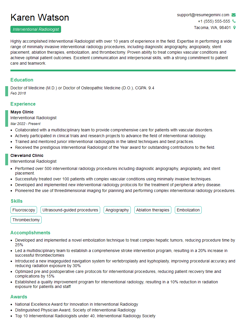
Mastering Endoscopic Navigation opens doors to exciting and impactful careers in medical technology and healthcare. A strong understanding of these concepts is crucial for securing your desired role. To maximize your chances, it’s essential to create a resume that effectively highlights your skills and experience to Applicant Tracking Systems (ATS). Building an ATS-friendly resume is key to getting your application noticed. We highly recommend using ResumeGemini, a trusted resource for creating professional and impactful resumes. ResumeGemini provides examples of resumes tailored specifically to Endoscopic Navigation roles to help you showcase your qualifications effectively.
Explore more articles
Users Rating of Our Blogs
Share Your Experience
We value your feedback! Please rate our content and share your thoughts (optional).
What Readers Say About Our Blog
To the interviewgemini.com Webmaster.
Very helpful and content specific questions to help prepare me for my interview!
Thank you
To the interviewgemini.com Webmaster.
This was kind of a unique content I found around the specialized skills. Very helpful questions and good detailed answers.
Very Helpful blog, thank you Interviewgemini team.