Feeling uncertain about what to expect in your upcoming interview? We’ve got you covered! This blog highlights the most important Veterinary endocrinology interview questions and provides actionable advice to help you stand out as the ideal candidate. Let’s pave the way for your success.
Questions Asked in Veterinary endocrinology Interview
Q 1. Describe the physiological regulation of glucose homeostasis.
Glucose homeostasis, the maintenance of stable blood glucose levels, is a tightly regulated process involving several hormones and organs. Think of it like a thermostat for your blood sugar.
Insulin, secreted by the pancreas in response to rising blood glucose (after a meal), facilitates glucose uptake by cells, primarily in muscle, liver, and adipose tissue. It’s the ‘cooling’ mechanism, lowering blood sugar.
Glucagon, also from the pancreas, is released when blood glucose is low. It stimulates the liver to break down glycogen (stored glucose) into glucose and release it into the bloodstream. This is the ‘heating’ mechanism, raising blood sugar.
Other hormones play supporting roles. Epinephrine (adrenaline) and cortisol, for instance, raise blood glucose levels during stress or fasting. The growth hormone also contributes to glucose regulation.
The liver acts as a central player, storing and releasing glucose as needed. It’s like the glucose warehouse.
The kidneys also play a role, excreting excess glucose in the urine when blood glucose levels are significantly elevated (above the renal threshold).
These components work in concert to maintain blood glucose within a narrow physiological range, ensuring a constant energy supply to the body’s cells.
Q 2. Explain the pathogenesis of diabetes mellitus in dogs and cats.
Diabetes mellitus in dogs and cats, much like in humans, is characterized by hyperglycemia (high blood glucose) due to a deficiency in insulin secretion (Type 1) or a reduced responsiveness of cells to insulin (Type 2, or insulin resistance).
Type 1 diabetes (more common in dogs) results from autoimmune destruction of the pancreatic beta cells, the cells responsible for insulin production. Imagine the ‘cooling’ system in our thermostat analogy failing completely.
Type 2 diabetes (more common in cats) is often associated with obesity and insulin resistance. The cells become less responsive to insulin, making it harder for glucose to enter the cells despite the presence of insulin. In our analogy, the ‘cooling’ system works but isn’t powerful enough to keep up with demand.
Both types lead to chronic hyperglycemia, causing damage to various organs, including the eyes, kidneys, and nerves.
Q 3. Discuss the diagnostic approach to hyperadrenocorticism in a dog.
Diagnosing hyperadrenocorticism (Cushing’s disease) in dogs requires a multi-step approach because clinical signs are often non-specific. It’s like solving a detective case, needing multiple clues to confirm the diagnosis.
Initial screening tests often include a low-dose dexamethasone suppression test (LDDS) and an ACTH stimulation test. These tests assess the adrenal gland’s response to different stimuli. We’re looking for over-production of cortisol.
Further investigations may include measuring urine cortisol-to-creatinine ratio (UFC) which measures cortisol levels in the urine; it’s a less invasive way to get a sense of overall cortisol levels.
Imaging techniques such as abdominal ultrasound or MRI can help identify the location and size of adrenal tumors.
It’s crucial to differentiate between pituitary-dependent hyperadrenocorticism (PDH, most common) and adrenal-dependent hyperadrenocorticism (ADH). This helps guide treatment strategies.
Q 4. Outline the treatment options for hypothyroidism in cats.
Treatment for feline hypothyroidism focuses on lifelong thyroid hormone replacement therapy with levothyroxine (T4). It’s like giving the thyroid gland a boost to correct its underproduction of hormones.
Dosage adjustment is crucial, based on regular monitoring of T4 levels and clinical response. We need to find the ‘sweet spot’ dose to bring the cat back to normal thyroid function. This is done through blood tests to check T4 levels. Too high a dose can lead to side-effects and not enough will not correct the underlying condition.
Monitoring for clinical improvement is critical; we expect to see increased activity levels, improved coat condition, and a return to a healthy body weight.
Lifestyle modifications can include weight management if obesity is a factor, addressing underlying conditions such as cardiac disease, and supportive care depending on individual symptoms.
Regular veterinary check-ups and blood tests are essential to ensure the effectiveness of treatment and prevent long-term complications.
Q 5. What are the common causes of hypercalcemia in dogs?
Hypercalcemia (high blood calcium) in dogs can stem from various causes, each requiring a different diagnostic and therapeutic approach.
Primary hyperparathyroidism: This is a relatively rare condition where an overactive parathyroid gland secretes excessive parathyroid hormone (PTH), leading to increased calcium levels.
Malignancy: Certain cancers, especially lymphoma, can produce substances (like parathyroid hormone-related protein) that increase calcium levels. This is often a serious condition.
Renal failure: Damaged kidneys may lose their ability to properly excrete calcium.
Vitamin D toxicity: Excessive intake of vitamin D can result in significantly raised calcium levels.
Bone diseases: Conditions affecting bone turnover, such as hypercalcemia of malignancy, can lead to the release of calcium into the bloodstream.
A thorough history, physical examination, and appropriate laboratory tests are essential to determine the underlying cause of hypercalcemia.
Q 6. Describe the clinical presentation of hypoparathyroidism in a dog.
Hypoparathyroidism in dogs is characterized by low levels of parathyroid hormone (PTH), leading to hypocalcemia (low blood calcium). The clinical presentation can vary greatly depending on the severity of the calcium deficiency.
Neuromuscular signs are common and can include muscle tremors, seizures, and tetany (muscle spasms). This is due to calcium’s essential role in nerve and muscle function. It’s like the body’s electrical system short-circuiting.
Cardiac abnormalities can occur due to calcium’s role in heart muscle contraction. We may observe arrhythmias.
Gastrointestinal signs such as vomiting and diarrhea can be present.
The severity of the clinical presentation reflects the degree of hypocalcemia. Prompt diagnosis and treatment are crucial to prevent life-threatening complications.
Q 7. Explain the role of insulin in regulating blood glucose levels.
Insulin is a crucial hormone that regulates blood glucose levels. It’s the body’s primary mechanism for lowering blood sugar after a meal.
Glucose uptake: Insulin binds to receptors on the surface of muscle, liver, and fat cells, triggering glucose transporters (GLUTs) to move to the cell membrane. This allows glucose to enter the cells from the bloodstream, effectively removing glucose from circulation.
Glycogen synthesis: In the liver and muscle, insulin promotes glycogen synthesis, the conversion of glucose into glycogen (stored glucose) for later use. It’s like storing energy for a rainy day.
Protein synthesis: Insulin also stimulates protein synthesis and inhibits protein breakdown, contributing to overall metabolic regulation.
Lipogenesis: Insulin promotes fat storage (lipogenesis) in adipose tissue. It prevents the breakdown of fat stores.
Without sufficient insulin, glucose cannot enter cells effectively, leading to hyperglycemia (high blood glucose).
Q 8. How would you diagnose and manage diabetic ketoacidosis in a dog?
Diabetic ketoacidosis (DKA) in dogs is a life-threatening emergency characterized by severe hyperglycemia (high blood sugar), dehydration, and the accumulation of ketone bodies. Diagnosis involves a thorough history, physical examination, and laboratory testing. We look for clinical signs like increased thirst and urination (polydipsia and polyuria), weight loss, lethargy, and potentially vomiting and diarrhea. Laboratory tests are crucial and include measuring blood glucose levels (expecting significantly elevated levels), blood ketone levels (to confirm ketoacidosis), electrolytes (to assess dehydration and electrolyte imbalances), and blood gas analysis (to evaluate acid-base balance). Treatment is intensive and focuses on fluid therapy (to correct dehydration and electrolyte imbalances), insulin administration (to lower blood glucose and ketone levels), and supportive care, including monitoring vital signs and addressing any concurrent infections or complications.
Imagine it like this: your dog’s body is running on empty and is starting to burn fat for energy instead of glucose. This process produces ketones, which make the blood acidic, causing severe problems. Treatment aims to provide glucose as fuel and stop the production of harmful ketones.
- Step 1: Immediate stabilization: Hospitalization is essential for close monitoring and aggressive fluid therapy.
- Step 2: Insulin administration: Low-dose continuous insulin infusion is typically used initially, adjusted based on frequent blood glucose monitoring.
- Step 3: Electrolyte correction: Potassium levels need careful monitoring and replacement as needed, as insulin administration can drive potassium into cells.
- Step 4: Ongoing monitoring: Frequent blood glucose and electrolyte checks are crucial for guiding treatment and preventing complications.
Q 9. What are the potential complications of Cushing’s disease in dogs?
Cushing’s disease, or hyperadrenocorticism, results from excessive cortisol production. The complications can be significant and affect multiple organ systems. Common problems include:
- Increased thirst and urination (polydipsia and polyuria): Due to the effect of cortisol on kidney function.
- Weight gain with thin skin and muscle loss: The redistribution of fat and muscle wasting are characteristic findings.
- Increased susceptibility to infections: Cortisol suppresses the immune system, making the dog more vulnerable to infections.
- Hepatomegaly (enlarged liver): Cortisol’s impact on liver metabolism can lead to liver enlargement.
- Pancreatitis: An increased risk of inflammation of the pancreas is observed in dogs with Cushing’s disease.
- Muscle weakness and thin skin: These can lead to bruising and easy wounding.
- Diabetes mellitus: High cortisol levels can contribute to insulin resistance and eventually diabetes.
- Behavioral changes: Lethargy, increased appetite, and changes in personality can occur.
Essentially, Cushing’s disease creates a hormonal imbalance that wreaks havoc throughout the body, leaving the dog vulnerable to various health issues. Managing these complications is a central part of treating Cushing’s disease.
Q 10. Discuss the importance of monitoring thyroid hormone levels during treatment of hypothyroidism.
Monitoring thyroid hormone levels during hypothyroidism treatment is critical for ensuring adequate replacement therapy and avoiding both under- and over-replacement. Hypothyroidism is characterized by insufficient thyroid hormone (T4 and T3), leading to a slowed metabolism. Treatment involves providing synthetic thyroid hormone (usually levothyroxine).
Regular blood tests measuring total T4 and sometimes free T4 (a more biologically active form) are essential. The goal is to achieve a T4 level within the reference range, reflecting normal thyroid function. Too little levothyroxine will leave the dog hypothyroid, with persistent symptoms, while too much can cause hyperthyroidism, with its own set of problems.
Think of it like carefully adjusting the dosage of a medication. We need to find the ‘sweet spot’ – enough to restore normal function but not so much that it causes problems.
Clinical signs such as lethargy, weight gain, and coat changes also help guide treatment, but laboratory monitoring provides objective measurements to ensure optimal therapeutic effect and adjust the dose as needed. Frequent monitoring, especially during initial treatment adjustments and any changes in the dog’s health status, is crucial for successful management.
Q 11. Explain the difference between primary and secondary hypothyroidism.
The difference between primary and secondary hypothyroidism lies in the location of the problem within the hypothalamic-pituitary-thyroid (HPT) axis. The HPT axis is a complex system where the hypothalamus signals the pituitary gland, which in turn stimulates the thyroid gland to produce thyroid hormones.
- Primary hypothyroidism: This is the most common type. It occurs when the thyroid gland itself is unable to produce sufficient thyroid hormone, usually due to autoimmune destruction or inflammation.
- Secondary hypothyroidism: This is less common. It occurs when there is a problem with the pituitary gland, which is unable to produce sufficient thyroid-stimulating hormone (TSH), the hormone that tells the thyroid gland to make thyroid hormones. This can result from pituitary tumors or other pituitary disorders.
In essence, primary hypothyroidism is a problem with the thyroid gland itself, whereas secondary hypothyroidism is a problem upstream, with the pituitary gland failing to stimulate the thyroid adequately. Diagnostic testing, including TSH and T4 levels, helps differentiate between the two, guiding treatment decisions.
Q 12. What are the key differences in the management of diabetes mellitus in cats vs. dogs?
While both dogs and cats develop diabetes mellitus (DM), there are key differences in management strategies:
- Insulin administration: Cats with DM are generally more insulin-sensitive, requiring smaller doses compared to dogs with similar body weights. The type of insulin used can also differ; cats often respond well to various formulations, whereas dogs may require specific types.
- Dietary management: Cats often require a higher-protein, lower-carbohydrate diet than dogs to control blood glucose. The frequency of feeding can also affect the choice of insulin.
- Monitoring: Home blood glucose monitoring is often more critical in cats due to their rapid responses to insulin. This may help refine the insulin dosing and prevent hypoglycemia. Regular veterinary check-ups are key in both cases.
- Frequency of treatment adjustments: In the case of cats, the dosage changes are more often adjusted based on home glucose monitoring.
- Disease presentation: Diabetes in cats is often diagnosed when already severely advanced, leading to more urgent management needs.
The differences reflect the varied metabolic responses of cats and dogs to insulin and dietary manipulation. A tailored approach is essential for successful diabetes management in both species, emphasizing individualized assessment and monitoring.
Q 13. Describe the diagnostic tests used to evaluate adrenal function.
Evaluating adrenal function involves a combination of tests designed to assess cortisol production and the responsiveness of the adrenal glands to stimulation. These tests help diagnose hyperadrenocorticism (Cushing’s disease) and hypoadrenocorticism (Addison’s disease).
- Low-dose dexamethasone suppression test (LDDS): A synthetic glucocorticoid (dexamethasone) is administered, and cortisol levels are measured to assess the adrenal gland’s response to suppression.
- ACTH stimulation test: ACTH (adrenocorticotropic hormone) is given intravenously, and cortisol levels are measured to evaluate adrenal gland responsiveness to stimulation.
- Cortisol measurements: Measuring basal cortisol levels in serum (blood) can provide an initial assessment, although it is not always diagnostic on its own.
- Endogenous ACTH concentration: Measuring the concentration of ACTH helps determine whether the problem originates in the pituitary or adrenal glands themselves.
The choice of test depends on the clinical suspicion and individual patient factors. Interpreting results requires careful consideration of other clinical findings and the patient’s overall health status. These tests offer valuable insights into the functioning of the adrenal glands.
Q 14. How would you differentiate between hyperthyroidism and hyperadrenocorticism in a cat?
Differentiating hyperthyroidism from hyperadrenocorticism in a cat can be challenging, as both conditions share some clinical signs like weight loss, increased thirst and urination, and increased appetite. However, key differences exist that help distinguish between the two:
- Hyperthyroidism: Characterized by an overactive thyroid gland, resulting in increased metabolic rate. Common clinical signs include weight loss despite increased appetite, increased activity, vomiting, and sometimes diarrhea. The physical exam may show a prominent thyroid gland. Laboratory testing demonstrates elevated T4 and T3 levels.
- Hyperadrenocorticism: Results from excessive cortisol production. Common signs include increased thirst and urination, increased appetite and weight gain (often with a pot-bellied appearance), muscle weakness, and thin skin. Laboratory testing involves the adrenal function tests described earlier.
Careful history taking, physical examination, and strategic use of laboratory tests, including thyroid hormone levels and adrenal function tests, are crucial for accurate diagnosis. Often, a combination of these helps to clearly distinguish the diseases.
A simple analogy: Hyperthyroidism is like having the engine revved too high, burning through energy. Hyperadrenocorticism is more like having a system overwhelmed by excessive hormones, leading to a broader range of effects.
Q 15. Discuss the use of insulin therapy in the management of diabetes mellitus.
Insulin therapy is crucial in managing diabetes mellitus, a condition where the body doesn’t produce enough insulin or can’t effectively use the insulin it produces. Insulin’s role is to facilitate glucose uptake from the bloodstream into cells, providing them with energy. In diabetic animals, this process is impaired, leading to high blood glucose levels (hyperglycemia).
Insulin therapy aims to mimic the body’s natural insulin production, regulating blood glucose levels and preventing the complications of diabetes. The type of insulin, dosage, and administration frequency are tailored to the individual patient, considering factors like their age, breed, body condition, and the severity of their diabetes. Common insulin types include short-acting, intermediate-acting, and long-acting insulins. Regular blood glucose monitoring is vital to adjust insulin doses and optimize therapy. For example, a newly diagnosed diabetic dog might start with a low dose of twice-daily insulin injections, with regular blood glucose checks to guide adjustments. If blood glucose remains high, the dose might be increased gradually. Conversely, if hypoglycemia (low blood glucose) occurs, the dose may need to be reduced.
A critical aspect is patient and owner education. Owners must understand the importance of consistent insulin administration, monitoring for signs of hypoglycemia (weakness, tremors, lethargy), and maintaining a regular feeding schedule to prevent blood glucose fluctuations. Success in managing diabetes often depends on a strong veterinary-client-patient relationship and excellent owner compliance.
Career Expert Tips:
- Ace those interviews! Prepare effectively by reviewing the Top 50 Most Common Interview Questions on ResumeGemini.
- Navigate your job search with confidence! Explore a wide range of Career Tips on ResumeGemini. Learn about common challenges and recommendations to overcome them.
- Craft the perfect resume! Master the Art of Resume Writing with ResumeGemini’s guide. Showcase your unique qualifications and achievements effectively.
- Don’t miss out on holiday savings! Build your dream resume with ResumeGemini’s ATS optimized templates.
Q 16. What are the potential side effects of glucocorticoid therapy?
Glucocorticoids, like prednisolone and dexamethasone, are powerful anti-inflammatory and immunosuppressive drugs frequently used in veterinary medicine. While effective, they can cause a range of side effects, some minor and others potentially serious. These side effects are often dose-dependent and duration-dependent, meaning higher doses and longer treatment durations increase the risk.
- Metabolic effects: Increased appetite and weight gain are common, along with increased thirst and urination (polydipsia and polyuria) due to their effect on glucose metabolism. They can also lead to increased blood sugar levels (hyperglycemia).
- Musculoskeletal effects: Glucocorticoids can weaken muscles, leading to muscle wasting (muscle atrophy), and weaken bones, increasing the risk of fractures (osteoporosis).
- Gastrointestinal effects: Gastritis and ulcers are potential complications.
- Immunosuppression: This is a major concern. Glucocorticoids suppress the immune system, making the animal more susceptible to infections.
- Behavioral changes: Some animals experience increased aggression or panting.
- Other effects: Cushing’s syndrome (hyperadrenocorticism), a condition caused by prolonged exposure to high glucocorticoid levels, can occur if these drugs are used inappropriately or for too long.
Careful monitoring and periodic blood work are necessary to minimize risks and assess potential side effects. For example, regular blood glucose monitoring is necessary in diabetic patients on glucocorticoids to manage potential hyperglycemia. We often recommend a gradual tapering of glucocorticoid dosage to minimize withdrawal effects. It’s crucial to weigh the benefits against the potential risks when using glucocorticoids.
Q 17. Describe the management of hypocalcemia in a dog.
Hypocalcemia, characterized by low blood calcium levels, can be life-threatening in dogs. Management involves identifying the underlying cause, providing supportive care, and correcting the calcium deficit. The approach is often multifaceted.
- Identify the cause: This is crucial. Causes can range from hypoparathyroidism (lack of parathyroid hormone) to kidney disease, pancreatitis, or certain toxins. A thorough history, physical examination, and blood tests are essential to determine the cause.
- Supportive care: This might involve intravenous fluids to support blood pressure and organ function if the dog is severely symptomatic (muscle tremors, seizures).
- Calcium replacement: This is the cornerstone of treatment. Calcium can be administered intravenously (IV) for rapid correction in emergencies or subcutaneously (SC) for more sustained correction. Oral calcium supplements may be used as well, but IV is faster in critical situations. The speed of correction is important to avoid re-bound effects.
- Address the underlying cause: Once the underlying cause is identified, specific therapies are implemented. For example, if hypoparathyroidism is the cause, hormone replacement therapy is necessary. If kidney disease is a factor, dietary adjustments and supportive measures are needed.
Imagine a dog presenting with tremors and seizures. We would immediately initiate IV calcium gluconate to rapidly correct the dangerously low calcium levels and then conduct further investigations to determine the underlying cause. This would likely include blood tests to assess kidney function, parathyroid hormone levels, and other indicators.
Q 18. Explain the role of parathyroid hormone in calcium regulation.
Parathyroid hormone (PTH) plays a central role in calcium homeostasis. It’s a hormone produced by the parathyroid glands, and its primary function is to maintain blood calcium levels within a narrow, tightly regulated range. PTH achieves this through several mechanisms.
- Increased bone resorption: PTH stimulates the breakdown of bone tissue, releasing calcium and phosphate into the bloodstream. This is a crucial mechanism for raising blood calcium levels when they are low.
- Increased intestinal calcium absorption: PTH indirectly increases calcium absorption from the intestines by stimulating the activation of vitamin D, which is essential for calcium absorption.
- Increased renal calcium reabsorption: PTH promotes the reabsorption of calcium by the kidneys, reducing calcium excretion in the urine.
Think of PTH as a calcium ‘thermostat.’ When blood calcium levels drop below the set point, PTH is released, initiating these mechanisms to bring calcium levels back up. Conversely, when calcium levels are too high, PTH secretion is suppressed.
Disruptions in PTH production or action can lead to significant problems. For example, hypoparathyroidism, characterized by low PTH levels, results in hypocalcemia. Conversely, hyperparathyroidism, where PTH levels are excessively high, can lead to hypercalcemia and bone disease.
Q 19. What are the common causes of hyperthyroidism in cats?
Hyperthyroidism, characterized by an overactive thyroid gland, is a common endocrine disorder in cats, predominantly affecting older cats. The vast majority of cases are caused by benign tumors of the thyroid gland (adenomas) resulting in excessive production of thyroid hormones (T3 and T4). Less commonly, thyroid carcinoma (cancer) can also be the cause.
The underlying cause is usually benign and originates within the thyroid glands themselves. These adenomas excessively produce thyroid hormone, leading to the clinical signs of hyperthyroidism. These signs usually manifest gradually, and the owner might not initially notice the changes.
While less frequent, other conditions can sometimes contribute to hyperthyroidism. It is important to consider and rule out other causes, such as the administration of exogenous thyroid hormones. A complete history, thorough physical examination, and specific diagnostic tests are crucial for a definitive diagnosis and to differentiate the causes.
Q 20. How would you manage a case of acute adrenal insufficiency?
Acute adrenal insufficiency (Addison’s disease) is a life-threatening emergency characterized by a sudden and severe deficiency of glucocorticoids and mineralocorticoids. This deficiency leads to a cascade of life-threatening symptoms. Management requires immediate action.
- Fluid therapy: Intravenous fluids are crucial to correct dehydration, restore blood pressure, and support organ function.
- Glucocorticoid replacement: Immediate administration of intravenous or intramuscular glucocorticoids, such as dexamethasone or hydrocortisone, is vital to replace the deficient hormones. This is often life-saving.
- Mineralocorticoid replacement: Mineralocorticoids, like desoxycorticosterone acetate (DOCA), are also necessary to regulate electrolyte imbalances, particularly sodium and potassium. This often requires an injection.
- Monitoring: Close monitoring of vital signs (heart rate, blood pressure, urine output), blood electrolyte levels (sodium, potassium), and blood glucose levels is essential.
- Long-term management: Once the acute crisis is over, long-term management with oral glucocorticoid and mineralocorticoid replacement therapy is necessary to prevent future episodes. This requires ongoing monitoring and regular blood work.
Imagine a dog presenting with collapse, severe weakness, and vomiting. Suspecting Addisonian crisis, we would immediately start intravenous fluids, inject dexamethasone and DOCA, and monitor blood pressure and electrolytes closely. This immediate intervention is critical to save the animal’s life. A rapid response is crucial because this condition can progress rapidly to death.
Q 21. Discuss the principles of interpreting endocrine laboratory results.
Interpreting endocrine laboratory results requires a holistic approach. It’s not simply about looking at individual values but considering them within the clinical context of the patient. This includes the animal’s history, physical examination findings, and other diagnostic tests. It is essential to consider the reference range provided by the laboratory, as this may vary slightly based on the methods and populations used to establish them.
- Reference ranges: Results should be interpreted in relation to the reference range provided by the laboratory. A value outside the reference range doesn’t always indicate disease, it simply suggests that further investigation is warranted. In some cases, values at the edge of the range can be clinically significant.
- Clinical correlation: The results must be integrated with the clinical picture. A slightly elevated thyroid hormone level without any clinical signs of hyperthyroidism might not be clinically significant, especially in an older cat. Conversely, a mild elevation in cortisol could be highly significant in a dog with suspected Addison’s disease.
- Sequential testing: Repeated testing is often necessary. A single abnormal result might be due to random variation or stress. Serial testing helps to establish trends and confirm diagnoses.
- Other tests: Endocrine testing often involves a combination of tests. For example, diagnosing hypothyroidism often involves measuring TSH and T4 levels.
For example, an elevated blood glucose alone doesn’t automatically diagnose diabetes. Other factors, such as the animal’s fructosamine levels, urine glucose, and clinical signs (polyuria, polydipsia, weight loss) must be considered. Similarly, a single low cortisol measurement requires confirmation with a stimulation test to diagnose Addison’s disease.
Q 22. Explain the importance of patient history in diagnosing endocrine disorders.
A thorough patient history is the cornerstone of diagnosing endocrine disorders. It’s like piecing together a puzzle; each piece of information, no matter how seemingly insignificant, contributes to the overall picture. We start by gathering information about the animal’s breed, age, and sex, as predispositions to certain endocrine diseases vary greatly. For example, certain breeds are more prone to hypothyroidism than others.
Next, we delve into the presenting clinical signs. Are they exhibiting weight changes, changes in appetite, thirst (polydipsia), urination (polyuria), lethargy, changes in coat, or skin conditions? These are all crucial clues. We then explore the animal’s diet, exercise routine, and any recent medications or environmental changes. A detailed history of previous illnesses is vital, as some underlying conditions can mimic endocrine diseases or exacerbate existing ones.
For instance, a dog presenting with lethargy, weight gain, and alopecia (hair loss) might initially seem like a simple case of obesity. However, a detailed history reveals that the owner recently changed its diet, and that lethargy has been progressing gradually, hinting towards hypothyroidism. Without this comprehensive history, we might miss the underlying endocrine issue.
Q 23. Describe the role of imaging techniques in evaluating endocrine diseases.
Imaging techniques play a significant supporting role in the diagnosis and management of endocrine diseases. They allow us to visualize the organs involved in endocrine function, assess their size and structure, and identify any abnormalities.
- Ultrasound: This is frequently used to evaluate the thyroid gland, adrenal glands, and abdominal organs. We can assess for size, shape, texture, and the presence of nodules or masses.
- Radiography (X-rays): While less specific than ultrasound, radiographs can reveal skeletal changes associated with certain endocrine disorders, such as the enlarged bones seen in hyperparathyroidism.
- Computed Tomography (CT) and Magnetic Resonance Imaging (MRI): These advanced imaging modalities provide high-resolution images, allowing for detailed evaluation of the pituitary gland, brain structures, and other organs. They are particularly helpful in cases of suspected pituitary tumors or other complex conditions.
For example, a cat with suspected hyperadrenocorticism (Cushing’s disease) might undergo abdominal ultrasound to assess the size and appearance of the adrenal glands. Enlarged adrenal glands can suggest hyperplasia (enlargement) or a tumor, supporting the diagnosis.
Q 24. What are the common complications of diabetes mellitus?
Diabetes mellitus, characterized by insufficient insulin production or action, carries a range of potential complications. These complications can affect various organ systems, impacting the animal’s quality of life and even leading to life-threatening situations.
- Diabetic Ketoacidosis (DKA): This life-threatening emergency occurs when the body breaks down fat for energy, leading to a buildup of ketones, causing acidosis (increased acidity in the blood). Signs include severe lethargy, dehydration, vomiting, and rapid breathing.
- Hyperosmolar Hyperglycemic State (HHS): Similar to DKA, but typically seen in less insulin-deficient animals. Characterized by extreme hyperglycemia (high blood sugar) and dehydration.
- Cataracts: High blood sugar levels can damage the lens of the eye, leading to cataracts and vision impairment.
- Hepatomegaly (Enlarged Liver): The liver plays a crucial role in glucose metabolism, and its function can be compromised in uncontrolled diabetes.
- Weight loss: This can occur despite increased appetite (polyphagia) due to poor glucose utilization.
- Increased susceptibility to infections: Impaired immune function is a common complication.
- Polyneuropathy: Damage to the nerves can lead to weakness, pain, and loss of sensation.
Effective management of diabetes through proper insulin therapy, diet, and monitoring is crucial to minimizing these complications and ensuring the best possible outcome for the patient.
Q 25. How do you approach a patient with unexplained weight loss and polydipsia?
Unexplained weight loss and polydipsia (increased thirst) are common presenting complaints that can signal a variety of endocrine and other medical problems. The approach requires a systematic evaluation to pinpoint the underlying cause.
- Thorough history and physical examination: We start by gathering a detailed history, focusing on the onset and progression of symptoms, diet, lifestyle, and any other concurrent health issues. A comprehensive physical exam helps assess the animal’s overall health status, including body condition score, palpation of abdominal organs, and examination of the skin and coat.
- Laboratory tests: Blood tests including a complete blood count (CBC), biochemistry profile, and urinalysis are essential. These tests can reveal abnormalities in kidney function, liver function, and blood glucose levels, helping us narrow down the possibilities. We’d also measure serum cortisol levels, TSH levels, and potentially other hormone levels depending on clinical suspicion.
- Imaging: Based on the initial findings, imaging studies such as abdominal ultrasound or radiographs may be indicated to assess the size and structure of organs such as the kidneys, liver, or adrenal glands.
- Additional diagnostic tests: Depending on the findings, further tests like ACTH stimulation tests (for Cushing’s disease), thyroid function tests (for hypothyroidism), or other endocrine function tests may be required.
For example, if a dog presents with weight loss, polydipsia, and elevated blood glucose, it could indicate diabetes mellitus. However, if weight loss and polydipsia are coupled with elevated serum cortisol levels, it could point towards hyperadrenocorticism (Cushing’s disease).
Q 26. Describe a challenging case you encountered in veterinary endocrinology and how you managed it.
One challenging case involved a young Golden Retriever presenting with severe hypercalcemia (high blood calcium). Initial tests ruled out common causes like hyperparathyroidism and cancer. The dog also showed signs of lethargy, polyuria, and polydipsia. The initial diagnostic workup was relatively unrevealing, and the high calcium levels persisted despite treatment with intravenous fluids.
The challenge lay in differentiating between various potential causes, including primary hyperparathyroidism, neoplasia (cancer), or even a less common disorder. After extensive investigation including advanced imaging (CT scan), we discovered a rare form of idiopathic hypercalcemia. It was a complex diagnostic journey and involved consultation with specialists to review all the data.
Management focused on careful monitoring of calcium levels and symptomatic treatment. We implemented a strict diet low in calcium and phosphorus. Regular blood tests were performed to monitor calcium levels, and adjustments to the treatment plan were made as needed. The outcome was positive, but it required prolonged management and close monitoring.
Q 27. What are the latest advances in the treatment of endocrine disorders?
Veterinary endocrinology is a constantly evolving field. Several exciting advances are shaping treatment approaches:
- Improved diagnostic techniques: Advanced imaging (MRI, CT) and more sensitive and specific hormone assays are improving our diagnostic capabilities and enabling earlier detection of endocrine disorders.
- Targeted therapies: We are seeing the development of more targeted therapies, such as specific medications that more precisely address the underlying mechanisms of endocrine diseases. This leads to better efficacy and fewer side effects.
- Personalized medicine: The move toward personalized medicine takes into account factors like breed, age, and individual response to tailor treatment plans for optimal outcomes.
- Novel drug delivery systems: Advances in drug delivery systems, such as implantable pumps for insulin delivery in diabetic animals, improve the convenience and effectiveness of long-term management.
- Regenerative medicine: Research into regenerative medicine and cell therapies shows promise in addressing certain endocrine diseases through tissue repair and regeneration.
These advances ultimately contribute to improved patient outcomes, enhanced quality of life, and a more refined approach to the management of endocrine diseases in veterinary medicine.
Q 28. What are your professional development goals related to veterinary endocrinology?
My professional development goals focus on staying at the forefront of advancements in veterinary endocrinology. I aim to enhance my expertise in advanced diagnostic techniques, particularly in the interpretation of advanced imaging studies. I’m also keen to expand my knowledge of novel therapeutic approaches and their practical application in clinical settings.
Furthermore, I plan to participate in continuing education courses and workshops to stay updated on the latest research and best practices. I am actively pursuing opportunities to collaborate with specialists in human endocrinology to leverage knowledge transfer and explore comparative aspects of endocrine disorders. Ultimately, my goal is to provide the highest quality care to my patients through continuous learning and professional growth.
Key Topics to Learn for Veterinary Endocrinology Interview
- Endocrine System Physiology: A thorough understanding of the hypothalamic-pituitary-adrenal (HPA) axis, the hypothalamic-pituitary-gonadal (HPG) axis, and the thyroid axis, including their intricate feedback mechanisms and hormonal regulation. Consider the physiological effects of hormone imbalances.
- Common Endocrine Disorders: Mastering the diagnostic and therapeutic approaches to conditions like hypothyroidism, hyperthyroidism, Cushing’s disease, Addison’s disease, diabetes mellitus in various species (canine, feline, equine etc.), and hypoadrenocorticism. Focus on clinical presentation, diagnostic testing (including interpretation of lab results), and treatment strategies.
- Diagnostic Imaging and Laboratory Interpretation: Develop proficiency in interpreting endocrine-related findings from various diagnostic tools, including blood work (complete blood counts, serum biochemistry profiles, hormone assays), urinalysis, and imaging techniques (radiography, ultrasound). Understand the limitations and strengths of each method.
- Pharmacology of Endocrine Medications: Gain a comprehensive knowledge of the mechanisms of action, therapeutic uses, potential side effects, and contraindications of commonly used endocrine medications. This includes glucocorticoids, mineralocorticoids, thyroid hormones, and insulin analogs.
- Surgical Management of Endocrine Diseases: Familiarize yourself with the surgical approaches used in the management of specific endocrine disorders, such as adrenalectomy for Cushing’s disease or thyroidectomy for hyperthyroidism. Understand pre- and postoperative care.
- Advanced Techniques and Research: Explore your knowledge of advanced diagnostic techniques (e.g., ACTH stimulation test, dexamethasone suppression test) and current research trends in veterinary endocrinology. Being able to discuss the latest advancements demonstrates your commitment to the field.
- Case Management and Problem-Solving: Practice applying your knowledge to various clinical scenarios. Focus on developing your ability to differentiate between endocrine disorders, formulate differential diagnoses, and create effective treatment plans based on patient presentation and test results.
Next Steps
Mastering veterinary endocrinology is crucial for career advancement, opening doors to specialized roles and increased earning potential. A strong foundation in this field showcases your expertise and commitment to providing the best possible care to your patients. To maximize your job prospects, crafting a well-structured, ATS-friendly resume is paramount. ResumeGemini is a trusted resource that can help you build a professional and impactful resume tailored to the specific requirements of veterinary endocrinology positions. Examples of resumes tailored to this specialty are available to guide you in showcasing your skills and experience effectively.
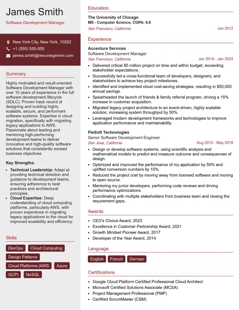
Check Out Resume Samples at ResumeGemini

Check Out Resume Samples at ResumeGemini

Check Out Resume Samples at ResumeGemini

Check Out Resume Samples at ResumeGemini

Check Out Resume Samples at ResumeGemini
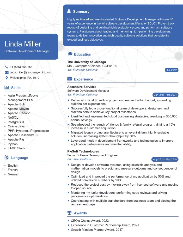
Check Out Resume Samples at ResumeGemini

Check Out Resume Samples at ResumeGemini
Explore more articles
Users Rating of Our Blogs
Share Your Experience
We value your feedback! Please rate our content and share your thoughts (optional).
What Readers Say About Our Blog
To the interviewgemini.com Webmaster.
Very helpful and content specific questions to help prepare me for my interview!
Thank you
To the interviewgemini.com Webmaster.
This was kind of a unique content I found around the specialized skills. Very helpful questions and good detailed answers.
Very Helpful blog, thank you Interviewgemini team.